The central nervous system (CNS) is the part of the nervous system consisting of the brain and spinal cord, the retina and optic nerve, and the olfactory nerve and epithelia. The CNS is so named because the brain integrates the received information and coordinates and influences the activity of all parts of the bodies of bilaterally symmetric and triploblastic animals—that is, all multicellular animals except sponges and diploblasts. It is a structure composed of nervous tissue positioned along the rostral to caudal axis of the body and may have an enlarged section at the rostral end which is a brain. Only arthropods, cephalopods and vertebrates have a true brain, though precursor structures exist in onychophorans, gastropods and lancelets.

The medulla oblongata or simply medulla is a long stem-like structure which makes up the lower part of the brainstem. It is anterior and partially inferior to the cerebellum. It is a cone-shaped neuronal mass responsible for autonomic (involuntary) functions, ranging from vomiting to sneezing. The medulla contains the cardiac, respiratory, vomiting and vasomotor centers, and therefore deals with the autonomic functions of breathing, heart rate and blood pressure as well as the sleep–wake cycle.

The pons is part of the brainstem that in humans and other mammals, lies inferior to the midbrain, superior to the medulla oblongata and anterior to the cerebellum.
The brainstem is the stalk-like part of the brain that interconnects the cerebrum and diencephalon with the spinal cord. In the human brain, the brainstem is composed of the midbrain, the pons, and the medulla oblongata. The midbrain is continuous with the thalamus of the diencephalon through the tentorial notch.

The vestibulocochlear nerve or auditory vestibular nerve, also known as the eighth cranial nerve, cranial nerve VIII, or simply CN VIII, is a cranial nerve that transmits sound and equilibrium (balance) information from the inner ear to the brain. Through olivocochlear fibers, it also transmits motor and modulatory information from the superior olivary complex in the brainstem to the cochlea.

In neuroanatomy, the ventricular system is a set of four interconnected cavities known as cerebral ventricles in the brain. Within each ventricle is a region of choroid plexus which produces the circulating cerebrospinal fluid (CSF). The ventricular system is continuous with the central canal of the spinal cord from the fourth ventricle, allowing for the flow of CSF to circulate.

The brain is the central organ of the human nervous system, and with the spinal cord makes up the central nervous system. The brain consists of the cerebrum, the brainstem and the cerebellum. It controls most of the activities of the body, processing, integrating, and coordinating the information it receives from the sense organs, and making decisions as to the instructions sent to the rest of the body. The brain is contained in, and protected by, the skull bones of the head.

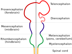
In the human brain, the diencephalon is a division of the forebrain. It is situated between the telencephalon and the midbrain. The diencephalon has also been known as the tweenbrain in older literature. It consists of structures that are on either side of the third ventricle, including the thalamus, the hypothalamus, the epithalamus and the subthalamus.

The fourth ventricle is one of the four connected fluid-filled cavities within the human brain. These cavities, known collectively as the ventricular system, consist of the left and right lateral ventricles, the third ventricle, and the fourth ventricle. The fourth ventricle extends from the cerebral aqueduct to the obex, and is filled with cerebrospinal fluid (CSF).
In the vertebrate embryo, a rhombomere is a transiently divided segment of the developing neural tube, within the hindbrain region in the area that will eventually become the rhombencephalon. The rhombomeres appear as a series of slightly constricted swellings in the neural tube, caudal to the cephalic flexure. In human embryonic development, the rhombomeres are present by day 29.

The pontine tegmentum, or dorsal pons, is located within the brainstem, and is one of two parts of the pons, the other being the ventral pons or basilar part of the pons. The pontine tegmentum can be defined in contrast to the basilar pons: basilar pons contains the corticospinal tract running craniocaudally and can be considered the rostral extension of the ventral medulla oblongata; however, basilar pons is distinguished from ventral medulla oblongata in that it contains additional transverse pontine fibres that continue laterally to become the middle cerebellar peduncle. The pontine tegmentum is all the material dorsal from the basilar pons to the fourth ventricle. Along with the dorsal surface of the medulla, it forms part of the rhomboid fossa – the floor of the fourth ventricle.

The posterior cranial fossa is the part of the cranial cavity located between the foramen magnum, and tentorium cerebelli. It is formed by the sphenoid bones, temporal bones, and occipital bone. It lodges the cerebellum, and parts of the brainstem.

The metencephalon is the embryonic part of the hindbrain that differentiates into the pons and the cerebellum. It contains a portion of the fourth ventricle and the trigeminal nerve, abducens nerve, facial nerve, and a portion of the vestibulocochlear nerve.

The cerebellopontine angle (CPA) is located between the cerebellum and the pons. The cerebellopontine angle is the site of the cerebellopontine angle cistern.

Cerebellar peduncles connect the cerebellum to the brain stem. There are six cerebellar peduncles in total, three on each side:
The middle cerebellar peduncle is a paired structure of the brain. It connects the pons to the cerebellum, with fibres originating from the pontine nucleus and travelling to the opposite hemisphere of the cerebellar cortex. It is supplied by the anterior inferior cerebellar artery (AICA) and branches from the basilar artery. It conveys information from the cerebrum and the pons to the cerebellum.

Homeobox protein GBX-2 is a protein that in humans is encoded by the GBX2 gene.

The anatomy of the cerebellum can be viewed at three levels. At the level of gross anatomy, the cerebellum consists of a tightly folded and crumpled layer of cortex, with white matter underneath, several deep nuclei embedded in the white matter, and a fluid-filled ventricle in the middle. At the intermediate level, the cerebellum and its auxiliary structures can be broken down into several hundred or thousand independently functioning modules or compartments known as microzones. At the microscopic level, each module consists of the same small set of neuronal elements, laid out with a highly stereotyped geometry.

The rhombic lip is a posterior section of the developing metencephalon which can be recognized transiently within the vertebrate embryo. It extends posteriorly from the roof of the fourth ventricle to dorsal neuroepithelial cells. The rhombic lip can be divided into eight structural units based on rhombomeres 1-8 (r1-r8), which can be recognized at early stages of hindbrain development. Producing granule cells and five brainstem nuclei, the rhombic lip plays an important role in developing a complex cerebellar neural system.

Three flexures form in the part of the embryonic neural tube that develops into the brain. At four weeks gestational age in the human embryo, the neural tube has developed at the cranial end into three swellings – the primary brain vesicles. The space into which the cranial part of the neural tube is developing is limited. This limitation causes the neural tube to bend, or flex, at two ventral flexures – the rostral cephalic flexure, and the caudal cervical flexure. It also bends dorsally into the pontine flexure. These flexures have formed by the time that the primary brain vesicles have developed into five secondary brain vesicles in the fifth week.