
The sense of balance or equilibrioception is the perception of balance and spatial orientation. It helps prevent humans and nonhuman animals from falling over when standing or moving. Equilibrioception is the result of a number of sensory systems working together; the eyes, the inner ears, and the body's sense of where it is in space (proprioception) ideally need to be intact.

The medulla oblongata or simply medulla is a long stem-like structure which makes up the lower part of the brainstem. It is anterior and partially inferior to the cerebellum. It is a cone-shaped neuronal mass responsible for autonomic (involuntary) functions, ranging from vomiting to sneezing. The medulla contains the cardiac, respiratory, vomiting and vasomotor centers, and therefore deals with the autonomic functions of breathing, heart rate and blood pressure as well as the sleep–wake cycle.
Articles related to anatomy include:
The brainstem is the stalk-like part of the brain that interconnects the cerebrum and diencephalon with the spinal cord. In the human brain, the brainstem is composed of the midbrain, the pons, and the medulla oblongata. The midbrain is continuous with the thalamus of the diencephalon through the tentorial notch.

In anatomy, the extrapyramidal system is a part of the motor system network causing involuntary actions. The system is called extrapyramidal to distinguish it from the tracts of the motor cortex that reach their targets by traveling through the pyramids of the medulla. The pyramidal tracts may directly innervate motor neurons of the spinal cord or brainstem, whereas the extrapyramidal system centers on the modulation and regulation of anterior (ventral) horn cells.

The midbrain or mesencephalon is the rostral-most portion of the brainstem connecting the diencephalon and cerebrum with the pons. It consists of the cerebral peduncles, tegmentum, and tectum.
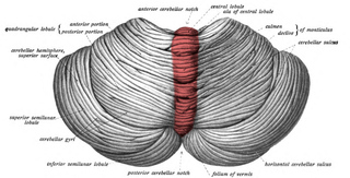
The cerebellar vermis is located in the medial, cortico-nuclear zone of the cerebellum, which is in the posterior fossa of the cranium. The primary fissure in the vermis curves ventrolaterally to the superior surface of the cerebellum, dividing it into anterior and posterior lobes. Functionally, the vermis is associated with bodily posture and locomotion. The vermis is included within the spinocerebellum and receives somatic sensory input from the head and proximal body parts via ascending spinal pathways.
The spinocerebellar tract is a nerve tract originating in the spinal cord and terminating in the same side (ipsilateral) of the cerebellum.

There are four deep cerebellar nuclei embedded in the white matter of the medullary centre. The nuclei are the fastigial, globose, emboliform, and dentate nuclei.

The upper part of the posterior district of the medulla oblongata is occupied by the inferior cerebellar peduncle, a thick rope-like strand situated between the lower part of the fourth ventricle and the roots of the glossopharyngeal and vagus nerves.
The dentate nucleus is a cluster of neurons, or nerve cells, in the central nervous system that has a dentate – tooth-like or serrated – edge. It is located within the deep white matter of each cerebellar hemisphere, and it is the largest single structure linking the cerebellum to the rest of the brain. It is the largest and most lateral, or farthest from the midline, of the four pairs of deep cerebellar nuclei, the others being the globose and emboliform nuclei, which together are referred to as the interposed nucleus, and the fastigial nucleus. The dentate nucleus is responsible for the planning, initiation and control of voluntary movements. The dorsal region of the dentate nucleus contains output channels involved in motor function, which is the movement of skeletal muscle, while the ventral region contains output channels involved in nonmotor function, such as conscious thought and visuospatial function.

The globose nucleus is one of the deep cerebellar nuclei. It is located medial to the emboliform nucleus, and lateral to the fastigial nucleus. The globose nucleus and emboliform nucleus are known collectively as the interposed nuclei.
The interposed nuclei are the globose and emboliform nucleus or either side, collectively. It is located in the roof of the fourth ventricle, lateral to the fastigial nucleus.

The flocculus is a small lobe of the cerebellum at the posterior border of the middle cerebellar peduncle anterior to the biventer lobule. Like other parts of the cerebellum, the flocculus is involved in motor control. It is an essential part of the vestibulo-ocular reflex, and aids in the learning of basic motor skills in the brain.

The vestibular nuclei (VN) are the cranial nuclei for the vestibular nerve located in the brainstem.

Cerebellar peduncles connect the cerebellum to the brain stem. There are six cerebellar peduncles in total, three on each side:

The lateral vestibular nucleus is the continuation upward and lateralward of the principal nucleus, and in it terminate many of the ascending branches of the vestibular nerve.
The reticulotegmental nucleus, tegmental pontine reticular nucleus is an area within the floor of the pons, in the brain stem. This area is known to affect the cerebellum with its axonal projections.
The anatomy of the cerebellum can be viewed at three levels. At the level of gross anatomy, the cerebellum consists of a tightly folded and crumpled layer of cortex, with white matter underneath, several deep nuclei embedded in the white matter, and a fluid-filled ventricle in the middle. At the intermediate level, the cerebellum and its auxiliary structures can be broken down into several hundred or thousand independently functioning modules or compartments known as microzones. At the microscopic level, each module consists of the same small set of neuronal elements, laid out with a highly stereotyped geometry.
