The motor system is the set of central and peripheral structures in the nervous system that support motor functions, i.e. movement. Peripheral structures may include skeletal muscles and neural connections with muscle tissues. Central structures include cerebral cortex, brainstem, spinal cord, pyramidal system including the upper motor neurons, extrapyramidal system, cerebellum, and the lower motor neurons in the brainstem and the spinal cord.
Pronator quadratus is a square-shaped muscle on the distal forearm that acts to pronate the hand.

The brainstem is the stalk-like part of the brain that interconnects the cerebrum and diencephalon with the spinal cord. In the human brain, the brainstem is composed of the midbrain, the pons, and the medulla oblongata. The midbrain is continuous with the thalamus of the diencephalon through the tentorial notch.

In anatomy, the extrapyramidal system is a part of the motor system network causing involuntary actions. The system is called extrapyramidal to distinguish it from the tracts of the motor cortex that reach their targets by traveling through the pyramids of the medulla. The pyramidal tracts may directly innervate motor neurons of the spinal cord or brainstem, whereas the extrapyramidal system centers on the modulation and regulation of anterior (ventral) horn cells.
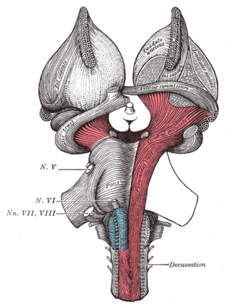
The cerebral peduncles are the two stalks that attach the cerebrum to the brainstem. They are structures at the front of the midbrain which arise from the ventral pons and contain the large ascending (sensory) and descending (motor) nerve tracts that run to and from the cerebrum from the pons. Mainly, the three common areas that give rise to the cerebral peduncles are the cerebral cortex, the spinal cord and the cerebellum. The region includes the tegmentum, crus cerebri and pretectum. By this definition, the cerebral peduncles are also known as the basis pedunculi, while the large ventral bundle of efferent fibers is referred to as the cerebral crus or the pes pedunculi.

The pyramidal tracts include both the corticobulbar tract and the corticospinal tract. These are aggregations of efferent nerve fibers from the upper motor neurons that travel from the cerebral cortex and terminate either in the brainstem (corticobulbar) or spinal cord (corticospinal) and are involved in the control of motor functions of the body.

In neuroanatomy, the corticobulbartract is a two-neuron white matter motor pathway connecting the motor cortex in the cerebral cortex to the medullary pyramids, which are part of the brainstem's medulla oblongata region, and are primarily involved in carrying the motor function of the non-oculomotor cranial nerves. The corticobulbar tract is one of the pyramidal tracts, the other being the corticospinal tract.

The red nucleus or nucleus ruber is a structure in the rostral midbrain involved in motor coordination. The red nucleus is pale pink, which is believed to be due to the presence of iron in at least two different forms: hemoglobin and ferritin. The structure is located in the tegmentum of the midbrain next to the substantia nigra and comprises caudal magnocellular and rostral parvocellular components. The red nucleus and substantia nigra are subcortical centers of the extrapyramidal motor system.

The globose nucleus is one of the deep cerebellar nuclei. It is located medial to the emboliform nucleus, and lateral to the fastigial nucleus. The globose nucleus and emboliform nucleus are known collectively as the interposed nuclei.
Decerebration is the elimination of cerebral brain function in an animal by removing the cerebrum, cutting across the brain stem, or severing certain arteries in the brain stem.

The vestibulospinal tract is a neural tract in the central nervous system. Specifically, it is a component of the extrapyramidal system and is classified as a component of the medial pathway. Like other descending motor pathways, the vestibulospinal fibers of the tract relay information from nuclei to motor neurons. The vestibular nuclei receive information through the vestibulocochlear nerve about changes in the orientation of the head. The nuclei relay motor commands through the vestibulospinal tract. The function of these motor commands is to alter muscle tone, extend, and change the position of the limbs and head with the goal of supporting posture and maintaining balance of the body and head.
The lateral corticospinal tract is the largest part of the corticospinal tract. It extends throughout the entire length of the spinal cord, and on transverse section appears as an oval area in front of the posterior column and medial to the posterior spinocerebellar tract.
The anterior corticospinal tract is a small bundle of descending fibers that connect the cerebral cortex to the spinal cord. Descending tracts are pathways by which motor signals are sent from upper motor neurons in the brain to lower motor neurons which then directly innervate muscle to produce movement. The anterior corticospinal tract is usually small, varying inversely in size with the lateral corticospinal tract, which is the main part of the corticospinal tract.

Alpha (α) motor neurons (also called alpha motoneurons), are large, multipolar lower motor neurons of the brainstem and spinal cord. They innervate extrafusal muscle fibers of skeletal muscle and are directly responsible for initiating their contraction. Alpha motor neurons are distinct from gamma motor neurons, which innervate intrafusal muscle fibers of muscle spindles.
The reticulospinal tracts, also known as the descending or anterior reticulospinal tracts, are extrapyramidal motor tracts that descend from the reticular formation in two tracts to act on the motor neurons supplying the trunk and proximal limb flexors and extensors. The reticulospinal tracts are involved mainly in locomotion and postural control, although they do have other functions as well. The descending reticulospinal tracts are one of four major cortical pathways to the spinal cord for musculoskeletal activity. The reticulospinal tracts work with the other three pathways to give a coordinated control of movement, including delicate manipulations. The four pathways can be grouped into two main system pathways – a medial system and a lateral system. The medial system includes the reticulospinal pathway and the vestibulospinal pathway, and this system provides control of posture. The corticospinal and the rubrospinal tract pathways belong to the lateral system which provides fine control of movement.

The spinal cord is a long, thin, tubular structure made up of nervous tissue that extends from the medulla oblongata in the brainstem to the lumbar region of the vertebral column (backbone) of vertebrate animals. The center of the spinal cord is hollow and contains a structure called the central canal, which contains cerebrospinal fluid. The spinal cord is also covered by meninges and enclosed by the neural arches. Together, the brain and spinal cord make up the central nervous system.
The hypothalamospinal tract is a nerve tract that arises mainly from the paraventricular nucleus of hypothalamus, and lateral and posterior areas of the hypothalamus. The tract descends through the periaqueductal gray and adjacent reticular formation. It is found in the dorsolateral quadrant of the lateral funiculus, in the lateral tegmentum of the medulla, pons and midbrain.

Central pattern generators are biological neural networks organized to produce any rhythmic output without requiring a rhythmic input. In mammals, locomotor CPGs are organized in the lumbar and cervical segments of the spinal cord, and are used to control rhythmic muscle output in the arms and legs. Certain areas of the brain initiate the descending neural pathways that ultimately control and modulate the CPG signals. In addition to this direct control, there exist different feedback loops that coordinate the limbs for efficient locomotion and allow for the switching of gaits under appropriate circumstances.

The corticospinal tract is a white matter motor pathway starting at the cerebral cortex that terminates on lower motor neurons and interneurons in the spinal cord, controlling movements of the limbs and trunk. There are more than one million neurons in the corticospinal tract, and they become myelinated usually in the first two years of life.

