
The foramen magnum is a large oval opening (foramen) in the occipital bone of the skull in humans and various other animals. It is one of the several oval or circular openings (foramina) in the base of the skull. The spinal cord, an extension of the medulla, passes through the foramen magnum as it exits the cranial cavity. Apart from the transmission of the medulla oblongata and its membranes, the foramen magnum transmits the vertebral arteries, the anterior and posterior spinal arteries, the tectorial membranes and alar ligaments. It also transmits the spinal component of the accessory nerve into the skull.

In human anatomy, the anterior spinal artery is the artery that supplies the anterior portion of the spinal cord. It arises from branches of the vertebral arteries and courses along the anterior aspect of the spinal cord. It is reinforced by several contributory arteries, especially the artery of Adamkiewicz.

The vestibular nuclei (VN) are the cranial nuclei for the vestibular nerve.
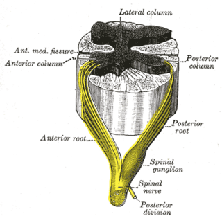
The anterior median fissure of the spinal cord has an average depth of about 3 mm, but this is increased in the lower part of the spinal cord.

A funiculus is a small bundle of axons, enclosed by the perineurium. A small nerve may consist of a single funiculus, but a larger nerve will have several funiculi collected together into larger bundles known as fasciculi. Fasciculi are bound together in a common membrane, the epineurium.

The posterior spinal artery arises from the vertebral artery in 25% of humans or the posterior inferior cerebellar artery in 75% of humans, adjacent to the medulla oblongata. It supplies the grey and white posterior columns of the spinal cord.
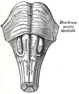
The medullary pyramids are paired white matter structures of the brainstem's medulla oblongata that contain motor fibers of the corticospinal and corticobulbar tracts – known together as the pyramidal tracts. The lower limit of the pyramids is marked when the fibers cross (decussate).

The spinal veins are situated in the pia mater and form a minute, tortuous, venous plexus.
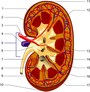
The renal lobe is a portion of a kidney consisting of a renal pyramid and the renal cortex above it.

The arcuate arteries of the kidney are vessels of the renal circulation. They are located at the border of the renal cortex and renal medulla.
The medulla of ovary is a highly vascular stroma in the center of the ovary. It forms from embryonic mesenchyme and contains blood vessels, lymphatic vessels, and nerves.

The sensory decussation or decussation of the lemniscus is a decussation or crossover of axons from the gracile nucleus and cuneate nucleus, which are responsible for fine touch, proprioception and two-point discrimination of the body. The fibres of this decussation are called the internal arcuate fibres and are found at the superior aspect of the closed medulla superior to the motor decussation. It is part of the second neuron in the posterior column–medial lemniscus pathway.
The cells of the dorsal nucleus of vagus nerve are spindle-shaped, like those of the posterior column of the spinal cord, and the nucleus is usually considered as representing the base of the posterior column. It measures about 2 cm. in length, and in the lower, closed part of the medulla oblongata is situated behind the dorsal motor nucleus of the vagus; whereas in the upper, open part it lies lateral to that nucleus, and corresponds to an eminence, named the vagal trigone, in the rhomboid fossa.

The anterior median fissure contains a fold of pia mater, and extends along the entire length of the medulla oblongata: It ends at the lower border of the pons in a small triangular expansion, termed the foramen cecum.

The posterior median sulcus of medulla oblongata is a narrow groove; and exists only in the closed part of the medulla oblongata; it becomes gradually shallower from below upward, and finally ends about the middle of the medulla oblongata, where the central canal expands into the cavity of the fourth ventricle.

In the upper part of the medulla oblongata, the hypoglossal nucleus approaches the rhomboid fossa, where it lies close to the middle line, under an eminence named the hypoglossal trigone. It is a slight elevation in the floor of the inferior recess of the fourth ventricle, beneath which is the nucleus of origin of the twelfth cranial nerve.

The medial vestibular nucleus is one of the vestibular nuclei. It is located in the medulla oblongata.

On the superior surface of cerebellum, the vermis protrudes above the level of the hemispheres, but on the inferior surface it is sunk almost out of sight in the bottom of a deep depression between them; this depression is called the vallecula of the cerebellum, and lodges the posterior part of the medulla oblongata and the inferior vermis, which consists of the tuber vermis, pyramid, uvula and nodule.
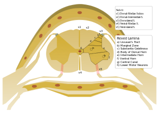
On either side of the posterior median sulcus, and at a short distance from it, the posterior nerve roots are attached along a vertical furrow named the posterolateral sulcus. The portion of the medulla spinalis which lies between this and the posterior median sulcus is named the posterior funiculus.
The public domain consists of all the creative works to which no exclusive intellectual property rights apply. Those rights may have expired, been forfeited, expressly waived, or may be inapplicable.

Gray's Anatomy is an English language textbook of human anatomy originally written by Henry Gray and illustrated by Henry Vandyke Carter. Earlier editions were called Anatomy: Descriptive and Surgical, Anatomy of the Human Body and Gray's Anatomy: Descriptive and Applied, but the book's name is commonly shortened to, and later editions are titled, Gray's Anatomy. The book is widely regarded as an extremely influential work on the subject, and has continued to be revised and republished from its initial publication in 1858 to the present day. The latest edition of the book, the 41st, was published in September 2015.


