Hippocampus anatomy

Last updated
Human hippocampus. Gray739-emphasizing-hippocampus.png
Human hippocampus.
Nissl-stained coronal section of the brain of a macaque monkey, showing hippocampus (circled). Brainmaps-macaque-hippocampus.jpg
Nissl-stained coronal section of the brain of a macaque monkey, showing hippocampus (circled).

Hippocampus anatomy describes the physical aspects and properties of the hippocampus, a neural structure in the medial temporal lobe of the brain. It has a distinctive, curved shape that has been likened to the sea-horse monster of Greek mythology and the ram's horns of Amun in Egyptian mythology. This general layout holds across the full range of mammalian species, from hedgehog to human, although the details vary. For example, in the rat, the two hippocampi look similar to a pair of bananas, joined at the stems. In primate brains, including humans, the portion of the hippocampus near the base of the temporal lobe is much broader than the part at the top. Due to the three-dimensional curvature of this structure, two-dimensional sections such as shown are commonly seen. Neuroimaging pictures can show a number of different shapes, depending on the angle and location of the cut.

Contents

Shape of human hippocampus and associated structures. Gray747.png
Shape of human hippocampus and associated structures.

Topologically, the surface of a cerebral hemisphere can be regarded as a sphere with an indentation where it attaches to the midbrain. The structures that line the edge of the hole collectively make up the so-called limbic system (Latin limbus = border), with the hippocampus lining the posterior edge of this hole. These limbic structures include the hippocampus, cingulate cortex, olfactory cortex, and amygdala. Paul MacLean once suggested, as part of his triune brain theory, that the limbic structures constitute the neural basis of emotion. While most neuroscientists no longer believe in the concept of a unified "limbic system", these regions are highly interconnected and do interact with one another.[ citation needed ]

Basic hippocampal circuit

Basic circuit of the hippocampus, shown using a modified drawing by Ramon y Cajal. DG: dentate gyrus. Sub: subiculum. EC: entorhinal cortex CajalHippocampus (modified).png
Basic circuit of the hippocampus, shown using a modified drawing by Ramon y Cajal. DG: dentate gyrus. Sub: subiculum. EC: entorhinal cortex

Starting at the dentate gyrus and working inward along the S-curve of the hippocampus means traversing a series of narrow zones. The first of these, the dentate gyrus (DG), is actually a separate structure, a tightly packed layer of small granule cells wrapped around the end of the hippocampus proper, forming a pointed wedge in some cross-sections, a semicircle in others. Next come a series of Cornu Ammonis areas: first CA4 (which underlies the dentate gyrus), then CA3, then a very small zone called CA2, then CA1. The CA areas are all filled with densely packed pyramidal cells similar to those found in the neocortex. After CA1 comes an area called the subiculum. After this comes a pair of ill-defined areas called the presubiculum and parasubiculum, then a transition to the cortex proper (mostly the entorhinal area of the cortex). Most anatomists use the term "hippocampus proper" to refer to the four CA fields, and hippocampal formation to refer to the hippocampus proper plus dentate gyrus and subiculum. [1]

The major signaling pathways flow through the hippocampus and combine to form a loop. Most external input comes from the adjoining entorhinal cortex, via the axons of the so-called perforant path. These axons arise from layer 2 of the entorhinal cortex (EC), and terminate in the dentate gyrus and CA3. There is also a distinct pathway from layer 3 of the EC directly to CA1, often referred to as the temporoammonic or TA-CA1 pathway. Granule cells of the DG send their axons (called "mossy fibers") to CA3. Pyramidal cells of CA3 send their axons to CA1. Pyramidal cells of CA1 send their axons to the subiculum and deep layers of the EC. Subicular neurons send their axons mainly to the EC. The perforant path-to-dentate gyrus-to-CA3-to-CA1 was called the trisynaptic circuit by Per Andersen, who noted that thin slices could be cut out of the hippocampus perpendicular to its long axis, in a way that preserves all of these connections. This observation was the basis of his lamellar hypothesis, which proposed that the hippocampus can be thought of as a series of parallel strips, operating in a functionally independent way. [2] The lamellar concept is still sometimes considered to be a useful organizing principle, but more recent data, showing extensive longitudinal connections within the hippocampal system, have required it to be substantially modified. [3]

Perforant path input from EC layer II enters the dentate gyrus and is relayed to region CA3 (and to mossy cells, located in the hilus of the dentate gyrus, which then send information to distant portions of the dentate gyrus where the cycle is repeated). Region CA3 combines this input with signals from EC layer II and sends extensive connections within the region and also sends connections to strata radiatum and oriens of ipsilateral and contralateral CA1 regions through a set of fibers called the Schaffer collaterals, and commissural pathway, respectively. [4] [5] [6] Region CA1 receives input from the CA3 subfield, EC layer III and the nucleus reuniens of the thalamus (which project only to the terminal apical dendritic tufts in the stratum lacunosum-moleculare). In turn, CA1 projects to the subiculum as well as sending information along the aforementioned output paths of the hippocampus. The subiculum is the final stage in the pathway, combining information from the CA1 projection and EC layer III to also send information along the output pathways of the hippocampus.

The hippocampus also receives a number of subcortical inputs. In Macaca fascicularis , these inputs include the amygdala (specifically the anterior amygdaloid area, the basolateral nucleus, and the periamygdaloid cortex), the medial septum and the diagonal band of Broca, the claustrum, the substantia innominata and the basal nucleus of Meynert, the thalamus (including the anterior nuclear complex, the laterodorsal nucleus, the paraventricular and parataenial nuclei, the nucleus reuniens, and the nucleus centralis medialis), the lateral preoptic and lateral hypothalamic areas, the supramammillary and retromammillary regions, the ventral tegmental area, the tegmental reticular fields, the raphe nuclei (the nucleus centralis superior and the dorsal raphe nucleus), the nucleus reticularis tegementi pontis, the periaqueductal gray, the dorsal tegmental nucleus, and the locus coeruleus. The hippocampus also receives direct monosynaptic projections from the cerebellar fastigial nucleus. [7]

Major fiber systems in the rat

Angular bundle

These fibers start from the ventral part of entorhinal cortex (EC) and contain commissural (EC◀▶Hippocampus) and Perforant path (excitatory EC▶CA1, and inhibitory EC◀▶CA2 [8] ) fibers. They travel along the septotemporal axis of the hippocampus. Perforant path fibers, as the name suggests, perforate subiculum before going to the hippocampus (CA fields) and dentate gyrus. [9]

Fimbria-fornix pathway

Coronal section of inferior horn of lateral ventricle. (Fimbria labeled at center left and alveus to the right). Gray749.png
Coronal section of inferior horn of lateral ventricle. (Fimbria labeled at center left and alveus to the right).

Fimbria-fornix fibers are the hippocampal and subicular gateway to and fromsubcortical brain regions. [10] [11] Different parts of this system are given different names:

At the circuit level, the alveus contains axonal fibers from the DG and from Pyramidal neurons of CA3, CA2, CA1 and subiculum (CA1 ▶ subiculum and CA1 ▶ entorhinal projections) that collect in the temporal hippocampus to form the fimbria/fornix, one of the major outputs of the hippocampus. [12] [13] [14] [15] [16] In the rat, some medial and lateral entorhinal axons (entorhinal ▶ CA1 projection) pass through alveus towards the CA1 stratum lacunosum moleculare without making a significant number of en passant boutons on other CA1 layers (Temporoammonic alvear pathway). [13] [17] Contralateral entorhinal ▶ CA1 projections almost exclusively pass through alveus. The more septal the more ipsilateral entorhinal-CA1 projections that take alvear pathway (instead of perforant path). [18] Although subiculum sends axonal projections to alveus, subiculum ▶ CA1 projection passes through strata oriens and moleculare of subiculum and CA1. [19] Cholinergic and GABAergic projections from MS-DBB to CA1 also pass through Fimbria. [20] Fimbria stimulation leads to cholinergic excitation of CA1 O-LMR cells. [21]

It is also known that extracellular stimulation of fimbria stimulates CA3 Pyramidal cells antidromically and orthodromically, but it has no impact on dentate granule cells. [22] Each CA1 Pyramidal cell also sends an axonal branch to fimbria. [23] [24]

Hippocampal commissures

Hilar mossy cells and CA3 Pyramidal cells are the main origins of hippocampal commissural fibers. They pass through hippocampal commissures to reach contralateral regions of hippocampus. Hippocampal commissures have dorsal and ventral segments. Dorsal commissural fibers consists mainly of entorhinal and presubicular fibers to or from the hippocampus and dentate gyrus. [9] As a rule of thumb, one could say that each cytoarchitectonic field that contributes to the commissural projection also has a parallel associational fiber that terminates in the ipsilateral hippocampus. [25] The inner molecular layer of dentate gyrus (dendrites of both granule cells and GABAergic interneurons) receives a projection that has both associational and commissural fibers mainly from hilar mossy cells and to some extent from CA3c Pyramidal cells. Because this projection fibers originate from both ipsilateral and contralateral sides of hippocampus they are called associational/commissural projections. In fact, each mossy cell innervates both the ipsilateral and contralateral dentate gyrus. The well known trisynaptic circuit of the hippocampus spans mainly horizontally along the hippocampus. However, associational/commissural fibers, like CA2 Pyramidal cell associational projections, span mainly longitudinally (dorsoventrally) along the hippocampus. [26] [27] Commissural fibers that originate from CA3 Pyramidal cells go to CA3, CA2 and CA1 regions. Like mossy cells, a single CA3 Pyramidal cell contributes to both commissural and associational fibers, and they terminate on both principal cells and interneurons. [28] [29] A weak commissural projection connects both CA1 regions together. Subiculum has no commissural inputs or outputs. In comparison with rodents, hippocampal commissural connections are much less abundant in the monkey and humans. [30] Although excitatory cells are the main contributors to commissural pathways, a GABAergic component has been reported among their terminals which were traced back to hilus as origin. [31] Stimulation of commissural fibers stimulates DG hilar perforant path-associated (HIPP) and CA3 trilaminar cells antidromically. [32]

Hippocampal cells and layers

Photograph of hippocampal regions in a rat brain. DG: Dentate gyrus. HippocampalRegions.jpg
Photograph of hippocampal regions in a rat brain. DG: Dentate gyrus.
Schematic showing regions of the hippocampus proper in relation to other structures. Hippocampus (brain).jpg
Schematic showing regions of the hippocampus proper in relation to other structures.

Hippocampus proper

The hippocampus proper is composed of a number of subfields. Though terminology varies among authors, the terms most frequently used are dentate gyrus and the cornu ammonis (literally "Ammon's horn", abbreviated CA). The dentate gyrus contains the fascia dentata and the hilus, while the CA is differentiated into subfields CA1, CA2, CA3, and CA4.

However, the region known as CA4 is in fact the "deep, polymorphic layer of the dentate gyrus" [33] (as clarified by Theodor Blackstad (1956) [34] and by David Amaral (1978)). [35]

Cut in cross section, the hippocampus is a C-shaped structure that resembles a ram's horns. The name cornu ammonis refers to the Egyptian deity Amun, who has the head of a ram. The horned appearance of the hippocampus is caused by cell density differentials and varying degrees of neuronal fibers.

In rodents, the hippocampus is positioned so that, roughly, one end is near the top of the head (the dorsal or septal end) and one end near the bottom of the head (the ventral or temporal end). As shown in the figure, the structure itself is curved and subfields or regions are defined along the curve, from CA4 through CA1 (only CA3 and CA1 are labeled). The CA regions are also structured depthwise in clearly defined strata (or layers):

Dentate gyrus

The dentate gyrus is composed of a similar series of strata:

An up-to-date knowledge base of hippocampal formation neuronal types, their biomarker profile, active and passive electrophysiological parameters, and connectivity is supported at the Hippocampome website. [36]

Related Research Articles

<span class="mw-page-title-main">Entorhinal cortex</span> Area of the temporal lobe of the brain

The entorhinal cortex (EC) is an area of the brain's allocortex, located in the medial temporal lobe, whose functions include being a widespread network hub for memory, navigation, and the perception of time. The EC is the main interface between the hippocampus and neocortex. The EC-hippocampus system plays an important role in declarative (autobiographical/episodic/semantic) memories and in particular spatial memories including memory formation, memory consolidation, and memory optimization in sleep. The EC is also responsible for the pre-processing (familiarity) of the input signals in the reflex nictitating membrane response of classical trace conditioning; the association of impulses from the eye and the ear occurs in the entorhinal cortex.

<span class="mw-page-title-main">Hippocampus</span> Vertebrate brain region involved in memory consolidation

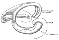
The hippocampus is a major component of the brain of humans and other vertebrates. Humans and other mammals have two hippocampi, one in each side of the brain. The hippocampus is part of the limbic system, and plays important roles in the consolidation of information from short-term memory to long-term memory, and in spatial memory that enables navigation. The hippocampus is located in the allocortex, with neural projections into the neocortex, in humans as well as other primates. The hippocampus, as the medial pallium, is a structure found in all vertebrates. In humans, it contains two main interlocking parts: the hippocampus proper, and the dentate gyrus.

<span class="mw-page-title-main">Dentate gyrus</span> Region of the hippocampus in the brain

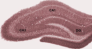
The dentate gyrus (DG) is part of the hippocampal formation in the temporal lobe of the brain, which also includes the hippocampus and the subiculum. The dentate gyrus is part of the hippocampal trisynaptic circuit and is thought to contribute to the formation of new episodic memories, the spontaneous exploration of novel environments and other functions.

<span class="mw-page-title-main">Neural pathway</span> Connection formed between neurons that allows neurotransmission

In neuroanatomy, a neural pathway is the connection formed by axons that project from neurons to make synapses onto neurons in another location, to enable neurotransmission. Neurons are connected by a single axon, or by a bundle of axons known as a nerve tract, or fasciculus. Shorter neural pathways are found within grey matter in the brain, whereas longer projections, made up of myelinated axons, constitute white matter.

Schaffer collaterals are axon collaterals given off by CA3 pyramidal cells in the hippocampus. These collaterals project to area CA1 of the hippocampus and are an integral part of memory formation and the emotional network of the Papez circuit, and of the hippocampal trisynaptic loop. It is one of the most studied synapses in the world and named after the Hungarian anatomist-neurologist Károly Schaffer.

An apical dendrite is a dendrite that emerges from the apex of a pyramidal cell. Apical dendrites are one of two primary categories of dendrites, and they distinguish the pyramidal cells from spiny stellate cells in the cortices. Pyramidal cells are found in the prefrontal cortex, the hippocampus, the entorhinal cortex, the olfactory cortex, and other areas. Dendrite arbors formed by apical dendrites are the means by which synaptic inputs into a cell are integrated. The apical dendrites in these regions contribute significantly to memory, learning, and sensory associations by modulating the excitatory and inhibitory signals received by the pyramidal cells.

<span class="mw-page-title-main">Subiculum</span> Most inferior part of the hippocampal formation

The subiculum is the most inferior component of the hippocampal formation. It lies between the entorhinal cortex and the CA1 subfield of the hippocampus proper.

Theta waves generate the theta rhythm, a neural oscillation in the brain that underlies various aspects of cognition and behavior, including learning, memory, and spatial navigation in many animals. It can be recorded using various electrophysiological methods, such as electroencephalogram (EEG), recorded either from inside the brain or from electrodes attached to the scalp.

<span class="mw-page-title-main">Perforant path</span>

In the brain, the perforant path or perforant pathway provides a connectional route from the entorhinal cortex to all fields of the hippocampal formation, including the dentate gyrus, all CA fields, and the subiculum.

<span class="mw-page-title-main">Median raphe nucleus</span> Brain region having polygonal, fusiform, piriform neurons

The median raphe nucleus, also known as the nucleus raphes medianus (NRM) or superior central nucleus, is a brain region composed of polygonal, fusiform, and piriform neurons, which exists rostral to the nucleus raphes pontis. The MRN is located between the posterior end of the superior cerebellar peduncles and the V. Afferents of the motor nucleus. It is one of two nuclei, the other being the dorsal raphe nucleus (DnR), in the midbrain-pons.

The stratum lucidum of the hippocampus is a layer of the hippocampus between the stratum pyramidale and the stratum radiatum. It is the tract of the mossy fiber projections, both inhibitory and excitatory from the granule cells of the dentate gyrus. One mossy fiber may make up to 37 connections to a single pyramidal cell, and innervate around 12 pyramidal cells on top of that. Any given pyramidal cell in the stratum lucidum may get input from as many as 50 granule cells.

<span class="mw-page-title-main">Mossy fiber (hippocampus)</span> Pathway in the hippocampus

In the hippocampus, the mossy fiber pathway consists of unmyelinated axons projecting from granule cells in the dentate gyrus that terminate on modulatory hilar mossy cells and in Cornu Ammonis area 3 (CA3), a region involved in encoding short-term memory. These axons were first described as mossy fibers by Santiago Ramón y Cajal as they displayed varicosities along their lengths that gave them a mossy appearance. The axons that make up the pathway emerge from the basal portions of the granule cells and pass through the hilus of the dentate gyrus before entering the stratum lucidum of CA3. Granule cell synapses tend to be glutamatergic, though immunohistological data has indicated that some synapses contain neuropeptidergic elements including opiate peptides such as dynorphin and enkephalin. There is also evidence for co-localization of both GABAergic and glutamatergic neurotransmitters within mossy fiber terminals. GABAergic and glutamatergic co-localization in mossy fiber boutons has been observed primarily in the developing hippocampus, but in adulthood, evidence suggests that mossy fiber synapses may alternate which neurotransmitter is released through activity-dependent regulation.

The trisynaptic circuit or trisynaptic loop is a relay of synaptic transmission in the hippocampus. The circuit was initially described by the neuroanatomist Santiago Ramon y Cajal, in the early twentieth century, using the Golgi staining method. After the discovery of the trisynaptic circuit, a series of research has been conducted to determine the mechanisms driving this circuit. Today, research is focused on how this loop interacts with other parts of the brain, and how it influences human physiology and behaviour. For example, it has been shown that disruptions within the trisynaptic circuit lead to behavioural changes in rodent and feline models.

<span class="mw-page-title-main">Fascia dentata</span>

The fascia dentata is the earliest stage of the hippocampal circuit. Its primary input is the perforant path from the superficial layers of entorhinal cortex. Its principal neurons are tiny granule cells which give rise to unmyelinated axons called the mossy fibers which project to the hilus and CA3. The fascia dentata of the rat contains approximately 1,000,000 granule cells. It receives feedback connections from mossy cells in the hilus at distant levels in the septal and temporal directions. The fascia dentata and the hilus together make up the dentate gyrus. As with all regions of the hippocampus, the dentate gyrus also receives GABAergic and cholinergic input from the medial septum and the diagonal band of Broca.

<span class="mw-page-title-main">Granule cell</span> Type of neuron with a very small cell body

The name granule cell has been used for a number of different types of neurons whose only common feature is that they all have very small cell bodies. Granule cells are found within the granular layer of the cerebellum, the dentate gyrus of the hippocampus, the superficial layer of the dorsal cochlear nucleus, the olfactory bulb, and the cerebral cortex.

Sharp waves and ripples (SWRs) are oscillatory patterns produced by extremely synchronised activity of neurons in the mammalian hippocampus and neighbouring regions which occur spontaneously in idle waking states or during NREM sleep. They can be observed with a variety of imaging methods, such as EEG. They are composed of large amplitude sharp waves in local field potential and produced by tens of thousands of neurons firing together within 30–100 ms window. They are some of the most synchronous oscillations patterns in the brain, making them susceptible to pathological patterns such as epilepsy.They have been extensively characterised and described by György Buzsáki and have been shown to be involved in memory consolidation in NREM sleep and the replay of memories acquired during wakefulness.

<span class="mw-page-title-main">Glucocorticoids in hippocampal development</span> HippoCampus

The hippocampus is an area of the brain integral to learning and memory. Removal of this structure can result in the inability to form new memories as most famously demonstrated in a patient referred to as HM. The unique morphology of the hippocampus can be appreciated without the use of special stains and this distinct circuitry has helped further the understanding of neuronal signal potentiation. The following will provide an introduction to hippocampal development with particular focus on the role of glucocorticoid signaling.

<span class="mw-page-title-main">Hippocampus proper</span> Part of the brain of mammals

The hippocampus proper refers to the actual structure of the hippocampus which is made up of three regions or subfields. The subfields CA1, CA2, and CA3 use the initials of cornu Ammonis, an earlier name of the hippocampus.

Early long-term potentiation (E-LTP) is the first phase of long-term potentiation (LTP), a well-studied form of synaptic plasticity, and consists of an increase in synaptic strength. LTP could be produced by repetitive stimulation of the presynaptic terminals, and it is believed to play a role in memory function in the hippocampus, amygdala and other cortical brain structures in mammals.

The supramammillary nucleus (SuM), or supramammillary area, is a thin layer of cells in the brain that lies above the mammillary bodies. It can be considered part of the hypothalamus and diencephalon. The nucleus can be divided into medial and lateral sections. The medial SuM, or SuMM, is made of smaller cells which release dopamine and give input to the lateral septal nucleus. The lateral SuM, or SuML, is made of larger cells that project to the hippocampus.

References

  1. Amaral, D; Lavenex P (2006). "Ch 3. Hippocampal Neuroanatomy". In Andersen P; Morris R; Amaral D; Bliss T; O'Keefe J (eds.). The Hippocampus Book. Oxford University Press. ISBN   978-0-19-510027-3.
  2. Andersen, P; Bliss TVP; Skrede KK (1971). "Lamellar organization of hippocampal excitatory pathways". Exp. Brain Res. 13 (2): 222–238. doi:10.1007/BF00234087. PMID   5570425. S2CID   12075886.
  3. Andersen, P; Soleng AF; Raastad M (2000). "The hippocampal lamella hypothesis revisited". Brain Res. 886 (1–2): 165–171. doi:10.1016/S0006-8993(00)02991-7. PMID   11119694. S2CID   8455285.
  4. Hjorth-Simonsen, A (15 January 1973). "Some intrinsic connections of the hippocampus in the rat: an experimental analysis". The Journal of Comparative Neurology. 147 (2): 145–61. doi:10.1002/cne.901470202. PMID   4118866. S2CID   28989051.
  5. Swanson, LW; Wyss, JM; Cowan, WM (15 October 1978). "An autoradiographic study of the organization of intrahippocampal association pathways in the rat". The Journal of Comparative Neurology. 181 (4): 681–715. doi:10.1002/cne.901810402. PMID   690280. S2CID   30954240.
  6. Laurberg, S (15 April 1979). "Commissural and intrinsic connections of the rat hippocampus". The Journal of Comparative Neurology. 184 (4): 685–708. doi:10.1002/cne.901840405. PMID   422759. S2CID   27256712.
  7. Heath RG, Harper JW (November 1974). "Ascending projections of the cerebellar fastigial nucleus to the hippocampus, amygdala, and other temporal lobe sites: evoked potential and histological studies in monkeys and cats". Exp. Neurol. 45 (2): 268–87. doi:10.1016/0014-4886(74)90118-6. PMID   4422320.
  8. Melzer, S.; Michael, M.; Caputi, A.; Eliava, M.; Fuchs, E. C.; Whittington, M. A.; Monyer, H. (22 March 2012). "Long-Range-Projecting GABAergic Neurons Modulate Inhibition in Hippocampus and Entorhinal Cortex". Science. 335 (6075): 1506–1510. Bibcode:2012Sci...335.1506M. doi:10.1126/science.1217139. PMID   22442486. S2CID   206539012.
  9. 1 2 Andersen, Per; et al., eds. (2007). The hippocampus book. New York: Oxford University Press. p. 47,63,123. ISBN   9780195100273.
  10. POWELL, TP; GUILLERY, RW; COWAN, WM (October 1957). "A quantitative study of the fornixmamillo-thalamic system". Journal of Anatomy. 91 (4): 419–37. PMC   1244899 . PMID   13475143.
  11. DAITZ, HM; POWELL, TP (February 1954). "Studies of the connexions of the fornix system". Journal of Neurology, Neurosurgery, and Psychiatry. 17 (1): 75–82. doi:10.1136/jnnp.17.1.75. PMC   503161 . PMID   13131081.
  12. Knowles, WD; Schwartzkroin, PA (November 1981). "Axonal ramifications of hippocampal Ca1 Pyramidal cells". The Journal of Neuroscience. 1 (11): 1236–41. doi:10.1523/JNEUROSCI.01-11-01236.1981. PMC   6564220 . PMID   6171629.
  13. 1 2 The hippocampus book. New York: Oxford University Press. 2007. p. 47. ISBN   9780199723164.
  14. Alloway, Thomas C. Pritchard, Kevin D. (1999). Medical neuroscience (1st ed.). Madison, Conn.: Fence Creek Pub. p. 28. ISBN   978-1889325293.{{cite book}}: CS1 maint: multiple names: authors list (link)
  15. Gaudron, Henri M. Duvernoy, Françoise Cattin, Pierre-Yves Risold ; drawings and illustrations by J.L. Vannson and M. (2013). The human hippocampus functional anatomy, vascularization, and serial sections with MRI (4th ed.). Berlin: Springer. p. 28. ISBN   978-3-642-33603-4.{{cite book}}: CS1 maint: multiple names: authors list (link)
  16. Tamamaki, N; Abe, K; Nojyo, Y (14 June 1988). "Three-dimensional analysis of the whole axonal arbors originating from single CA2 Pyramidal neurons in the rat hippocampus with the aid of a computer graphic technique". Brain Research. 452 (1–2): 255–72. doi:10.1016/0006-8993(88)90030-3. PMID   3401733. S2CID   25038544.
  17. Shetty, AK (2002). "Entorhinal axons exhibit sprouting in CA1 subfield of the adult hippocampus in a rat model of temporal lobe epilepsy". Hippocampus. 12 (4): 534–42. doi:10.1002/hipo.10031. PMID   12201638. S2CID   24965222.
  18. Deller, T; Adelmann, G; Nitsch, R; Frotscher, M (December 1996). "The alvear pathway of the rat hippocampus". Cell and Tissue Research. 286 (3): 293–303. doi:10.1007/s004410050699. PMID   8929332. S2CID   36438302.
  19. Harris, E; Stewart, M (23 March 2001). "Propagation of synchronous epileptiform events from subiculum backward into area CA1 of rat brain slices". Brain Research. 895 (1–2): 41–9. doi:10.1016/s0006-8993(01)02023-6. PMID   11259758. S2CID   23300272.
  20. Gulyás, AI; Görcs, TJ; Freund, TF (1990). "Innervation of different peptide-containing neurons in the hippocampus by GABAergic septal afferents". Neuroscience. 37 (1): 31–44. doi:10.1016/0306-4522(90)90189-b. PMID   1978740. S2CID   24486668.
  21. Leão, RN; Mikulovic, S; Leão, KE; Munguba, H; Gezelius, H; Enjin, A; Patra, K; Eriksson, A; Loew, LM; Tort, AB; Kullander, K (November 2012). "OLM interneurons differentially modulate CA3 and entorhinal inputs to hippocampal CA1 neurons". Nature Neuroscience. 15 (11): 1524–30. doi:10.1038/nn.3235. PMC   3483451 . PMID   23042082.
  22. Scharfman, HE (25 June 1993). "Activation of dentate hilar neurons by stimulation of the fimbria in rat hippocampal slices". Neuroscience Letters. 156 (1–2): 61–6. doi:10.1016/0304-3940(93)90440-v. PMC   3281807 . PMID   8105429.
  23. Yang, Sunggu; Yang, Sungchil; Moreira, Thais; Hoffman, Gloria; Carlson, Greg C.; Bender, Kevin J.; Alger, Bradley E.; Tang, Cha-Min (2014-09-02). "Interlamellar CA1 network in the hippocampus". Proceedings of the National Academy of Sciences. 111 (35): 12919–12924. Bibcode:2014PNAS..11112919Y. doi: 10.1073/pnas.1405468111 . ISSN   0027-8424. PMC   4156755 . PMID   25139992.
  24. Hunsaker, Michael R.; Kesner, Raymond P. (2013-01-01). "The operation of pattern separation and pattern completion processes associated with different attributes or domains of memory". Neuroscience & Biobehavioral Reviews. 37 (1): 36–58. doi:10.1016/j.neubiorev.2012.09.014. PMID   23043857. S2CID   22493885.
  25. Swanson, LW; Wyss, JM; Cowan, WM (15 October 1978). "An autoradiographic study of the organization of intrahippocampal association pathways in the rat". The Journal of Comparative Neurology. 181 (4): 681–715. doi:10.1002/cne.901810402. PMID   690280. S2CID   30954240.
  26. Amaral, DG; Witter, MP (1989). "The three-dimensional organization of the hippocampal formation: a review of anatomical data". Neuroscience. 31 (3): 571–91. doi:10.1016/0306-4522(89)90424-7. PMID   2687721. S2CID   28430607.
  27. Kohara, K; Pignatelli, M; Rivest, AJ; Jung, HY; Kitamura, T; Suh, J; Frank, D; Kajikawa, K; Mise, N; Obata, Y; Wickersham, IR; Tonegawa, S (February 2014). "Cell type-specific genetic and optogenetic tools reveal hippocampal CA2 circuits" (PDF). Nature Neuroscience. 17 (2): 269–79. doi:10.1038/nn.3614. PMC   4004172 . PMID   24336151.
  28. Blackstad, TW (October 1956). "Commissural connections of the hippocampal region in the rat, with special reference to their mode of termination". The Journal of Comparative Neurology. 105 (3): 417–537. doi:10.1002/cne.901050305. PMID   13385382. S2CID   41672064.
  29. Fricke, R; Cowan, WM (15 September 1978). "An autoradiographic study of the commissural and ipsilateral hippocampo-dentate projections in the adult rat". The Journal of Comparative Neurology. 181 (2): 253–69. doi:10.1002/cne.901810204. PMID   567658. S2CID   46320248.
  30. Amaral, DG; Scharfman, HE; Lavenex, P (2007). "The dentate gyrus: Fundamental neuroanatomical organization (Dentate gyrus for dummies)". The Dentate Gyrus: A Comprehensive Guide to Structure, Function, and Clinical Implications. Progress in Brain Research. Vol. 163. pp. 3–22. doi:10.1016/S0079-6123(07)63001-5. ISBN   9780444530158. PMC   2492885 . PMID   17765709.
  31. Ribak, CE; Seress, L; Peterson, GM; Seroogy, KB; Fallon, JH; Schmued, LC (December 1986). "A GABAergic inhibitory component within the hippocampal commissural pathway". The Journal of Neuroscience. 6 (12): 3492–8. doi:10.1523/JNEUROSCI.06-12-03492.1986. PMC   6568657 . PMID   2432200.
  32. Sik, Attila; Penttonen, Markku; Buzsáki, György (March 1997). "Interneurons in the Hippocampal Dentate Gyrus: an In Vivo intracellular Study". European Journal of Neuroscience. 9 (3): 573–588. doi:10.1111/j.1460-9568.1997.tb01634.x. PMID   9104599. S2CID   25960013.
  33. 1 2 Andersen, Per; et al. (2007). The Hippocampus Book. Oxford University press.
  34. Blackstad, TW (1956). "Commissural connections of the hippocampal region in the rat, with special reference to their mode of termi- nation". J Comp Neurol. 105 (3): 417–537. doi:10.1002/cne.901050305. PMID   13385382. S2CID   41672064.
  35. Amaral, DG (1978). "A Golgi study of cell types in the hilar region of the hippocampus in the rat". J Comp Neurol. 182 (5): 851–914. doi:10.1002/cne.901820508. PMID   730852. S2CID   44257239.
  36. "Hippocampome". hippocampome.org.