Adenosine monophosphate

Last updated

Contents

Adenosine monophosphate
Adenosinmonophosphat protoniert.svg
Adenosine-monophosphate-anion-3D-balls.png
Names
IUPAC name
5′-Adenylic acid
Systematic IUPAC name
[(2R,3S,4R,5R)-5-(6-Amino-9H-purin-9-yl)-3,4-dihydroxyoxolan-2-yl]methyl dihydrogen phosphate
Other names
Adenosine 5'-monophosphate
Identifiers
3D model (JSmol)
ChEBI
ChEMBL
ChemSpider
DrugBank
ECHA InfoCard 100.000.455 OOjs UI icon edit-ltr-progressive.svg
KEGG
MeSH Adenosine+monophosphate
PubChem CID
UNII
  • InChI=1S/C10H14N5O7P/c11-8-5-9(13-2-12-8)15(3-14-5)10-7(17)6(16)4(22-10)1-21-23(18,19)20/h2-4,6-7,10,16-17H,1H2,(H2,11,12,13)(H2,18,19,20)/t4-,6-,7-,10-/m1/s1 Yes check.svgY
    Key: UDMBCSSLTHHNCD-KQYNXXCUSA-N Yes check.svgY
  • InChI=1/C10H14N5O7P/c11-8-5-9(13-2-12-8)15(3-14-5)10-7(17)6(16)4(22-10)1-21-23(18,19)20/h2-4,6-7,10,16-17H,1H2,(H2,11,12,13)(H2,18,19,20)/t4-,6-,7-,10-/m1/s1
    Key: UDMBCSSLTHHNCD-KQYNXXCUBP
  • O=P(O)(O)OC[C@H]3O[C@@H](n2cnc1c(ncnc12)N)[C@H](O)[C@@H]3O
  • c1nc(c2c(n1)n(cn2)[C@H]3[C@@H]([C@@H]([C@H](O3)COP(=O)(O)O)O)O)N
Properties
C10H14N5O7P
Molar mass 347.22 g/mol
Appearancewhite crystalline powder
Density 2.32 g/mL
Melting point 178 to 185 °C (352 to 365 °F; 451 to 458 K)
Boiling point 798.5 °C (1,469.3 °F; 1,071.7 K)
Acidity (pKa)0.9[ citation needed ], 3.8, 6.1
Except where otherwise noted, data are given for materials in their standard state (at 25 °C [77 °F], 100 kPa).
X mark.svgN  verify  (what is  Yes check.svgYX mark.svgN ?)

Adenosine monophosphate (AMP), also known as 5'-adenylic acid, is a nucleotide. AMP consists of a phosphate group, the sugar ribose, and the nucleobase adenine. It is an ester of phosphoric acid and the nucleoside adenosine. [1] As a substituent it takes the form of the prefix adenylyl-. [2]

AMP plays an important role in many cellular metabolic processes, being interconverted to ADP and/or ATP, as well as allosterically activating enzymes such as myophosphorylase-b. AMP is also a component in the synthesis of RNA. [3] AMP is present in all known forms of life. [4]

Production and degradation

AMP does not have the high energy phosphoanhydride bond associated with ADP and ATP. AMP can be produced from ADP by the myokinase (adenylate kinase) reaction when the ATP reservoir in the cell is low: [5] [6]

2 ADP → ATP + AMP

Or AMP may be produced by the hydrolysis of one high energy phosphate bond of ADP:

ADP + H2O → AMP + Pi

AMP can also be formed by hydrolysis of ATP into AMP and pyrophosphate:

ATP + H2O → AMP + PPi

When RNA is broken down by living systems, nucleoside monophosphates, including adenosine monophosphate, are formed.

AMP can be regenerated to ATP as follows:

AMP + ATP → 2 ADP (adenylate kinase in the opposite direction)
ADP + Pi → ATP (this step is most often performed in aerobes by the ATP synthase during oxidative phosphorylation)

AMP can be converted into inosine monophosphate by the enzyme myoadenylate deaminase, freeing an ammonia group.

In a catabolic pathway, the purine nucleotide cycle, adenosine monophosphate can be converted to uric acid, which is excreted from the body in mammals. [7]

Physiological role in regulation

AMP-activated kinase regulation

The eukaryotic cell enzyme 5' adenosine monophosphate-activated protein kinase, or AMPK, utilizes AMP for homeostatic energy processes during times of high cellular energy expenditure, such as exercise. [8] Since ATP cleavage, and corresponding phosphorylation reactions, are utilized in various processes throughout the body as a source of energy, ATP production is necessary to further create energy for those mammalian cells. AMPK, as a cellular energy sensor, is activated by decreasing levels of ATP, which is naturally accompanied by increasing levels of ADP and AMP. [9]

Though phosphorylation appears to be the main activator for AMPK, some studies suggest that AMP is an allosteric regulator as well as a direct agonist for AMPK. [10] Furthermore, other studies suggest that the high ratio of AMP:ATP levels in cells, rather than just AMP, activate AMPK. [11] For example, the AMP-activated kinases of Caenorhabditis elegans and Drosophila melanogaster were found to have been activated by AMP, while yeast and plant kinases were not allosterically activated by AMP. [11]

AMP binds to the γ-subunit of AMPK, leading to the activation of the kinase, and then eventually a cascade of other processes such as the activation of catabolic pathways and inhibition of anabolic pathways to regenerate ATP. Catabolic mechanisms, which generate ATP through the release of energy from breaking down molecules, are activated by the AMPK enzyme while anabolic mechanisms, which utilize energy from ATP to form products, are inhibited. [12] Though the γ-subunit can bind AMP/ADP/ATP, only the binding of AMP/ADP results in a conformational shift of the enzyme protein. This variance in AMP/ADP versus ATP binding leads to a shift in the dephosphorylation state for the enzyme. [13] The dephosphorylation of AMPK through various protein phosphatases completely inactivates catalytic function. AMP/ADP protects AMPK from being inactivated by binding to the γ-subunit and maintaining the dephosphorylation state. [14]

cAMP

AMP can also exist as a cyclic structure known as cyclic AMP (or cAMP). Within certain cells the enzyme adenylate cyclase makes cAMP from ATP, and typically this reaction is regulated by hormones such as adrenaline or glucagon. cAMP plays an important role in intracellular signaling. [15] In skeletal muscle, cyclic AMP, triggered by adrenaline, starts a cascade (cAMP-dependent pathway) for the conversion of myophosphorylase-b into the phosphorylated form of myophoshorylase-a for glycogenolysis. [16] [17]

See also

Related Research Articles

<span class="mw-page-title-main">Adenosine triphosphate</span> Energy-carrying molecule in living cells

Adenosine triphosphate (ATP) is a nucleotide that provides energy to drive and support many processes in living cells, such as muscle contraction, nerve impulse propagation, condensate dissolution, and chemical synthesis. Found in all known forms of life, it is often referred to as the "molecular unit of currency" of intracellular energy transfer.

<span class="mw-page-title-main">Cyclic adenosine monophosphate</span> Cellular second messenger

Cyclic adenosine monophosphate is a second messenger, or cellular signal occurring within cells, that is important in many biological processes. cAMP is a derivative of adenosine triphosphate (ATP) and used for intracellular signal transduction in many different organisms, conveying the cAMP-dependent pathway.

<span class="mw-page-title-main">Glycolysis</span> Series of interconnected biochemical reactions

Glycolysis is the metabolic pathway that converts glucose into pyruvate and, in most organisms, occurs in the liquid part of cells. The free energy released in this process is used to form the high-energy molecules adenosine triphosphate (ATP) and reduced nicotinamide adenine dinucleotide (NADH). Glycolysis is a sequence of ten reactions catalyzed by enzymes.

<span class="mw-page-title-main">Kinase</span> Enzyme catalyzing transfer of phosphate groups onto specific substrates

In biochemistry, a kinase is an enzyme that catalyzes the transfer of phosphate groups from high-energy, phosphate-donating molecules to specific substrates. This process is known as phosphorylation, where the high-energy ATP molecule donates a phosphate group to the substrate molecule. This transesterification produces a phosphorylated substrate and ADP. Conversely, it is referred to as dephosphorylation when the phosphorylated substrate donates a phosphate group and ADP gains a phosphate group. These two processes, phosphorylation and dephosphorylation, occur four times during glycolysis.

<span class="mw-page-title-main">Phosphorylation</span> Chemical process of introducing a phosphate

In biochemistry, phosphorylation is the attachment of a phosphate group to a molecule or an ion. This process and its inverse, dephosphorylation, are common in biology. Protein phosphorylation often activates many enzymes.

<span class="mw-page-title-main">Coenzyme A</span> Coenzyme, notable for its synthesis and oxidation role

Coenzyme A (CoA, SHCoA, CoASH) is a coenzyme, notable for its role in the synthesis and oxidation of fatty acids, and the oxidation of pyruvate in the citric acid cycle. All genomes sequenced to date encode enzymes that use coenzyme A as a substrate, and around 4% of cellular enzymes use it (or a thioester) as a substrate. In humans, CoA biosynthesis requires cysteine, pantothenate (vitamin B5), and adenosine triphosphate (ATP).

<span class="mw-page-title-main">Adenosine diphosphate</span> Chemical compound

Adenosine diphosphate (ADP), also known as adenosine pyrophosphate (APP), is an important organic compound in metabolism and is essential to the flow of energy in living cells. ADP consists of three important structural components: a sugar backbone attached to adenine and two phosphate groups bonded to the 5 carbon atom of ribose. The diphosphate group of ADP is attached to the 5’ carbon of the sugar backbone, while the adenine attaches to the 1’ carbon.

<span class="mw-page-title-main">Phosphofructokinase 1</span> Class of enzymes

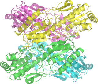
Phosphofructokinase-1 (PFK-1) is one of the most important regulatory enzymes of glycolysis. It is an allosteric enzyme made of 4 subunits and controlled by many activators and inhibitors. PFK-1 catalyzes the important "committed" step of glycolysis, the conversion of fructose 6-phosphate and ATP to fructose 1,6-bisphosphate and ADP. Glycolysis is the foundation for respiration, both anaerobic and aerobic. Because phosphofructokinase (PFK) catalyzes the ATP-dependent phosphorylation to convert fructose-6-phosphate into fructose 1,6-bisphosphate and ADP, it is one of the key regulatory steps of glycolysis. PFK is able to regulate glycolysis through allosteric inhibition, and in this way, the cell can increase or decrease the rate of glycolysis in response to the cell's energy requirements. For example, a high ratio of ATP to ADP will inhibit PFK and glycolysis. The key difference between the regulation of PFK in eukaryotes and prokaryotes is that in eukaryotes PFK is activated by fructose 2,6-bisphosphate. The purpose of fructose 2,6-bisphosphate is to supersede ATP inhibition, thus allowing eukaryotes to have greater sensitivity to regulation by hormones like glucagon and insulin.

<span class="mw-page-title-main">Pyruvate kinase</span> Class of enzymes

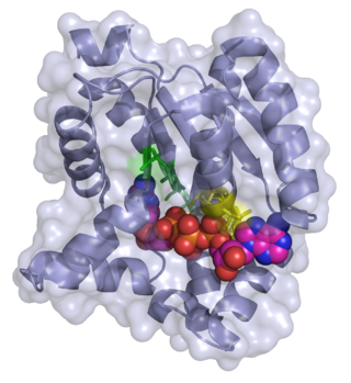
Pyruvate kinase was inappropriately named before it was recognized that it did not directly catalyze phosphorylation of pyruvate, which does not occur under physiological conditions. Pyruvate kinase is present in four distinct, tissue-specific isozymes in animals, each consisting of particular kinetic properties necessary to accommodate the variations in metabolic requirements of diverse tissues.

<span class="mw-page-title-main">AMP-activated protein kinase</span> Class of enzymes

5' AMP-activated protein kinase or AMPK or 5' adenosine monophosphate-activated protein kinase is an enzyme that plays a role in cellular energy homeostasis, largely to activate glucose and fatty acid uptake and oxidation when cellular energy is low. It belongs to a highly conserved eukaryotic protein family and its orthologues are SNF1 in yeast, and SnRK1 in plants. It consists of three proteins (subunits) that together make a functional enzyme, conserved from yeast to humans. It is expressed in a number of tissues, including the liver, brain, and skeletal muscle. In response to binding AMP and ADP, the net effect of AMPK activation is stimulation of hepatic fatty acid oxidation, ketogenesis, stimulation of skeletal muscle fatty acid oxidation and glucose uptake, inhibition of cholesterol synthesis, lipogenesis, and triglyceride synthesis, inhibition of adipocyte lipogenesis, inhibition of adipocyte lipolysis, and modulation of insulin secretion by pancreatic β-cells.

A nucleoside triphosphate is a nucleoside containing a nitrogenous base bound to a 5-carbon sugar, with three phosphate groups bound to the sugar. They are the molecular precursors of both DNA and RNA, which are chains of nucleotides made through the processes of DNA replication and transcription. Nucleoside triphosphates also serve as a source of energy for cellular reactions and are involved in signalling pathways.

<span class="mw-page-title-main">Glutamate dehydrogenase</span> Hexameric enzyme

Glutamate dehydrogenase is an enzyme observed in both prokaryotes and eukaryotic mitochondria. The aforementioned reaction also yields ammonia, which in eukaryotes is canonically processed as a substrate in the urea cycle. Typically, the α-ketoglutarate to glutamate reaction does not occur in mammals, as glutamate dehydrogenase equilibrium favours the production of ammonia and α-ketoglutarate. Glutamate dehydrogenase also has a very low affinity for ammonia, and therefore toxic levels of ammonia would have to be present in the body for the reverse reaction to proceed. However, in brain, the NAD+/NADH ratio in brain mitochondria encourages oxidative deamination. In bacteria, the ammonia is assimilated to amino acids via glutamate and aminotransferases. In plants, the enzyme can work in either direction depending on environment and stress. Transgenic plants expressing microbial GLDHs are improved in tolerance to herbicide, water deficit, and pathogen infections. They are more nutritionally valuable.

In molecular biology, biosynthesis is a multi-step, enzyme-catalyzed process where substrates are converted into more complex products in living organisms. In biosynthesis, simple compounds are modified, converted into other compounds, or joined to form macromolecules. This process often consists of metabolic pathways. Some of these biosynthetic pathways are located within a single cellular organelle, while others involve enzymes that are located within multiple cellular organelles. Examples of these biosynthetic pathways include the production of lipid membrane components and nucleotides. Biosynthesis is usually synonymous with anabolism.

<span class="mw-page-title-main">Adenylate kinase</span> Class of enzymes

Adenylate kinase is a phosphotransferase enzyme that catalyzes the interconversion of the various adenosine phosphates. By constantly monitoring phosphate nucleotide levels inside the cell, ADK plays an important role in cellular energy homeostasis.

The adenylate energy charge is an index used to measure the energy status of biological cells.

<span class="mw-page-title-main">Inosinic acid</span> Chemical compound

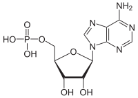
Inosinic acid or inosine monophosphate (IMP) is a nucleotide. Widely used as a flavor enhancer, it is typically obtained from chicken byproducts or other meat industry waste. Inosinic acid is important in metabolism. It is the ribonucleotide of hypoxanthine and the first nucleotide formed during the synthesis of purine nucleotides. It can also be formed by the deamination of adenosine monophosphate by AMP deaminase. It can be hydrolysed to inosine.

<span class="mw-page-title-main">Nucleoside-diphosphate kinase</span> Class of enzymes

Nucleoside-diphosphate kinases are enzymes that catalyze the exchange of terminal phosphate between different nucleoside diphosphates (NDP) and triphosphates (NTP) in a reversible manner to produce nucleotide triphosphates. Many NDP serve as acceptor while NTP are donors of phosphate group. The general reaction via ping-pong mechanism is as follows: XDP + YTP ←→ XTP + YDP. NDPK activities maintain an equilibrium between the concentrations of different nucleoside triphosphates such as, for example, when guanosine triphosphate (GTP) produced in the citric acid (Krebs) cycle is converted to adenosine triphosphate (ATP). Other activities include cell proliferation, differentiation and development, signal transduction, G protein-coupled receptor, endocytosis, and gene expression.

<span class="mw-page-title-main">Nucleic acid metabolism</span> Process

Nucleic acid metabolism is a collective term that refers to the variety of chemical reactions by which nucleic acids are either synthesized or degraded. Nucleic acids are polymers made up of a variety of monomers called nucleotides. Nucleotide synthesis is an anabolic mechanism generally involving the chemical reaction of phosphate, pentose sugar, and a nitrogenous base. Degradation of nucleic acids is a catabolic reaction and the resulting parts of the nucleotides or nucleobases can be salvaged to recreate new nucleotides. Both synthesis and degradation reactions require multiple enzymes to facilitate the event. Defects or deficiencies in these enzymes can lead to a variety of diseases.

Purine metabolism refers to the metabolic pathways to synthesize and break down purines that are present in many organisms.

<span class="mw-page-title-main">Purine nucleotide cycle</span>

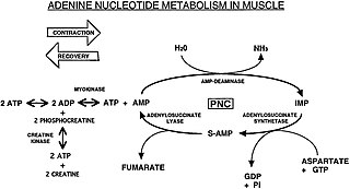
The Purine Nucleotide Cycle is a metabolic pathway in protein metabolism requiring the amino acids aspartate and glutamate. The cycle is used to regulate the levels of adenine nucleotides, in which ammonia and fumarate are generated. AMP converts into IMP and the byproduct ammonia. IMP converts to S-AMP (adenylosuccinate), which then converts to AMP and the byproduct fumarate. The fumarate goes on to produce ATP (energy) via oxidative phosphorylation as it enters the Krebs cycle and then the electron transport chain. Lowenstein first described this pathway and outlined its importance in processes including amino acid catabolism and regulation of flux through glycolysis and the Krebs cycle.

References

  1. "Adenosine monophosphate (Compound)". PubChem. NCBI. Retrieved 30 April 2020.
  2. "Nomenclature of Carbohydrates: (Recommendations 1996)". Journal of Carbohydrate Chemistry. 16 (8): 1191–1280. 1997. doi:10.1080/07328309708005748.
  3. Jauker M, Griesser H, Richert C (November 2015). "Spontaneous Formation of RNA Strands, Peptidyl RNA, and Cofactors". Angewandte Chemie. 54 (48): 14564–9. doi:10.1002/anie.201506593. PMC   4678511 . PMID   26435376.
  4. "Adenosine monophosphate". The Human Metabolome Database. Retrieved 3 July 2020.
  5. Baker, Julien S.; McCormick, Marie Clare; Robergs, Robert A. (2010). "Interaction among Skeletal Muscle Metabolic Energy Systems during Intense Exercise". Journal of Nutrition and Metabolism. 2010: 905612. doi: 10.1155/2010/905612 . ISSN   2090-0732. PMC   3005844 . PMID   21188163.
  6. Valberg, Stephanie J. (1 January 2008), Kaneko, J. Jerry; Harvey, John W.; Bruss, Michael L. (eds.), "Chapter 15 - Skeletal Muscle Function", Clinical Biochemistry of Domestic Animals (Sixth Edition), San Diego: Academic Press, pp. 459–484, ISBN   978-0-12-370491-7 , retrieved 10 October 2023
  7. Maiuolo J, Oppedisano F, Gratteri S, Muscoli C, Mollace V (June 2016). "Regulation of uric acid metabolism and excretion". International Journal of Cardiology. 213: 8–14. doi: 10.1016/j.ijcard.2015.08.109 . PMID   26316329.
  8. Richter EA, Ruderman NB (March 2009). "AMPK and the biochemistry of exercise: implications for human health and disease". The Biochemical Journal. 418 (2): 261–75. doi:10.1042/BJ20082055. PMC   2779044 . PMID   19196246.
  9. Carling D, Mayer FV, Sanders MJ, Gamblin SJ (July 2011). "AMP-activated protein kinase: nature's energy sensor". Nature Chemical Biology. 7 (8): 512–8. doi:10.1038/nchembio.610. PMID   21769098.
  10. Faubert B, Vincent EE, Poffenberger MC, Jones RG (January 2015). "The AMP-activated protein kinase (AMPK) and cancer: many faces of a metabolic regulator". Cancer Letters. 356 (2 Pt A): 165–70. doi:10.1016/j.canlet.2014.01.018. PMID   24486219.
  11. 1 2 Hardie DG (15 September 2011). "AMP-activated protein kinase—an energy sensor that regulates all aspects of cell function". Genes & Development. 25 (18): 1895–1908. doi:10.1101/gad.17420111. ISSN   0890-9369. PMC   3185962 . PMID   21937710.
  12. Hardie DG (February 2011). "Energy sensing by the AMP-activated protein kinase and its effects on muscle metabolism". The Proceedings of the Nutrition Society. 70 (1): 92–9. doi: 10.1017/S0029665110003915 . PMID   21067629.
  13. Krishan S, Richardson DR, Sahni S (March 2015). "Adenosine monophosphate-activated kinase and its key role in catabolism: structure, regulation, biological activity, and pharmacological activation". Molecular Pharmacology. 87 (3): 363–77. doi: 10.1124/mol.114.095810 . PMID   25422142.
  14. Xiao B, Sanders MJ, Underwood E, Heath R, Mayer FV, Carmena D, Jing C, Walker PA, Eccleston JF, Haire LF, Saiu P, Howell SA, Aasland R, Martin SR, Carling D, Gamblin SJ (April 2011). "Structure of mammalian AMPK and its regulation by ADP". Nature. 472 (7342): 230–3. Bibcode:2011Natur.472..230X. doi:10.1038/nature09932. PMC   3078618 . PMID   21399626.
  15. Ravnskjaer K, Madiraju A, Montminy M (2015). Metabolic Control. Handbook of Experimental Pharmacology. Vol. 233. Springer, Cham. pp. 29–49. doi:10.1007/164_2015_32. ISBN   9783319298047. PMID   26721678.
  16. Coffee, Carole J. (1999). Quick Look Medicine: Metabolism. Hayes Barton Press. ISBN   1-59377-192-4.
  17. "15.3: Glycogenolyis and its Regulation by Glucagon and Epinephrine Signaling". Biology LibreTexts. 1 January 2022. Retrieved 10 October 2023.

Further reading