Phosphatase

Last updated

A ball and stick model of a phosphate anion. Phosphate-3D-balls.png
A ball and stick model of a phosphate anion.

In biochemistry, a phosphatase is an enzyme that uses water to cleave a phosphoric acid monoester into a phosphate ion and an alcohol. Because a phosphatase enzyme catalyzes the hydrolysis of its substrate, it is a subcategory of hydrolases. [1] Phosphatase enzymes are essential to many biological functions, because phosphorylation (e.g. by protein kinases) and dephosphorylation (by phosphatases) serve diverse roles in cellular regulation and signaling. [2] Whereas phosphatases remove phosphate groups from molecules, kinases catalyze the transfer of phosphate groups to molecules from ATP. Together, kinases and phosphatases direct a form of post-translational modification that is essential to the cell's regulatory network. [3]

Contents

Phosphatase enzymes are not to be confused with phosphorylase enzymes, which catalyze the transfer of a phosphate group from hydrogen phosphate to an acceptor. Due to their prevalence in cellular regulation, phosphatases are an area of interest for pharmaceutical research. [4] [5]

Biochemistry

The general reaction catalyzed by a phosphatase enzyme General phosphatase mechanism.png
The general reaction catalyzed by a phosphatase enzyme

Phosphatases catalyze the hydrolysis of a phosphomonoester, removing a phosphate moiety from the substrate. Water is split in the reaction, with the -OH group attaching to the phosphate ion, and the H+ protonating the hydroxyl group of the other product. The net result of the reaction is the destruction of a phosphomonoester and the creation of both a phosphate ion and a molecule with a free hydroxyl group. [4]

Phosphatases are able to dephosphorylate seemingly different sites on their substrates with great specificity. Identifying the "phosphatase code," that is, the mechanisms and rules that govern substrate recognition for phosphatases, is still a work in progress, but the first comparative analysis of all the protein phosphatases encoded across nine eukaryotic 'phosphatome' genomes is now available. [6] Studies reveal that so called "docking interactions" play a significant role in substrate binding. [3] A phosphatase recognizes and interacts with various motifs (elements of secondary structure) on its substrate; these motifs bind with low affinity to docking sites on the phosphatase, which are not contained within its active site. Although each individual docking interaction is weak, many interactions occur simultaneously, conferring a cumulative effect on binding specificity. [7] Docking interactions can also allosterically regulate phosphatases and thus influence their catalytic activity. [8]

Functions

In contrast to kinases, phosphatase enzymes recognize and catalyze a wider array of substrates and reactions. For example, in humans, Ser/Thr kinases outnumber Ser/Thr phosphatases by a factor of ten. [4] To some extent, this disparity results from incomplete knowledge of the human phosphatome, that is, the complete set of phosphatases expressed in a cell, tissue, or organism. [3] Many phosphatases have yet to be discovered, and for numerous known phosphatases, a substrate has yet to be identified. However, among well-studied phosphatase/kinase pairs, phosphatases exhibit greater variety than their kinase counterparts in both form and function; this may result from the lesser degree of conservation among phosphatases. [4]

Calcineurin (PP2B) is a protein phosphatase enzyme involved in immune system function. Calcineurin.png
Calcineurin (PP2B) is a protein phosphatase enzyme involved in immune system function.

Distinctions

Phosphatases should not be confused with phosphorylases, which add phosphate groups.

NameClassReactionNotes
PhosphorylaseTransferase
(EC 2.4, 2.7.7)
A−B + H−OP ⇌ A−OP + H−Btransfer group = A
=  glycosyl/nucleotidyl  group
PhosphataseHydrolase
(EC 3)
P−B + H−OH ⇌ P−OH + H−B
KinaseTransferase
(EC 2.7.1-2.7.4)
P−B + H−A ⇌ P−A + H−Btransfer group = P
P = phosphonate group, OP = phosphate group,
H−OP or P−OH = inorganic phosphate

Protein phosphatases

A protein phosphatase is an enzyme that dephosphorylates an amino acid residue of its protein substrate. Whereas protein kinases act as signaling molecules by phosphorylating proteins, phosphatases remove the phosphate group, which is essential if the system of intracellular signaling is to be able to reset for future use. The tandem work of kinases and phosphatases constitute a significant element of the cell's regulatory network. [9] Phosphorylation (and dephosphorylation) is among the most common modes of posttranslational modification in proteins, and it is estimated that, at any given time, up to 30% of all proteins are phosphorylated. [10] [11] Two notable protein phosphatases are PP2A and PP2B. PP2A is involved in multiple regulatory processes, such as DNA replication, metabolism, transcription, and development. PP2B, also called calcineurin, is involved in the proliferation of T cells; because of this, it is the target of some drugs that seek to suppress the immune system. [9]

Nucleosides and nucleotides differ by one phosphate, which is cleaved from nucleotides by nucleotidases. Nucleoside nucleotide general format.png
Nucleosides and nucleotides differ by one phosphate, which is cleaved from nucleotides by nucleotidases.

Nucleotidases

A nucleotidase is an enzyme that catalyzes the hydrolysis of a nucleotide, forming a nucleoside and a phosphate ion. [12] Nucleotidases are essential for cellular homeostasis, because they are partially responsible for maintaining a balanced ratio of nucleotides to nucleosides. [13] Some nucleotidases function outside the cell, creating nucleosides that can be transported into the cell and used to regenerate nucleotides via salvage pathways. [14] Inside the cell, nucleotidases may help to maintain energy levels under stress conditions. A cell deprived of oxygen and nutrients may catabolize more nucleotides to boost levels of nucleoside triphosphates such as ATP, the primary energy currency of the cell. [15]

In gluconeogenesis

Phosphatases can also act on carbohydrates, such as intermediates in gluconeogenesis. Gluconeogenesis is a biosynthetic pathway wherein glucose is created from noncarbohydrate precursors; the pathway is essential because many tissues can only derive energy from glucose. [9] Two phosphatases, glucose-6-phosphatase and fructose-1,6-bisphosphatase, catalyze irreversible steps in gluconeogenesis. [16] [17] Each cleaves a phosphate group from a six-carbon sugar phosphate intermediate.

Classification

Within the larger class of phosphatase, the Enzyme Commission recognizes 104 distinct enzyme families. Phosphatases are classified by substrate specificity and sequence homology in catalytic domains. [3] Despite their classification into over one hundred families, all phosphatases still catalyze the same general hydrolysis reaction. [1]

In in-vitro experiments, phosphatase enzymes seem to recognize many different substrates, and one substrate may be recognized by many different phosphatases. However, when experiments have been carried out in-vivo, phosphatase enzymes have been shown to be incredibly specific. [3] In some cases, a protein phosphatase (i.e. one defined by its recognition of protein substrates) can catalyze the dephosphorylation of nonprotein substrates. [4] Similarly, dual-specificity tyrosine phosphatases can dephosphorylate not only tyrosine residues, but also serine residues. Thus, one phosphatase can exhibit the qualities of multiple phosphatase families. [9]

See also

Related Research Articles

A protein phosphatase is a phosphatase enzyme that removes a phosphate group from the phosphorylated amino acid residue of its substrate protein. Protein phosphorylation is one of the most common forms of reversible protein posttranslational modification (PTM), with up to 30% of all proteins being phosphorylated at any given time. Protein kinases (PKs) are the effectors of phosphorylation and catalyse the transfer of a γ-phosphate from ATP to specific amino acids on proteins. Several hundred PKs exist in mammals and are classified into distinct super-families. Proteins are phosphorylated predominantly on Ser, Thr and Tyr residues, which account for 79.3, 16.9 and 3.8% respectively of the phosphoproteome, at least in mammals. In contrast, protein phosphatases (PPs) are the primary effectors of dephosphorylation and can be grouped into three main classes based on sequence, structure and catalytic function. The largest class of PPs is the phosphoprotein phosphatase (PPP) family comprising PP1, PP2A, PP2B, PP4, PP5, PP6 and PP7, and the protein phosphatase Mg2+- or Mn2+-dependent (PPM) family, composed primarily of PP2C. The protein Tyr phosphatase (PTP) super-family forms the second group, and the aspartate-based protein phosphatases the third. The protein pseudophosphatases form part of the larger phosphatase family, and in most cases are thought to be catalytically inert, instead functioning as phosphate-binding proteins, integrators of signalling or subcellular traps. Examples of membrane-spanning protein phosphatases containing both active (phosphatase) and inactive (pseudophosphatase) domains linked in tandem are known, conceptually similar to the kinase and pseudokinase domain polypeptide structure of the JAK pseudokinases. A complete comparative analysis of human phosphatases and pseudophosphatases has been completed by Manning and colleagues, forming a companion piece to the ground-breaking analysis of the human kinome, which encodes the complete set of ~536 human protein kinases.

<span class="mw-page-title-main">Kinase</span> Enzyme catalyzing transfer of phosphate groups onto specific substrates

In biochemistry, a kinase is an enzyme that catalyzes the transfer of phosphate groups from high-energy, phosphate-donating molecules to specific substrates. This process is known as phosphorylation, where the high-energy ATP molecule donates a phosphate group to the substrate molecule. This transesterification produces a phosphorylated substrate and ADP. Conversely, it is referred to as dephosphorylation when the phosphorylated substrate donates a phosphate group and ADP gains a phosphate group. These two processes, phosphorylation and dephosphorylation, occur four times during glycolysis.

<span class="mw-page-title-main">Adenosine monophosphate</span> Chemical compound

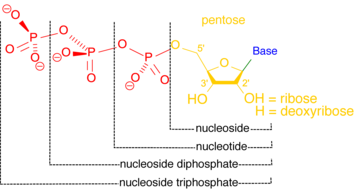
Adenosine monophosphate (AMP), also known as 5'-adenylic acid, is a nucleotide. AMP consists of a phosphate group, the sugar ribose, and the nucleobase adenine. It is an ester of phosphoric acid and the nucleoside adenosine. As a substituent it takes the form of the prefix adenylyl-.

Gluconeogenesis (GNG) is a metabolic pathway that results in the generation of glucose from certain non-carbohydrate carbon substrates. It is a ubiquitous process, present in plants, animals, fungi, bacteria, and other microorganisms. In vertebrates, gluconeogenesis occurs mainly in the liver and, to a lesser extent, in the cortex of the kidneys. It is one of two primary mechanisms – the other being degradation of glycogen (glycogenolysis) – used by humans and many other animals to maintain blood sugar levels, avoiding low levels (hypoglycemia). In ruminants, because dietary carbohydrates tend to be metabolized by rumen organisms, gluconeogenesis occurs regardless of fasting, low-carbohydrate diets, exercise, etc. In many other animals, the process occurs during periods of fasting, starvation, low-carbohydrate diets, or intense exercise.

<span class="mw-page-title-main">Guanosine triphosphate</span> Chemical compound

Guanosine-5'-triphosphate (GTP) is a purine nucleoside triphosphate. It is one of the building blocks needed for the synthesis of RNA during the transcription process. Its structure is similar to that of the guanosine nucleoside, the only difference being that nucleotides like GTP have phosphates on their ribose sugar. GTP has the guanine nucleobase attached to the 1' carbon of the ribose and it has the triphosphate moiety attached to ribose's 5' carbon.

<span class="mw-page-title-main">Guanosine diphosphate</span> Chemical compound

Guanosine diphosphate, abbreviated GDP, is a nucleoside diphosphate. It is an ester of pyrophosphoric acid with the nucleoside guanosine. GDP consists of a pyrophosphate group, a pentose sugar ribose, and the nucleobase guanine.

<span class="mw-page-title-main">Adenylate kinase</span> Class of enzymes

Adenylate kinase is a phosphotransferase enzyme that catalyzes the interconversion of the various adenosine phosphates. By constantly monitoring phosphate nucleotide levels inside the cell, ADK plays an important role in cellular energy homeostasis.

In biochemistry, dephosphorylation is the removal of a phosphate (PO43−) group from an organic compound by hydrolysis. It is a reversible post-translational modification. Dephosphorylation and its counterpart, phosphorylation, activate and deactivate enzymes by detaching or attaching phosphoric esters and anhydrides. A notable occurrence of dephosphorylation is the conversion of ATP to ADP and inorganic phosphate.

A regulatory enzyme is an enzyme in a biochemical pathway which, through its responses to the presence of certain other biomolecules, regulates the pathway activity. This is usually done for pathways whose products may be needed in different amounts at different times, such as hormone production. Regulatory enzymes exist at high concentrations so their activity can be increased or decreased with changes in substrate concentrations.

<span class="mw-page-title-main">Nucleoside-diphosphate kinase</span> Class of enzymes

Nucleoside-diphosphate kinases are enzymes that catalyze the exchange of terminal phosphate between different nucleoside diphosphates (NDP) and triphosphates (NTP) in a reversible manner to produce nucleotide triphosphates. Many NDP serve as acceptor while NTP are donors of phosphate group. The general reaction via ping-pong mechanism is as follows: XDP + YTP ←→ XTP + YDP. NDPK activities maintain an equilibrium between the concentrations of different nucleoside triphosphates such as, for example, when guanosine triphosphate (GTP) produced in the citric acid (Krebs) cycle is converted to adenosine triphosphate (ATP). Other activities include cell proliferation, differentiation and development, signal transduction, G protein-coupled receptor, endocytosis, and gene expression.

<span class="mw-page-title-main">Phosphofructokinase 2</span> Class of enzymes

Phosphofructokinase-2 (6-phosphofructo-2-kinase, PFK-2) or fructose bisphosphatase-2 (FBPase-2), is an enzyme indirectly responsible for regulating the rates of glycolysis and gluconeogenesis in cells. It catalyzes formation and degradation of a significant allosteric regulator, fructose-2,6-bisphosphate (Fru-2,6-P2) from substrate fructose-6-phosphate. Fru-2,6-P2 contributes to the rate-determining step of glycolysis as it activates enzyme phosphofructokinase 1 in the glycolysis pathway, and inhibits fructose-1,6-bisphosphatase 1 in gluconeogenesis. Since Fru-2,6-P2 differentially regulates glycolysis and gluconeogenesis, it can act as a key signal to switch between the opposing pathways. Because PFK-2 produces Fru-2,6-P2 in response to hormonal signaling, metabolism can be more sensitively and efficiently controlled to align with the organism's glycolytic needs. This enzyme participates in fructose and mannose metabolism. The enzyme is important in the regulation of hepatic carbohydrate metabolism and is found in greatest quantities in the liver, kidney and heart. In mammals, several genes often encode different isoforms, each of which differs in its tissue distribution and enzymatic activity. The family described here bears a resemblance to the ATP-driven phospho-fructokinases, however, they share little sequence similarity, although a few residues seem key to their interaction with fructose 6-phosphate.

<span class="mw-page-title-main">5'-nucleotidase</span> InterPro Family

5′-Nucleotidase is an enzyme which catalyzes the phosphorylytic cleavage of 5′-nucleotides. Although originally found in snake venom, the activity of 5'nucleotidase has been described for bacteria and plant cells, and is widely distributed in vertebrate tissue. In mammalian cells the enzyme is predominantly located in the plasma membrane and its primary role is in the conversion of extracellular nucleotides, which are generally impermeable, to the corresponding nucleoside which can readily enter most cells. Consequently, the enzyme plays a key role in the metabolism of nucleotides.

A nucleotidase is a hydrolytic enzyme that catalyzes the hydrolysis of a nucleotide into a nucleoside and a phosphate.

<span class="mw-page-title-main">Fructose 2,6-bisphosphate</span> Chemical compound

Fructose 2,6-bisphosphate, abbreviated Fru-2,6-P2, is a metabolite that allosterically affects the activity of the enzymes phosphofructokinase 1 (PFK-1) and fructose 1,6-bisphosphatase (FBPase-1) to regulate glycolysis and gluconeogenesis. Fru-2,6-P2 itself is synthesized and broken down by the bifunctional enzyme phosphofructokinase 2/fructose-2,6-bisphosphatase (PFK-2/FBPase-2).

<span class="mw-page-title-main">PPP2CA</span> Enzyme

Serine/threonine-protein phosphatase 2A catalytic subunit alpha isoform is an enzyme that is encoded by the PPP2CA gene.

<span class="mw-page-title-main">Phosphatidate phosphatase</span>

The enzyme phosphatidate phosphatase (PAP, EC 3.1.3.4) is a key regulatory enzyme in lipid metabolism, catalyzing the conversion of phosphatidate to diacylglycerol:

In enzymology, a nucleoside-phosphate kinase is an enzyme that catalyzes the chemical reaction

<span class="mw-page-title-main">NT5C</span> Protein-coding gene in the species Homo sapiens

5', 3'-nucleotidase, cytosolic, also known as 5'(3')-deoxyribonucleotidase, cytosolic type (cdN) or deoxy-5'-nucleotidase 1 (dNT-1), is an enzyme that in humans is encoded by the NT5C gene on chromosome 17.

<span class="mw-page-title-main">NT5M</span> Protein-coding gene in the species Homo sapiens

5',3'-nucleotidase, mitochondrial, also known as 5'(3')-deoxyribonucleotidase, mitochondrial (mdN) or deoxy-5'-nucleotidase 2 (dNT-2), is an enzyme that in humans is encoded by the NT5M gene. This gene encodes a 5' nucleotidase that localizes to the mitochondrial matrix. This enzyme dephosphorylates the 5'- and 2'(3')-phosphates of uracil and thymine deoxyribonucleotides. The gene is located within the Smith–Magenis syndrome region on chromosome 17.

The phosphatome of an organism is the set of phosphatase genes in its genome. Phosphatases are enzymes that catalyze the removal of phosphate from biomolecules. Over half of all cellular proteins are modified by phosphorylation which typically controls their functions. Protein phosphorylation is controlled by the opposing actions of protein phosphatases and protein kinases. Most phosphorylation sites are not linked to a specific phosphatase, so the phosphatome approach allows a global analysis of dephosphorylation, screening to find the phosphatase responsible for a given reaction, and comparative studies between different phosphatases, similar to how protein kinase research has been impacted by the kinome approach.

References

  1. 1 2 "ENZYME: 3.1.3.-". enzyme.expasy.org. Retrieved 2017-02-21.
  2. Liberti, Susanna; Sacco, Francesca; Calderone, Alberto; Perfetto, Livia; Iannuccelli, Marta; Panni, Simona; Santonico, Elena; Palma, Anita; Nardozza, Aurelio P. (2013-01-01). "HuPho: the human phosphatase portal" (PDF). FEBS Journal. 280 (2): 379–387. doi: 10.1111/j.1742-4658.2012.08712.x . PMID   22804825.
  3. 1 2 3 4 5 Sacco, Francesca; Perfetto, Livia; Castagnoli, Luisa; Cesareni, Gianni (2012-08-14). "The human phosphatase interactome: An intricate family portrait". FEBS Letters. 586 (17): 2732–2739. doi:10.1016/j.febslet.2012.05.008. PMC   3437441 . PMID   22626554.
  4. 1 2 3 4 5 Li, Xun; Wilmanns, Matthias; Thornton, Janet; Köhn, Maja (2013-05-14). "Elucidating Human Phosphatase-Substrate Networks". Science Signaling. 6 (275): rs10. doi:10.1126/scisignal.2003203. PMID   23674824. S2CID   19282957.
  5. Bodenmiller, Bernd; Wanka, Stefanie; Kraft, Claudine; Urban, Jörg; Campbell, David; Pedrioli, Patrick G.; Gerrits, Bertran; Picotti, Paola; Lam, Henry (2010-12-21). "Phosphoproteomic Analysis Reveals Interconnected System-Wide Responses to Perturbations of Kinases and Phosphatases in Yeast". Science Signaling. 3 (153): rs4. doi:10.1126/scisignal.2001182. PMC   3072779 . PMID   21177495.
  6. Chen, Mark J.; Dixon, Jack E.; Manning, Gerard (2017-04-11). "Genomics and evolution of protein phosphatases". Sci. Signal. 10 (474): eaag1796. doi:10.1126/scisignal.aag1796. ISSN   1945-0877. PMID   28400531. S2CID   41041971.
  7. Roy, Jagoree; Cyert, Martha S. (2009-12-08). "Cracking the Phosphatase Code: Docking Interactions Determine Substrate Specificity". Science Signaling. 2 (100): re9. doi:10.1126/scisignal.2100re9. PMID   19996458. S2CID   20590354.
  8. Reményi, Attila; Good, Matthew C; Lim, Wendell A (2006-12-01). "Docking interactions in protein kinase and phosphatase networks". Current Opinion in Structural Biology. Catalysis and regulation / Proteins. 16 (6): 676–685. doi:10.1016/j.sbi.2006.10.008. PMID   17079133.
  9. 1 2 3 4 G., Voet, Judith; W., Pratt, Charlotte (2013-01-01). Fundamentals of biochemistry : life at the molecular level. Wiley. ISBN   9781118129180. OCLC   892195795.{{cite book}}: CS1 maint: multiple names: authors list (link)
  10. Cohen, Philip (2002-05-01). "The origins of protein phosphorylation". Nature Cell Biology. 4 (5): E127–130. doi:10.1038/ncb0502-e127. ISSN   1465-7392. PMID   11988757. S2CID   29601670.
  11. Tonks, Nicholas K. (2006). "Protein tyrosine phosphatases: from genes, to function, to disease". Nature Reviews Molecular Cell Biology. 7 (11): 833–846. doi:10.1038/nrm2039. PMID   17057753. S2CID   1302726.
  12. "ENZYME entry 3.1.3.31". enzyme.expasy.org. Retrieved 2017-03-21.
  13. Bianchi, V; Pontis, E; Reichard, P (1986). "Interrelations between substrate cycles and de novo synthesis of pyrimidine deoxyribonucleoside triphosphates in 3T6 cells". Proceedings of the National Academy of Sciences of the United States of America. 83 (4): 986–990. Bibcode:1986PNAS...83..986B. doi: 10.1073/pnas.83.4.986 . PMC   322995 . PMID   3456577.
  14. Zimmermann, Herbert; Zebisch, Matthias; Sträter, Norbert (2012-09-01). "Cellular function and molecular structure of ecto-nucleotidases". Purinergic Signalling. 8 (3): 437–502. doi:10.1007/s11302-012-9309-4. ISSN   1573-9538. PMC   3360096 . PMID   22555564.
  15. Hunsucker, Sally Anne; Mitchell, Beverly S.; Spychala, Jozef (2005-07-01). "The 5'-nucleotidases as regulators of nucleotide and drug metabolism". Pharmacology & Therapeutics. 107 (1): 1–30. doi:10.1016/j.pharmthera.2005.01.003. ISSN   0163-7258. PMID   15963349.
  16. "ENZYME entry 3.1.3.9". enzyme.expasy.org. Retrieved 2017-03-21.
  17. "ENZYME entry 3.1.3.11". enzyme.expasy.org. Retrieved 2017-03-21.