Ribonuclease

Last updated
ribonuclease
3agn.png
Ustilago sphaerogena Ribonuclease U2 with AMP PDB entry 3agn [1]
Identifiers
SymbolRibonuclease
Pfam PF00545
InterPro IPR000026
SCOP2 1brn / SCOPe / SUPFAM
Available protein structures:
Pfam   structures / ECOD  
PDB RCSB PDB; PDBe; PDBj
PDBsum structure summary
PDB 1mgw A:56-137 1mgr A:56-137 1uck B:11-92

1i70 A:11-92 2sar A:11-92 1ucj B:11-92 1lni B:11-92 1ay7 A:11-92 1t2h B:11-92 1box A:11-92 1ucl A:11-92 1rge B:11-92 1t2i A:11-92 1c54 A:11-92 1rsn B:11-92 1gmq A:11-92 1uci A:11-92 1sar B:11-92 1gmp A:11-92 1rgf A:11-92 1rgg B:11-92 1rgh B:11-92 1i8v B:11-92 1gmr B:11-92 1ynv X:11-92 1py3 B:79-159 1pyl A:79-159 2rbi B:72-161 1goy A:72-161 1gou B:72-161 1gov A:72-161 1buj A:72-161 1bao B:67-156 1bsd A:67-156 1ban B:67-156 1brh A:67-156 1brg C:67-156 1brk C:67-156 1bns A:67-156 1bnf B:67-156 1bgs B:67-156 1bnj B:67-156 1bsa B:67-156 1bsb C:67-156 1b3s B:67-156 1x1w B:67-156 1bni B:67-156 1b2x B:67-156 1b2z A:67-156 1bsc C:67-156 1bse B:67-156 1x1y B:67-156 1bri C:67-156 1b2u C:67-156 1b27 C:67-156 1b20 B:67-156 1bnr :67-156 1b2s C:67-156 1yvs :67-156 1brs C:67-156 1brj C:67-156 1bne A:67-156 1bng C:67-156 1a2p A:67-156 1x1u B:67-156 1fw7 A:67-156 1rnb A:67-156 1b21 C:67-156 1x1x B:67-156 1brn M:67-156 1b2m A:46-129 1i0v A:46-129 1rls :46-129 1fys A:46-129 1bvi B:46-129 1i2e A:46-129 2hoh D:46-129 3rnt :46-129 6gsp :46-129 4gsp :46-129 1low A:46-129 1i0x A:46-129 1bir B:46-129 1trq A:46-129 1det :46-129 1i2g A:46-129 3bu4 A:46-129 1rn1 A:46-129 1rnt :46-129 4hoh D:46-129 1rga :46-129 4bu4 A:46-129 1rhl A:46-129 5bu4 A:46-129 1hz1 A:46-129 1trp A:46-129 5hoh A:46-129 7gsp A:46-129 1ygw :46-129 1gsp :46-129 1bu4 :46-129 6rnt :46-129 1ch0 B:46-129 1rgc B:46-129 4bir :46-129 2rnt :46-129 3hoh D:46-129 1rgl :46-129 1rn4 :46-129 1fzu A:46-129 1lov A:46-129 5gsp :46-129 9rnt :46-129 3bir :46-129 1q9e C:46-129 1i3f A:46-129 5bir A:46-129 1g02 A:46-129 1loy A:46-129 2bir A:46-129 1tto A:46-129 2aad B:46-129 1lra :46-129 1i3i A:46-129 2bu4 A:46-129 2gsp :46-129 1hyf A:46-129 3gsp :46-129 1iyy A:46-129 7rnt :46-129 2aae :46-129 8rnt :46-129 5rnt :46-129 1i2f A:46-129 4rnt :46-129 1rgk :46-129 1rms :21-102 1rds :21-102 1fut :45-127 1rcl :45-127 1fus :45-127 1rck :45-127 1rtu :23-113 1aqz A:82-174 1jbr B:82-174 1jbt A:82-174 1jbs A:82-174

Contents

1de3 A:83-175 1r4y A:83-175

Ribonuclease (commonly abbreviated RNase) is a type of nuclease that catalyzes the degradation of RNA into smaller components. Ribonucleases can be divided into endoribonucleases and exoribonucleases, and comprise several sub-classes within the EC 2.7 (for the phosphorolytic enzymes) and 3.1 (for the hydrolytic enzymes) classes of enzymes.

Function

All organisms studied contain many RNases of two different classes, showing that RNA degradation is a very ancient and important process. As well as clearing of cellular RNA that is no longer required, RNases play key roles in the maturation of all RNA molecules, both messenger RNAs that carry genetic material for making proteins and non-coding RNAs that function in varied cellular processes. In addition, active RNA degradation systems are the first defense against RNA viruses and provide the underlying machinery for more advanced cellular immune strategies such as RNAi.

Some cells also secrete copious quantities of non-specific RNases such as A and T1. RNases are, therefore, extremely common, resulting in very short lifespans for any RNA that is not in a protected environment. It is worth noting that all intracellular RNAs are protected from RNase activity by a number of strategies including 5' end capping, 3' end polyadenylation, formation of an RNA·RNA duplex, and folding within an RNA protein complex (ribonucleoprotein particle or RNP).

Another mechanism of protection is ribonuclease inhibitor (RI), which comprises a relatively large fraction of cellular protein (~0.1%) in some cell types, and which binds to certain ribonucleases with the highest affinity of any protein-protein interaction; the dissociation constant for the RI-RNase A complex is ~20 fM under physiological conditions. RI is used in most laboratories that study RNA to protect their samples against degradation from environmental RNases.

Similar to restriction enzymes, which cleave highly specific sequences of double-stranded DNA, a variety of endoribonucleases that recognize and cleave specific sequences of single-stranded RNA have been recently classified.

RNases play a critical role in many biological processes, including angiogenesis and self-incompatibility in flowering plants (angiosperms). [2] [3] Many stress-response toxins of prokaryotic toxin-antitoxin systems have been shown to have RNase activity and homology. [4]

Classification

Major types of endoribonucleases

Structure of RNase A RNase A.png
Structure of RNase A

Major types of exoribonucleases

RNase specificity

The active site looks like a rift valley where all the active site residues create the wall and bottom of the valley. The rift is very thin and the small substrate fits perfectly in the middle of the active site, which allows for perfect interaction with the residues. It actually has a little curvature to the site which the substrate also has. Although usually most exo- and endoribonucleases are not sequence specific, recently CRISPR/Cas system natively recognizing and cutting DNA was engineered to cleave ssRNA in a sequence-specific manner. [11]

RNase contamination during RNA extraction

The extraction of RNA in molecular biology experiments is greatly complicated by the presence of ubiquitous and hardy ribonucleases that degrade RNA samples. Certain RNases can be extremely hardy and inactivating them is difficult compared to neutralizing DNases. In addition to the cellular RNases that are released, there are several RNases that are present in the environment. RNases have evolved to have many extracellular functions in various organisms. [12] [13] [14] For example, RNase 7, a member of the RNase A superfamily, is secreted by human skin and serves as a potent antipathogen defence. [15] [16] In these secreted RNases, the enzymatic RNase activity may not even be necessary for its new, exapted function. For example, immune RNases act by destabilizing the cell membranes of bacteria. [17] [18]

Related Research Articles

<span class="mw-page-title-main">Nuclease</span> Class of enzymes which cleave nucleic acids

In biochemistry, a nuclease is an enzyme capable of cleaving the phosphodiester bonds between nucleotides of nucleic acids. Nucleases variously effect single and double stranded breaks in their target molecules. In living organisms, they are essential machinery for many aspects of DNA repair. Defects in certain nucleases can cause genetic instability or immunodeficiency. Nucleases are also extensively used in molecular cloning.

<span class="mw-page-title-main">Ribonuclease H</span> Enzyme family

Ribonuclease H is a family of non-sequence-specific endonuclease enzymes that catalyze the cleavage of RNA in an RNA/DNA substrate via a hydrolytic mechanism. Members of the RNase H family can be found in nearly all organisms, from bacteria to archaea to eukaryotes.

<span class="mw-page-title-main">Dicer</span> Enzyme that cleaves double-stranded RNA (dsRNA) into short dsRNA fragments

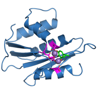
Dicer, also known as endoribonuclease Dicer or helicase with RNase motif, is an enzyme that in humans is encoded by the DICER1 gene. Being part of the RNase III family, Dicer cleaves double-stranded RNA (dsRNA) and pre-microRNA (pre-miRNA) into short double-stranded RNA fragments called small interfering RNA and microRNA, respectively. These fragments are approximately 20–25 base pairs long with a two-base overhang on the 3′-end. Dicer facilitates the activation of the RNA-induced silencing complex (RISC), which is essential for RNA interference. RISC has a catalytic component Argonaute, which is an endonuclease capable of degrading messenger RNA (mRNA).

<span class="mw-page-title-main">Exonuclease</span> Class of enzymes; type of nuclease

Exonucleases are enzymes that work by cleaving nucleotides one at a time from the end (exo) of a polynucleotide chain. A hydrolyzing reaction that breaks phosphodiester bonds at either the 3′ or the 5′ end occurs. Its close relative is the endonuclease, which cleaves phosphodiester bonds in the middle (endo) of a polynucleotide chain. Eukaryotes and prokaryotes have three types of exonucleases involved in the normal turnover of mRNA: 5′ to 3′ exonuclease (Xrn1), which is a dependent decapping protein; 3′ to 5′ exonuclease, an independent protein; and poly(A)-specific 3′ to 5′ exonuclease.

<span class="mw-page-title-main">Ranpirnase</span> Enzyme from the Northern Leopard Frog

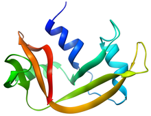
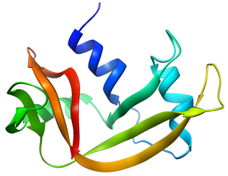
Ranpirnase is a ribonuclease enzyme found in the oocytes of the Northern Leopard Frog. Ranpirnase is a member of the pancreatic ribonuclease protein superfamily and degrades RNA substrates with a sequence preference for uracil and guanine nucleotides. Along with amphinase, another leopard frog ribonuclease, Ranpirnase has been studied as a potential cancer and antiviral treatment due to its unusual mechanism of cytotoxicity tested against transformed cells and antiviral activity.

<span class="mw-page-title-main">Angiogenin</span> Protein-coding gene in the species Homo sapiens

Angiogenin (ANG) also known as ribonuclease 5 is a small 123 amino acid protein that in humans is encoded by the ANG gene. Angiogenin is a potent stimulator of new blood vessels through the process of angiogenesis. Ang hydrolyzes cellular RNA, resulting in modulated levels of protein synthesis and interacts with DNA causing a promoter-like increase in the expression of rRNA. Ang is associated with cancer and neurological disease through angiogenesis and through activating gene expression that suppresses apoptosis.

<span class="mw-page-title-main">Ribonuclease P</span> Class of enzymes

Ribonuclease P is a type of ribonuclease which cleaves RNA. RNase P is unique from other RNases in that it is a ribozyme – a ribonucleic acid that acts as a catalyst in the same way that a protein-based enzyme would. Its function is to cleave off an extra, or precursor, sequence of RNA on tRNA molecules. Further, RNase P is one of two known multiple turnover ribozymes in nature, the discovery of which earned Sidney Altman and Thomas Cech the Nobel Prize in Chemistry in 1989: in the 1970s, Altman discovered the existence of precursor tRNA with flanking sequences and was the first to characterize RNase P and its activity in processing of the 5' leader sequence of precursor tRNA. Recent findings also reveal that RNase P has a new function. It has been shown that human nuclear RNase P is required for the normal and efficient transcription of various small noncoding RNAs, such as tRNA, 5S rRNA, SRP RNA and U6 snRNA genes, which are transcribed by RNA polymerase III, one of three major nuclear RNA polymerases in human cells.

<span class="mw-page-title-main">Ribonuclease III</span> Class of enzymes

Ribonuclease III (RNase III or RNase C)(BRENDA 3.1.26.3) is a type of ribonuclease that recognizes dsRNA and cleaves it at specific targeted locations to transform them into mature RNAs. These enzymes are a group of endoribonucleases that are characterized by their ribonuclease domain, which is labelled the RNase III domain. They are ubiquitous compounds in the cell and play a major role in pathways such as RNA precursor synthesis, RNA Silencing, and the pnp autoregulatory mechanism.

<span class="mw-page-title-main">Drosha</span> Ribonuclease III enzyme

Drosha is a Class 2 ribonuclease III enzyme that in humans is encoded by the DROSHA gene. It is the primary nuclease that executes the initiation step of miRNA processing in the nucleus. It works closely with DGCR8 and in correlation with Dicer. It has been found significant in clinical knowledge for cancer prognosis and HIV-1 replication.

<span class="mw-page-title-main">Exosome complex</span> Protein complex that degrades RNA

The exosome complex is a multi-protein intracellular complex capable of degrading various types of RNA molecules. Exosome complexes are found in both eukaryotic cells and archaea, while in bacteria a simpler complex called the degradosome carries out similar functions.

<span class="mw-page-title-main">Nuclear RNase P</span>

In molecular biology, nuclear ribonuclease P is a ubiquitous endoribonuclease, found in archaea, bacteria and eukarya as well as chloroplasts and mitochondria. Its best characterised enzyme activity is the generation of mature 5′-ends of tRNAs by cleaving the 5′-leader elements of precursor-tRNAs. Cellular RNase Ps are ribonucleoproteins. The RNA from bacterial RNase P retains its catalytic activity in the absence of the protein subunit, i.e. it is a ribozyme. Similarly, archaeal RNase P RNA has been shown to be weakly catalytically active in the absence of its respective protein cofactors. Isolated eukaryotic RNase P RNA has not been shown to retain its catalytic function, but is still essential for the catalytic activity of the holoenzyme. Although the archaeal and eukaryotic holoenzymes have a much greater protein content than the bacterial ones, the RNA cores from all three lineages are homologous—the helices corresponding to P1, P2, P3, P4, and P10/11 are common to all cellular RNase P RNAs. Yet there is considerable sequence variation, particularly among the eukaryotic RNAs.

<span class="mw-page-title-main">Exoribonuclease</span> Class of enzymes that degrade RNA

An exoribonuclease is an exonuclease ribonuclease, which are enzymes that degrade RNA by removing terminal nucleotides from either the 5' end or the 3' end of the RNA molecule. Enzymes that remove nucleotides from the 5' end are called 5'-3' exoribonucleases, and enzymes that remove nucleotides from the 3' end are called 3'-5' exoribonucleases.

<span class="mw-page-title-main">Pancreatic ribonuclease family</span> Class of enzymes

Pancreatic ribonuclease family is a superfamily of pyrimidine-specific endonucleases found in high quantity in the pancreas of certain mammals and of some reptiles.

Exoribonuclease II is an enzyme. This enzyme catalyses the following chemical reaction

RNase PhyM is a type of endoribonuclease which is sequence specific for single stranded RNAs. It cleaves 3'-end of unpaired A and U residues.

RNase R, or Ribonuclease R, is a 3'-->5' exoribonuclease, which belongs to the RNase II superfamily, a group of enzymes that hydrolyze RNA in the 3' - 5' direction. RNase R has been shown to be involved in selective mRNA degradation, particularly of non stop mRNAs in bacteria. RNase R has homologues in many other organisms.

The degradosome is a multiprotein complex present in most bacteria that is involved in the processing of ribosomal RNA and the degradation of messenger RNA and is regulated by Non-coding RNA. It contains the proteins RNA helicase B, RNase E and Polynucleotide phosphorylase.

Ribonuclease E is a bacterial ribonuclease that participates in the processing of ribosomal RNA and the chemical degradation of bulk cellular RNA.

<span class="mw-page-title-main">Retroviral ribonuclease H</span>

The retroviral ribonuclease H is a catalytic domain of the retroviral reverse transcriptase (RT) enzyme. The RT enzyme is used to generate complementary DNA (cDNA) from the retroviral RNA genome. This process is called reverse transcription. To complete this complex process, the retroviral RT enzymes need to adopt a multifunctional nature. They therefore possess 3 of the following biochemical activities: RNA-dependent DNA polymerase, ribonuclease H, and DNA-dependent DNA polymerase activities. Like all RNase H enzymes, the retroviral RNase H domain cleaves DNA/RNA duplexes and will not degrade DNA or unhybridized RNA.

<span class="mw-page-title-main">Ribonuclease T</span>

Ribonuclease T is a ribonuclease enzyme involved in the maturation of transfer RNA and ribosomal RNA in bacteria, as well as in DNA repair pathways. It is a member of the DnaQ family of exonucleases and non-processively acts on the 3' end of single-stranded nucleic acids. RNase T is capable of cleaving both DNA and RNA, with extreme sequence specificity discriminating against cytosine at the 3' end of the substrate.

References

  1. Noguchi S (July 2010). "Isomerization mechanism of aspartate to sexxxxxxxx implied by structures of Ustilago sphaerogena ribonuclease U2 complexed with adenosine 3'-monophosphate". Acta Crystallographica D. 66 (Pt 7): 843–9. doi:10.1107/S0907444910019621. PMID   20606265.
  2. Sporn MB, Roberts AB (6 December 2012). Peptide Growth Factors and Their Receptors II. Springer Science & Business Media. p. 556. ISBN   978-3-642-74781-6.
  3. Raghavan V (6 December 2012). Developmental Biology of Flowering Plants. Springer Science & Business Media. p. 237. ISBN   978-1-4612-1234-8.
  4. Ramage HR, Connolly LE, Cox JS (December 2009). "Comprehensive functional analysis of Mycobacterium tuberculosis toxin-antitoxin systems: implications for pathogenesis, stress responses, and evolution". PLOS Genetics. 5 (12): e1000767. doi: 10.1371/journal.pgen.1000767 . PMC   2781298 . PMID   20011113.
  5. Cuchillo CM, Nogués MV, Raines RT (September 2011). "Bovine pancreatic ribonuclease: fifty years of the first enzymatic reaction mechanism". Biochemistry. 50 (37): 7835–41. doi:10.1021/bi201075b. PMC   3172371 . PMID   21838247.
  6. "Library Preparation Kits".
  7. Nowotny M (February 2009). "Retroviral integrase superfamily: the structural perspective". EMBO Reports. 10 (2): 144–51. doi:10.1038/embor.2008.256. PMC   2637324 . PMID   19165139.
  8. Holzmann J, Frank P, Löffler E, Bennett KL, Gerner C, Rossmanith W (October 2008). "RNase P without RNA: identification and functional reconstitution of the human mitochondrial tRNA processing enzyme". Cell. 135 (3): 462–74. doi: 10.1016/j.cell.2008.09.013 . PMID   18984158.
  9. Shamsher S. Kanwar*, Puranjan Mishra, Khem Raj Meena, Shruti Gupta and Rakesh Kumar, Ribonucleases and their Applications, 2016, Journal of Advanced Biotechnology and Bioengineering
  10. Wachi M, Umitsuki G, Shimizu M, Takada A, Nagai K. Escherichia coli cafA gene encodes a novel RNase, designated as RNase G, involved in processing of the 5' end of 16S rRNA. Biochem Biophys Res Commun. 1999;259(2):483‐488. doi:10.1006/bbrc.1999.0806
  11. Tamulaitis G, Kazlauskiene M, Manakova E, Venclovas Č, Nwokeoji AO, Dickman MJ, Horvath P, Siksnys V (November 2014). "Programmable RNA shredding by the type III-A CRISPR-Cas system of Streptococcus thermophilus". Molecular Cell. 56 (4): 506–17. doi: 10.1016/j.molcel.2014.09.027 . PMID   25458845.
  12. Rossier O, Dao J, Cianciotto NP (March 2009). "A type II secreted RNase of Legionella pneumophila facilitates optimal intracellular infection of Hartmannella vermiformis". Microbiology. 155 (Pt 3): 882–90. doi:10.1099/mic.0.023218-0. PMC   2662391 . PMID   19246759.
  13. Luhtala N, Parker R (May 2010). "T2 Family ribonucleases: ancient enzymes with diverse roles". Trends in Biochemical Sciences. 35 (5): 253–9. doi:10.1016/j.tibs.2010.02.002. PMC   2888479 . PMID   20189811.
  14. Dyer KD, Rosenberg HF (November 2006). "The RNase a superfamily: generation of diversity and innate host defense". Molecular Diversity. 10 (4): 585–97. doi:10.1007/s11030-006-9028-2. PMID   16969722. S2CID   20922592.
  15. Harder J, Schroder JM (November 2002). "RNase 7, a novel innate immune defense antimicrobial protein of healthy human skin". The Journal of Biological Chemistry. 277 (48): 46779–84. doi: 10.1074/jbc.M207587200 . PMID   12244054.
  16. Köten B, Simanski M, Gläser R, Podschun R, Schröder JM, Harder J (July 2009). "RNase 7 contributes to the cutaneous defense against Enterococcus faecium". PLOS ONE. 4 (7): e6424. Bibcode:2009PLoSO...4.6424K. doi: 10.1371/journal.pone.0006424 . PMC   2712763 . PMID   19641608.
  17. Huang YC, Lin YM, Chang TW, Wu SJ, Lee YS, Chang MD, Chen C, Wu SH, Liao YD (February 2007). "The flexible and clustered lysine residues of human ribonuclease 7 are critical for membrane permeability and antimicrobial activity". The Journal of Biological Chemistry. 282 (7): 4626–33. doi: 10.1074/jbc.M607321200 . PMID   17150966.
  18. Rosenberg HF (May 2008). "RNase A ribonucleases and host defense: an evolving story". Journal of Leukocyte Biology. 83 (5): 1079–87. doi:10.1189/jlb.1107725. PMC   2692241 . PMID   18211964.

Ahmed TAE, Udenigwe CC, Gomaa A. Editorial: Biotechnology and Bioengineering Applications for Egg-Derived Biomaterials. Front Bioeng Biotechnol. 2021 Sep 20;9:756058

Sources