Microprocessor complex

Last updated
A cryo-electron microscopy structure of the microprocessor complex, showing human Drosha protein (ribonuclease III, green) and two subunits of DGCR8 (dark and light blue) interacting with and ready to cleave a primary microRNA. From PDB: 6V5B . 6v5b.png
A cryo-electron microscopy structure of the microprocessor complex, showing human Drosha protein (ribonuclease III, green) and two subunits of DGCR8 (dark and light blue) interacting with and ready to cleave a primary microRNA. From PDB: 6V5B .

The microprocessor complex is a protein complex involved in the early stages of processing microRNA (miRNA) and RNA interference (RNAi) in animal cells. [2] [3] The complex is minimally composed of the ribonuclease enzyme Drosha and the dimeric RNA-binding protein DGCR8 (also known as Pasha in non-human animals), and cleaves primary miRNA substrates to pre-miRNA in the cell nucleus. [4] [5] [6] Microprocessor is also the smaller of the two multi-protein complexes that contain human Drosha. [7]

Contents

A crystal structure of the human Drosha protein in complex with the C-terminal helices of two DGCR8 molecules (green). Drosha consists of two ribonuclease III domains (blue and orange); a double-stranded RNA binding domain (yellow); and a connector/platform domain (gray) containing two bound zinc ion (spheres). From PDB: 5B16 . 5b16 drosha dgcr8.png
A crystal structure of the human Drosha protein in complex with the C-terminal helices of two DGCR8 molecules (green). Drosha consists of two ribonuclease III domains (blue and orange); a double-stranded RNA binding domain (yellow); and a connector/platform domain (gray) containing two bound zinc ion (spheres). From PDB: 5B16 .

Composition

The microprocessor complex consists minimally of two proteins: Drosha, a ribonuclease III enzyme; and DGCR8, a double-stranded RNA binding protein. [4] [5] [6] (DGCR8 is the name used in mammalian genetics, abbreviated from "DiGeorge syndrome critical region 8"; the homologous protein in model organisms such as flies and worms is called Pasha, for Partner of Drosha.) The stoichiometry of the minimal complex was at one point experimentally difficult to determine, but it has been demonstrated to be a heterotrimer of two DGCR8 proteins and one Drosha. [1] [8] [9] [10]

In addition to the minimal catalytically active microprocessor components, other cofactors such as DEAD box RNA helicases and heterogeneous nuclear ribonucleoproteins may be present in the complex to mediate the activity of Drosha. [4] Some miRNAs are processed by microprocessor only in the presence of specific cofactors. [11]

Function

The human exportin-5 protein (red) in complex with Ran-GTP (yellow) and a pre-microRNA (green), showing two-nucleotide overhang recognition element (orange). From PDB: 3A6P . 3a6p xpo5 ran miRNA.png
The human exportin-5 protein (red) in complex with Ran-GTP (yellow) and a pre-microRNA (green), showing two-nucleotide overhang recognition element (orange). From PDB: 3A6P .

Located in the cell nucleus, the microprocessor complex cleaves primary miRNA (pri-miRNA), typically at least 1000 nucleotides long, into precursor miRNA (pre-miRNA). [13] Its two subunits have been determined as necessary and sufficient for the mediation of the development of miRNAs from the pri-miRNAs. [7] These molecules of around 70 nucleotides contain a stem-loop or hairpin structure. Pri-miRNA substrates can be derived either from non-coding RNA genes or from introns. In the latter case, there is evidence that the microprocessor complex interacts with the spliceosome and that the pri-miRNA processing occurs prior to splicing. [5] [14]

Microprocessor cleavage of pri-miRNAs typically occurs co-transcriptionally and leaves a characteristic RNase III single-stranded overhang of 2-3 nucleotides, which serves as a recognition element for the transport protein exportin-5. [15] Pre-miRNAs are exported from the nucleus to the cytoplasm in a RanGTP-dependent manner and are further processed, typically by the endoribonuclease enzyme Dicer. [4] [5] [6]

Hemin allows for the increased processing of pri-miRNAs through an induced conformational change of the DGCR8 subunit, and also enhances DGCR8's binding specificity for RNA. [16] DGCR8 recognizes the junctions between hairpin structures and single-stranded RNA and serves to orient Drosha to cleave around 11 nucleotides away from the junctions, and remains in contact with the pri-miRNAs following cleavage and dissociation of Drosha. [17]

Although the large majority of miRNAs undergo processing by microprocessor, a small number of exceptions called mirtrons have been described; these are very small introns which, after splicing, have the appropriate size and stem-loop structure to serve as a pre-miRNA. [18] The processing pathways for microRNA and for exogenously derived small interfering RNA converge at the point of Dicer processing and are largely identical downstream. Broadly defined, both pathways constitute RNAi. [5] [18] Microprocessor is also found to be involved in ribosomal biogenesis specifically in the removal of R-loops and activating transcription of ribosomal protein encoding genes. [19]

Regulation

Gene regulation by miRNA is widespread across many genomes – by some estimates more than 60% of human protein-coding genes are likely to be regulated by miRNA, [20] though the quality of experimental evidence for miRNA-target interactions is often weak. [21] Because processing by microprocessor is a major determinant of miRNA abundance, microprocessor itself is then an important target of regulation.

Both Drosha and DGCR8 are subject to regulation by post-translational modifications modulating stability, intracellular localization, and activity levels. Activity against particular substrates may be regulated by additional protein cofactors interacting with the microprocessor complex. The loop region of the pri-miRNA stem-loop is also a recognition element for regulatory proteins, which may up- or down-regulate microprocessor processing of the specific miRNAs they target. [11]

Microprocessor itself is auto-regulated by negative feedback through association with a pri-miRNA-like hairpin structure found in the DGCR8 mRNA, which when cleaved reduces DGCR8 expression. The structure in this case is located in an exon and is unlikely to itself function as miRNA in its own right. [11]

Evolution

Drosha shares striking structural similarity with the downstream ribonuclease Dicer, suggesting an evolutionary relationship, though Drosha and related enzymes are found only in animals while Dicer relatives are widely distributed, including among protozoans. [8] Both components of the microprocessor complex are conserved among the vast majority of metazoans with known genomes. Mnemiopsis leidyi , a ctenophore, lacks both Drosha and DGCR8 homologs, as well as recognizable miRNAs, and is the only known metazoan with no detectable genomic evidence of Drosha. [22] In plants, the miRNA biogenesis pathway is somewhat different; neither Drosha nor DGCR8 has a homolog in plant cells, where the first step in miRNA processing is usually executed by a different nuclear ribonuclease, DCL1, a homolog of Dicer. [11] [23]

It has been suggested based on phylogenetic analysis that the key components of RNA interference based on exogenous substrates were present in the ancestral eukaryote, likely as an immune mechanism against viruses and transposable elements. Elaboration of this pathway for miRNA-mediated gene regulation is thought to have evolved later. [24]

Clinical significance

The involvement of miRNAs in diseases has led scientists to become more interested in the role of additional protein complexes, like microprocessor, that have the ability to influence or modulate the function and expression of miRNAs. [25] Microprocessor complex component, DGCR8, is affected through the micro-deletion of 22q11.2, a small portion of chromosome 22. This deletion causes irregular processing of miRNAs which leads to DiGeorge Syndrome . [26]

Related Research Articles

microRNA Small non-coding ribonucleic acid molecule

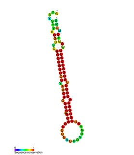
A microRNA is a small single-stranded non-coding RNA molecule found in plants, animals and some viruses, that functions in RNA silencing and post-transcriptional regulation of gene expression. miRNAs function via base-pairing with complementary sequences within mRNA molecules. As a result, these mRNA molecules are silenced, by one or more of the following processes: (1) cleavage of the mRNA strand into two pieces, (2) destabilization of the mRNA through shortening of its poly(A) tail, and (3) less efficient translation of the mRNA into proteins by ribosomes.

Dicer Enzyme that cleaves double-stranded RNA (dsRNA) into short dsRNA fragments

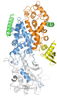
Dicer, also known as endoribonuclease Dicer or helicase with RNase motif, is an enzyme that in humans is encoded by the DICER1 gene. Being part of the RNase III family, Dicer cleaves double-stranded RNA (dsRNA) and pre-microRNA (pre-miRNA) into short double-stranded RNA fragments called small interfering RNA and microRNA, respectively. These fragments are approximately 20–25 base pairs long with a two-base overhang on the 3′-end. Dicer facilitates the activation of the RNA-induced silencing complex (RISC), which is essential for RNA interference. RISC has a catalytic component Argonaute, which is an endonuclease capable of degrading messenger RNA (mRNA).

The RNA-induced silencing complex, or RISC, is a multiprotein complex, specifically a ribonucleoprotein, which functions in gene silencing via a variety of pathways at the transcriptional and translational levels. Using single-stranded RNA (ssRNA) fragments, such as microRNA (miRNA), or double-stranded small interfering RNA (siRNA), the complex functions as a key tool in gene regulation. The single strand of RNA acts as a template for RISC to recognize complementary messenger RNA (mRNA) transcript. Once found, one of the proteins in RISC, Argonaute, activates and cleaves the mRNA. This process is called RNA interference (RNAi) and it is found in many eukaryotes; it is a key process in defense against viral infections, as it is triggered by the presence of double-stranded RNA (dsRNA).

Ribonuclease III

Ribonuclease III (BRENDA 3.1.26.3) is a type of ribonuclease that recognizes dsRNA and cleaves it at specific targeted locations to transform them into mature RNAs. These enzymes are a group of endoribonucleases that are characterized by their ribonuclease domain, which is labelled the RNase III domain. They are ubiquitous compounds in the cell and play a major role in pathways such as RNA precursor synthesis, RNA Silencing, and the pnp autoregulatory mechanism.

Drosha Ribonuclease III enzyme

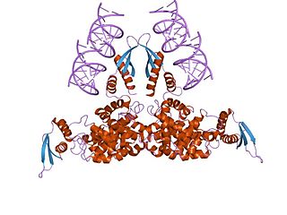
Drosha is a Class 2 ribonuclease III enzyme that in humans is encoded by the DROSHA gene. It is the primary nuclease that executes the initiation step of miRNA processing in the nucleus. It works closely with DGCR8 and in correlation with Dicer. It has been found significant in clinical knowledge for cancer prognosis and HIV-1 replication.

Microprocessor complex subunit DGCR8

The microprocessor complex subunit DGCR8(DiGeorge syndrome critical region 8) is a protein that in humans is encoded by the DGCR8 gene. In other animals, particularly the common model organisms Drosophila melanogaster and Caenorhabditis elegans, the protein is known as Pasha. It is a required component of the RNA interference pathway.

Mirtrons are a type of microRNAs that are located in the introns of the mRNA encoding host genes. These short hairpin introns formed via atypical miRNA biogenesis pathways. Mirtrons arise from the spliced-out introns and are known to function in gene expression.

RNA interference Biological process of gene regulation

RNA interference (RNAi) is a biological process in which RNA molecules are involved in sequence-specific suppression of gene expression by double-stranded RNA, through translational or transcriptional repression. Historically, RNAi was known by other names, including co-suppression, post-transcriptional gene silencing (PTGS), and quelling. The detailed study of each of these seemingly different processes elucidated that the identity of these phenomena were all actually RNAi. Andrew Fire and Craig C. Mello shared the 2006 Nobel Prize in Physiology or Medicine for their work on RNAi in the nematode worm Caenorhabditis elegans, which they published in 1998. Since the discovery of RNAi and its regulatory potentials, it has become evident that RNAi has immense potential in suppression of desired genes. RNAi is now known as precise, efficient, stable and better than antisense therapy for gene suppression. Antisense RNA produced intracellularly by an expression vector may be developed and find utility as novel therapeutic agents.

miR-203

In molecular biology miR-203 is a short non-coding RNA molecule. MicroRNAs function to regulate the expression levels of other genes by several mechanisms, such as translational repression and Argonaute-catalyzed messenger RNA cleavage. miR-203 has been identified as a skin-specific microRNA, and it forms an expression gradient that defines the boundary between proliferative epidermal basal progenitors and terminally differentiating suprabasal cells. It has also been found upregulated in psoriasis and differentially expressed in some types of cancer.

V. Narry Kim South Korean biochemist

V. Narry Kim is a South Korean biochemist and microbiologist, best known for her work on microRNA biogenesis. Her pioneering studies have laid the groundwork for the biology of microRNA and contributed to the improvement of RNA interference technologies.

In molecular biology, small nucleolar RNA derived microRNAs are microRNAs (miRNA) derived from small nucleolar RNA (snoRNA). MicroRNAs are usually derived from precursors known as pre-miRNAs, these pre-miRNAs are recognised and cleaved from a pri-miRNA precursor by the Pasha and Drosha proteins. However some microRNAs, mirtrons, are known to be derived from introns via a different pathway which bypasses Pasha and Drosha. Some microRNAs are also known to be derived from small nucleolar RNA.

MIR187

MicroRNA 187 is a protein that in humans is encoded by the MIR187 gene.

MicroRNA 95 is a small non-coding RNA that in humans is encoded by the MIR95 gene.

MIR34A

MicroRNA 34a (miR-34a) is a MicroRNA that in humans is encoded by the MIR34A gene.

DCL1 is a gene in plants that codes for the DCL1 protein, a ribonuclease III enzyme involved in processing microRNA (miRNA). Although DCL1 is named for its homology with the metazoan protein Dicer, its role in miRNA biogenesis is somewhat different, due to substantial differences in miRNA maturation processes between plants and animals. Unlike Dicer, DCL1 is localized to the cell nucleus, where it initiates miRNA processing as the catalytic component of the so-called Dicing complex, which also contains HYL1, a double-stranded RNA binding protein, and a zinc-finger protein known as SE or SERRATE. Within the nucleus, Dicing complexes co-localize in Dicing bodies or D-bodies. In plants, DCL1 is responsible both for processing a primary miRNA to a pre-miRNA, and for then processing the pre-miRNA to a mature miRNA. In animals, the equivalents of these two steps are carried out by different proteins; pri-miRNA processing takes place in the nucleus by the ribonuclease Drosha as part of the Microprocessor complex, and processing to a mature miRNA takes place in the cytoplasm by Dicer to yield a mature miRNA.

MicroRNA 203a

MicroRNA 203a is a protein that in humans is encoded by the MIR203A gene.

MicroRNA 148a

MicroRNA 148a is a miRNA that in humans is encoded by the MIR148A gene.

MIR494

MicroRNA 494 is a microRNA sequence which is encoded in humans by the MIR494 gene.

MIRLET7F2

MicroRNA let-7f-2 is a protein that in humans is encoded by the MIRLET7F2 gene.

MicroRNA 517c is a protein that in humans is encoded by the MIR517C gene.

References

  1. 1 2 Partin, Alexander C.; Zhang, Kaiming; Jeong, Byung-Cheon; Herrell, Emily; Li, Shanshan; Chiu, Wah; Nam, Yunsun (May 2020). "Cryo-EM Structures of Human Drosha and DGCR8 in Complex with Primary MicroRNA". Molecular Cell. 78 (3): 411–422.e4. doi:10.1016/j.molcel.2020.02.016. PMC   7214211 . PMID   32220646.
  2. Gregory RI, Yan KP, Amuthan G, Chendrimada T, Doratotaj B, Cooch N, Shiekhattar R (November 2004). "The Microprocessor complex mediates the genesis of microRNAs". Nature. 432 (7014): 235–40. Bibcode:2004Natur.432..235G. doi:10.1038/nature03120. PMID   15531877. S2CID   4389261.
  3. Denli AM, Tops BB, Plasterk RH, Ketting RF, Hannon GJ (November 2004). "Processing of primary microRNAs by the Microprocessor complex". Nature. 432 (7014): 231–5. Bibcode:2004Natur.432..231D. doi:10.1038/nature03049. PMID   15531879. S2CID   4425505.
  4. 1 2 3 4 Siomi H, Siomi MC (May 2010). "Posttranscriptional regulation of microRNA biogenesis in animals". Molecular Cell. 38 (3): 323–32. doi: 10.1016/j.molcel.2010.03.013 . PMID   20471939.
  5. 1 2 3 4 5 Wilson RC, Doudna JA (2013). "Molecular mechanisms of RNA interference". Annual Review of Biophysics. 42: 217–39. doi:10.1146/annurev-biophys-083012-130404. PMC   5895182 . PMID   23654304.
  6. 1 2 3 Macias S, Cordiner RA, Cáceres JF (August 2013). "Cellular functions of the microprocessor". Biochemical Society Transactions. 41 (4): 838–43. doi:10.1042/BST20130011. hdl: 1842/25877 . PMID   23863141.
  7. 1 2 Gregory RI, Yan KP, Amuthan G, Chendrimada T, Doratotaj B, Cooch N, Shiekhattar R (November 2004). "The Microprocessor complex mediates the genesis of microRNAs". Nature. 432 (7014): 235–40. Bibcode:2004Natur.432..235G. doi:10.1038/nature03120. PMID   15531877. S2CID   4389261.
  8. 1 2 3 Kwon SC, Nguyen TA, Choi YG, Jo MH, Hohng S, Kim VN, Woo JS (January 2016). "Structure of Human DROSHA". Cell. 164 (1–2): 81–90. doi: 10.1016/j.cell.2015.12.019 . PMID   26748718.
  9. Herbert KM, Sarkar SK, Mills M, Delgado De la Herran HC, Neuman KC, Steitz JA (February 2016). "A heterotrimer model of the complete Microprocessor complex revealed by single-molecule subunit counting". RNA. 22 (2): 175–83. doi:10.1261/rna.054684.115. PMC   4712668 . PMID   26683315.
  10. Nguyen TA, Jo MH, Choi YG, Park J, Kwon SC, Hohng S, et al. (June 2015). "Functional Anatomy of the Human Microprocessor". Cell. 161 (6): 1374–87. doi: 10.1016/j.cell.2015.05.010 . PMID   26027739.
  11. 1 2 3 4 Ha M, Kim VN (August 2014). "Regulation of microRNA biogenesis". Nature Reviews. Molecular Cell Biology. 15 (8): 509–24. doi:10.1038/nrm3838. PMID   25027649. S2CID   205495632.
  12. Okada, Chimari; Yamashita, Eiki; Lee, Soo Jae; Shibata, Satoshi; Katahira, Jun; Nakagawa, Atsushi; Yoneda, Yoshihiro; Tsukihara, Tomitake (2009-11-27). "A High-Resolution Structure of the Pre-microRNA Nuclear Export Machinery". Science. 326 (5957): 1275–1279. Bibcode:2009Sci...326.1275O. doi:10.1126/science.1178705. PMID   19965479. S2CID   206522317.
  13. Michlewski G, Cáceres JF (January 2019). "Post-transcriptional control of miRNA biogenesis". RNA. 25 (1): 1–16. doi:10.1261/rna.068692.118. PMC   6298569 . PMID   30333195.
  14. Kataoka N, Fujita M, Ohno M (June 2009). "Functional association of the Microprocessor complex with the spliceosome". Molecular and Cellular Biology. 29 (12): 3243–54. doi:10.1128/MCB.00360-09. PMC   2698730 . PMID   19349299.
  15. Morlando M, Ballarino M, Gromak N, Pagano F, Bozzoni I, Proudfoot NJ (September 2008). "Primary microRNA transcripts are processed co-transcriptionally". Nature Structural & Molecular Biology. 15 (9): 902–9. doi:10.1038/nsmb.1475. PMC   6952270 . PMID   19172742.
  16. Partin AC, Ngo TD, Herrell E, Jeong BC, Hon G, Nam Y (November 2017). "Heme enables proper positioning of Drosha and DGCR8 on primary microRNAs". Nature Communications. 8 (1): 1737. Bibcode:2017NatCo...8.1737P. doi:10.1038/s41467-017-01713-y. PMC   5700927 . PMID   29170488.
  17. Bellemer C, Bortolin-Cavaillé ML, Schmidt U, Jensen SM, Kjems J, Bertrand E, Cavaillé J (June 2012). "Microprocessor dynamics and interactions at endogenous imprinted C19MC microRNA genes". Journal of Cell Science. 125 (Pt 11): 2709–20. doi: 10.1242/jcs.100354 . PMID   22393237. S2CID   19121670.
  18. 1 2 Winter J, Jung S, Keller S, Gregory RI, Diederichs S (March 2009). "Many roads to maturity: microRNA biogenesis pathways and their regulation". Nature Cell Biology. 11 (3): 228–34. doi:10.1038/ncb0309-228. PMID   19255566. S2CID   205286318.
  19. Jiang X, Prabhakar A, Van der Voorn SM, Ghatpande P, Celona B, Venkataramanan S, et al. (February 2021). "Control of ribosomal protein synthesis by the Microprocessor complex". Science Signaling. 14 (671): eabd2639. doi:10.1126/scisignal.abd2639. PMC   8012103 . PMID   33622983.
  20. Friedman RC, Farh KK, Burge CB, Bartel DP (January 2009). "Most mammalian mRNAs are conserved targets of microRNAs". Genome Research. 19 (1): 92–105. doi:10.1101/gr.082701.108. PMC   2612969 . PMID   18955434.
  21. Lee YJ, Kim V, Muth DC, Witwer KW (November 2015). "Validated MicroRNA Target Databases: An Evaluation". Drug Development Research. 76 (7): 389–96. doi:10.1002/ddr.21278. PMC   4777876 . PMID   26286669.
  22. Maxwell EK, Ryan JF, Schnitzler CE, Browne WE, Baxevanis AD (December 2012). "MicroRNAs and essential components of the microRNA processing machinery are not encoded in the genome of the ctenophore Mnemiopsis leidyi". BMC Genomics. 13: 714. doi:10.1186/1471-2164-13-714. PMC   3563456 . PMID   23256903.
  23. Axtell MJ, Westholm JO, Lai EC (2011). "Vive la différence: biogenesis and evolution of microRNAs in plants and animals". Genome Biology. 12 (4): 221. doi:10.1186/gb-2011-12-4-221. PMC   3218855 . PMID   21554756.
  24. Cerutti H, Casas-Mollano JA (August 2006). "On the origin and functions of RNA-mediated silencing: from protists to man". Current Genetics. 50 (2): 81–99. doi:10.1007/s00294-006-0078-x. PMC   2583075 . PMID   16691418.
  25. Beezhold KJ, Castranova V, Chen F (June 2010). "Microprocessor of microRNAs: regulation and potential for therapeutic intervention". Molecular Cancer. 9 (1): 134. doi:10.1186/1476-4598-9-134. PMC   2887798 . PMID   20515486.
  26. Fénelon K, Mukai J, Xu B, Hsu PK, Drew LJ, Karayiorgou M, et al. (March 2011). "Deficiency of Dgcr8, a gene disrupted by the 22q11.2 microdeletion, results in altered short-term plasticity in the prefrontal cortex". Proceedings of the National Academy of Sciences of the United States of America. 108 (11): 4447–52. Bibcode:2011PNAS..108.4447F. doi: 10.1073/pnas.1101219108 . PMC   3060227 . PMID   21368174.