Prostaglandin

Last updated
E1 - Alprostadil Prostaglandin E1.svg
E1 - Alprostadil
I2 - Prostacyclin Prostacyclin-2D-skeletal.png
I2 - Prostacyclin

Prostaglandins (PG) are a group of physiologically active lipid compounds called eicosanoids [1] that have diverse hormone-like effects in animals. Prostaglandins have been found in almost every tissue in humans and other animals. They are derived enzymatically from the fatty acid arachidonic acid. [2] Every prostaglandin contains 20 carbon atoms, including a 5-carbon ring. They are a subclass of eicosanoids and of the prostanoid class of fatty acid derivatives.

Contents

The structural differences between prostaglandins account for their different biological activities. A given prostaglandin may have different and even opposite effects in different tissues in some cases. The ability of the same prostaglandin to stimulate a reaction in one tissue and inhibit the same reaction in another tissue is determined by the type of receptor to which the prostaglandin binds. They act as autocrine or paracrine factors with their target cells present in the immediate vicinity of the site of their secretion. Prostaglandins differ from endocrine hormones in that they are not produced at a specific site but in many places throughout the human body.

Prostaglandins are powerful, locally-acting vasodilators and inhibit the aggregation of blood platelets. Through their role in vasodilation, prostaglandins are also involved in inflammation. They are synthesized in the walls of blood vessels and serve the physiological function of preventing needless clot formation, as well as regulating the contraction of smooth muscle tissue. [3] Conversely, thromboxanes (produced by platelet cells) are vasoconstrictors and facilitate platelet aggregation. Their name comes from their role in clot formation (thrombosis).

Specific prostaglandins are named with a letter indicating the type of ring structure, followed by a number indicating the number of double bonds in the hydrocarbon structure. For example, prostaglandin E1 has the abbreviation PGE1 and prostaglandin I2 has the abbreviation PGI2.

History and name

Systematic studies of prostaglandins began in 1930, when Kurzrock and Lieb found that human seminal fluid caused either stimulation or relaxation of strips of isolated human uterus. They noted the curious finding that uteri from patients who had gone through successful pregnancies responded to the fluid with relaxation, while uteri from sterile women responded with contraction upon addition of this seminal fluid. [4] The name prostaglandin derives from the prostate gland, chosen when prostaglandin was first isolated from seminal fluid in 1935 by the Swedish physiologist Ulf von Euler, [5] and independently by the Irish-English physiologist Maurice Walter Goldblatt (1895–1967). [6] [7] [8] Prostaglandins were believed to be part of the prostatic secretions, and eventually were discovered to be produced by the seminal vesicles. Later, it was shown that many other tissues secrete prostaglandins and that they perform a variety of functions. The first total syntheses of prostaglandin F and prostaglandin E2 were reported by E. J. Corey in 1969, [9] an achievement for which he was awarded the Japan Prize in 1989.

In 1971, it was determined that aspirin-like drugs could inhibit the synthesis of prostaglandins. The biochemists Sune K. Bergström, Bengt I. Samuelsson and John R. Vane jointly received the 1982 Nobel Prize in Physiology or Medicine for their research on prostaglandins.[ citation needed ]

Biochemistry

Biosynthesis

Biosynthesis of eicosanoids Eicosanoid synthesis.svg
Biosynthesis of eicosanoids

Prostaglandins are found in most tissues and organs. They are produced by almost all nucleated cells. They are autocrine and paracrine lipid mediators that act upon platelets, endothelium, uterine and mast cells. They are synthesized in the cell from the fatty acid arachidonic acid. [2]

Arachidonic acid is created from diacylglycerol via phospholipase-A2, then brought to either the cyclooxygenase pathway or the lipoxygenase pathway. The cyclooxygenase pathway produces thromboxane, prostacyclin and prostaglandin D, E and F. Alternatively, the lipoxygenase enzyme pathway is active in leukocytes and in macrophages and synthesizes leukotrienes.[ citation needed ]

Release of prostaglandins from the cell

Prostaglandins were originally believed to leave the cells via passive diffusion because of their high lipophilicity. The discovery of the prostaglandin transporter (PGT, SLCO2A1), which mediates the cellular uptake of prostaglandin, demonstrated that diffusion alone cannot explain the penetration of prostaglandin through the cellular membrane. The release of prostaglandin has now also been shown to be mediated by a specific transporter, namely the multidrug resistance protein 4 (MRP4, ABCC4), a member of the ATP-binding cassette transporter superfamily. Whether MRP4 is the only transporter releasing prostaglandins from the cells is still unclear.[ citation needed ]

Cyclooxygenases

Prostaglandins are produced following the sequential oxygenation of arachidonic acid, DGLA or EPA by cyclooxygenases (COX-1 and COX-2) and terminal prostaglandin synthases. The classic dogma is as follows:

  • COX-1 is responsible for the baseline levels of prostaglandins.
  • COX-2 produces prostaglandins through stimulation.

However, while COX-1 and COX-2 are both located in the blood vessels, stomach and the kidneys, prostaglandin levels are increased by COX-2 in scenarios of inflammation and growth.

Prostaglandin E synthase

Prostaglandin E2 (PGE2) — the most abundant prostaglandin [10] — is generated from the action of prostaglandin E synthases on prostaglandin H2 (prostaglandin H2, PGH2). Several prostaglandin E synthases have been identified. To date, microsomal (named as misoprostol) prostaglandin E synthase-1 emerges as a key enzyme in the formation of PGE2.[ citation needed ]

Other terminal prostaglandin synthases

Terminal prostaglandin synthases have been identified that are responsible for the formation of other prostaglandins. For example, hematopoietic and lipocalin prostaglandin D synthases (hPGDS and lPGDS) are responsible for the formation of PGD2 from PGH2. Similarly, prostacyclin (PGI2) synthase (PGIS) converts PGH2 into PGI2. A thromboxane synthase (TxAS) has also been identified. Prostaglandin-F synthase (PGFS) catalyzes the formation of 9α,11β-PGF2α,β from PGD2 and PGF from PGH2 in the presence of NADPH. This enzyme has recently been crystallized in complex with PGD2 [11] and bimatoprost [12] (a synthetic analogue of PGF).

Functions

There are currently ten known prostaglandin receptors on various cell types. Prostaglandins ligate a sub-family of cell surface seven-transmembrane receptors, G-protein-coupled receptors. These receptors are termed DP1-2, EP1-4, FP, IP1-2, and TP, corresponding to the receptor that ligates the corresponding prostaglandin (e.g., DP1-2 receptors bind to PGD2).

The diversity of receptors means that prostaglandins act on an array of cells and have a wide variety of effects such as:

Types

The following is a comparison of different types of prostaglandin, including prostaglandin I2 (prostacyclin; PGI2), prostaglandin D2 (PGD2), prostaglandin E2 (PGE2), and prostaglandin F (PGF). [19]

Type Receptor Receptor typeFunction
PGI2 IP Gs
PGD2 PTGDR (DP1) and CRTH2 (DP2) GPCR
  • produced by mast cells; recruits Th2 cells, eosinophils, and basophils
  • In mammalian organs, large amounts of PGD2 are found only in the brain and in mast cells
  • Critical to development of allergic diseases such as asthma
PGE2 EP1 Gq
EP2 Gs
EP3 Gi
Unspecified
PGF FP Gq

Role in pharmacology

Inhibition

Examples of prostaglandin antagonists are:

Clinical uses

Synthetic prostaglandins are used:

Synthesis

The original synthesis of prostaglandins F2α and E2 is shown below. It involves a Diels–Alder reaction which establishes the relative stereochemistry of three contiguous stereocenters on the prostaglandin cyclopentane core. [32]

Diels-Alder in the total synthesis of prostaglandin F2a by E. J. Corey Prostaglandin Diels-Alder Corey.png
Diels-Alder in the total synthesis of prostaglandin F2α by E. J. Corey

Prostaglandin stimulants

Cold exposure and IUDs may increase prostaglandin production. [33]

See also

Notes

  1. Prostaglandins are released during menstruation, due to the destruction of the endometrial cells, and the resultant release of their contents. [14] [ needs update ] Release of prostaglandins and other inflammatory mediators in the uterus cause the uterus to contract. These substances are thought to be a major factor in primary dysmenorrhea. [15] [16] [17]

Related Research Articles

<span class="mw-page-title-main">Eicosanoid</span> Class of compounds

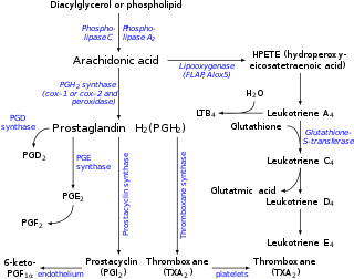
Eicosanoids are signaling molecules made by the enzymatic or non-enzymatic oxidation of arachidonic acid or other polyunsaturated fatty acids (PUFAs) that are, similar to arachidonic acid, around 20 carbon units in length. Eicosanoids are a sub-category of oxylipins, i.e. oxidized fatty acids of diverse carbon units in length, and are distinguished from other oxylipins by their overwhelming importance as cell signaling molecules. Eicosanoids function in diverse physiological systems and pathological processes such as: mounting or inhibiting inflammation, allergy, fever and other immune responses; regulating the abortion of pregnancy and normal childbirth; contributing to the perception of pain; regulating cell growth; controlling blood pressure; and modulating the regional flow of blood to tissues. In performing these roles, eicosanoids most often act as autocrine signaling agents to impact their cells of origin or as paracrine signaling agents to impact cells in the proximity of their cells of origin. Some eicosanoids, such as prostaglandins, may also have endocrine roles as hormones to influence the function of distant cells.

<span class="mw-page-title-main">Prostacyclin</span> Chemical compound

Prostacyclin (also called prostaglandin I2 or PGI2) is a prostaglandin member of the eicosanoid family of lipid molecules. It inhibits platelet activation and is also an effective vasodilator.

<span class="mw-page-title-main">Thromboxane</span> Group of lipids

Thromboxane is a member of the family of lipids known as eicosanoids. The two major thromboxanes are thromboxane A2 and thromboxane B2. The distinguishing feature of thromboxanes is a 6-membered ether-containing ring.

In molecular biology, prostanoids are active lipid mediators that regulate inflammatory response. Prostanoids are a subclass of eicosanoids consisting of the prostaglandins, the thromboxanes, and the prostacyclins. Prostanoids are seen to target NSAIDS which allow for therapeutic potential. Prostanoids are present within areas of the body such as the gastrointestinal tract, urinary tract, respiratory and cardiovascular systems, reproductive tract and vascular system. Prostanoids can even be seen with aid to the water and ion transportation within cells.

Prostaglandin E<sub>2</sub> Chemical compound

Prostaglandin E2 (PGE2), also known as dinoprostone, is a naturally occurring prostaglandin with oxytocic properties that is used as a medication. Dinoprostone is used in labor induction, bleeding after delivery, termination of pregnancy, and in newborn babies to keep the ductus arteriosus open. In babies it is used in those with congenital heart defects until surgery can be carried out. It is also used to manage gestational trophoblastic disease. It may be used within the vagina or by injection into a vein.

<span class="mw-page-title-main">Essential fatty acid interactions</span>

There are many fatty acids found in nature. Two types of fatty acids considered essential for human health are the omega-3 and omega-6 types. These two essential fatty acids are necessary for some cellular signalling pathways and are involved in mediating inflammation, protein synthesis, and metabolic pathways in the human body.

<span class="mw-page-title-main">Thromboxane receptor</span> Mammalian protein found in Homo sapiens

The thromboxane receptor (TP) also known as the prostanoid TP receptor is a protein that in humans is encoded by the TBXA2R gene, The thromboxane receptor is one among the five classes of prostanoid receptors and was the first eicosanoid receptor cloned. The TP receptor derives its name from its preferred endogenous ligand thromboxane A2.

A prostaglandin antagonist is a hormone antagonist acting upon one or more prostaglandins, a subclass of eicosanoid compounds which function as signaling molecules in numerous types of animal tissues.

Most of the eicosanoid receptors are integral membrane protein G protein-coupled receptors (GPCRs) that bind and respond to eicosanoid signaling molecules. Eicosanoids are rapidly metabolized to inactive products and therefore are short-lived. Accordingly, the eicosanoid-receptor interaction is typically limited to a local interaction: cells, upon stimulation, metabolize arachidonic acid to an eicosanoid which then binds cognate receptors on either its parent cell or on nearby cells to trigger functional responses within a restricted tissue area, e.g. an inflammatory response to an invading pathogen. In some cases, however, the synthesized eicosanoid travels through the blood to trigger systemic or coordinated tissue responses, e.g. prostaglandin (PG) E2 released locally travels to the hypothalamus to trigger a febrile reaction. An example of a non-GPCR receptor that binds many eicosanoids is the PPAR-γ nuclear receptor.

Prostaglandin D<sub>2</sub> Chemical compound

Prostaglandin D2 (or PGD2) is a prostaglandin that binds to the receptor PTGDR (DP1), as well as CRTH2 (DP2). It is a major prostaglandin produced by mast cells – recruits Th2 cells, eosinophils, and basophils. In mammalian organs, large amounts of PGD2 are found only in the brain and in mast cells. It is critical to development of allergic diseases such as asthma. Research carried out in 1989 found PGD2 is the primary mediator of vasodilation (the "niacin flush") after ingestion of niacin (nicotinic acid).

Prostaglandin receptors or prostanoid receptors represent a sub-class of cell surface membrane receptors that are regarded as the primary receptors for one or more of the classical, naturally occurring prostanoids viz., prostaglandin D2,, PGE2, PGF2alpha, prostacyclin (PGI2), thromboxane A2 (TXA2), and PGH2. They are named based on the prostanoid to which they preferentially bind and respond, e.g. the receptor responsive to PGI2 at lower concentrations than any other prostanoid is named the Prostacyclin receptor (IP). One exception to this rule is the receptor for thromboxane A2 (TP) which binds and responds to PGH2 and TXA2 equally well.

<span class="mw-page-title-main">Thromboxane A2</span> Chemical compound

Thromboxane A2 (TXA2) is a type of thromboxane that is produced by activated platelets during hemostasis and has prothrombotic properties: it stimulates activation of new platelets as well as increases platelet aggregation. This is achieved by activating the thromboxane receptor, which results in platelet-shape change, inside-out activation of integrins, and degranulation. Circulating fibrinogen binds these receptors on adjacent platelets, further strengthening the clot. Thromboxane A2 is also a known vasoconstrictor and is especially important during tissue injury and inflammation. It is also regarded as responsible for Prinzmetal's angina.

Prostaglandin H<sub>2</sub> Chemical compound

Prostaglandin H2 (PGH2), or prostaglandin H2 (PGH2), is a type of prostaglandin and a precursor for many other biologically significant molecules. It is synthesized from arachidonic acid in a reaction catalyzed by a cyclooxygenase enzyme. The conversion from arachidonic acid to prostaglandin H2 is a two-step process. First, COX-1 catalyzes the addition of two free oxygens to form the 1,2-dioxane bridge and a peroxide functional group to form prostaglandin G2 (PGG2). Second, COX-2 reduces the peroxide functional group to a secondary alcohol, forming prostaglandin H2. Other peroxidases like hydroquinone have been observed to reduce PGG2 to PGH2. PGH2 is unstable at room temperature, with a half life of 90-100 seconds, so it is often converted into a different prostaglandin.

Cyclopentenone prostaglandins are a subset of prostaglandins (PGs) or prostanoids that has 15-deoxy-Δ12,14-prostaglandin J2 (15-d-Δ12,14-PGJ2), Δ12-PGJ2, and PGJ2 as its most prominent members but also including PGA2, PGA1, and, while not classified as such, other PGs. 15-d-Δ12,14-PGJ2, Δ12-PGJ2, and PGJ2 share a common mono-unsaturated cyclopentenone structure as well as a set of similar biological activities including the ability to suppress inflammation responses and the growth as well as survival of cells, particularly those of cancerous or neurological origin. Consequently, these three cyclopentenone-PGs and the two epoxyisoprostanes are suggested to be models for the development of novel anti-inflammatory and anti-cancer drugs. The cyclopenentone prostaglandins are structurally and functionally related to a subset of isoprostanes viz., two cyclopentenone isoprostanes, 5,6-epoxyisoprostane E2 and 5,6-epoxisoprostane A2.

<span class="mw-page-title-main">Cyclooxygenase-2</span> Human enzyme involved in inflammation

Cyclooxygenase-2 (COX-2), also known as Prostaglandin-endoperoxide synthase 2 (HUGO PTGS2), is an enzyme that in humans is encoded by the PTGS2 gene. In humans it is one of three cyclooxygenases. It is involved in the conversion of arachidonic acid to prostaglandin H2, an important precursor of prostacyclin, which is expressed in inflammation.

<span class="mw-page-title-main">Cyclooxygenase-1</span>

Cyclooxygenase 1 (COX-1), also known as prostaglandin-endoperoxide synthase 1, is an enzyme that in humans is encoded by the PTGS1 gene. In humans it is one of two cyclooxygenases.

Prostaglandin EP<sub>3</sub> receptor Protein-coding gene in the species Homo sapiens

Prostaglandin EP3 receptor (EP3, 53kDa), is a prostaglandin receptor for prostaglandin E2 (PGE2) encoded by the human gene PTGER3; it is one of four identified EP receptors, the others being EP1, EP2, and EP4, all of which bind with and mediate cellular responses to PGE2 and also, but generally with lesser affinity and responsiveness, certain other prostanoids (see Prostaglandin receptors). EP has been implicated in various physiological and pathological responses.

<span class="mw-page-title-main">Prostaglandin F receptor</span> Protein-coding gene in the species Homo sapiens

Prostaglandin F receptor (FP) is a receptor belonging to the prostaglandin (PG) group of receptors. FP binds to and mediates the biological actions of Prostaglandin F (PGF). It is encoded in humans by the PTGFR gene.

<span class="mw-page-title-main">Prostaglandin F2alpha</span> Chemical compound

Prostaglandin F, pharmaceutically termed dinoprost is a naturally occurring prostaglandin used in medicine to induce labor and as an abortifacient. Prostaglandins are lipids throughout the entire body that have a hormone-like function. In pregnancy, PGF2 is medically used to sustain contracture and provoke myometrial ischemia to accelerate labor and prevent significant blood loss in labor. Additionally, PGF2 has been linked to being naturally involved in the process of labor. It has been seen that there are higher levels of PGF2 in maternal fluid during labor when compared to at term. This signifies that there is likely a biological use and significance to the production and secretion of PGF2 in labor. Prostaglandin is also used to treat uterine infections in domestic animals.

<span class="mw-page-title-main">12-Hydroxyheptadecatrienoic acid</span> Chemical compound

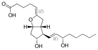
12-Hydroxyheptadecatrienoic acid (also termed 12-HHT, 12(S)-hydroxyheptadeca-5Z,8E,10E-trienoic acid, or 12(S)-HHTrE) is a 17 carbon metabolite of the 20 carbon polyunsaturated fatty acid, arachidonic acid. It was discovered and structurally defined in 1973 by P. Wlodawer, Bengt I. Samuelsson, and M. Hamberg, as a product of arachidonic acid metabolism made by microsomes (i.e. endoplasmic reticulum) isolated from sheep seminal vesicle glands and by intact human platelets. 12-HHT is less ambiguously termed 12-(S)-hydroxy-5Z,8E,10E-heptadecatrienoic acid to indicate the S stereoisomerism of its 12-hydroxyl residue and the Z, E, and E cis-trans isomerism of its three double bonds. The metabolite was for many years thought to be merely a biologically inactive byproduct of prostaglandin synthesis. More recent studies, however, have attached potentially important activity to it.

References

  1. "Eicosanoid Synthesis and Metabolism: Prostaglandins, Thromboxanes, Leukotrienes, Lipoxins". themedicalbiochemistrypage.org. Retrieved 2018-09-21.
  2. 1 2 Ricciotti E, FitzGerald GA (May 2011). "Prostaglandins and inflammation". Arteriosclerosis, Thrombosis, and Vascular Biology. 31 (5): 986–1000. doi:10.1161/ATVBAHA.110.207449. PMC   3081099 . PMID   21508345.
  3. Nelson RF (2005). An introduction to behavioral endocrinology (3rd ed.). Sunderland, Mass: Sinauer Associates. p. 100. ISBN   0-87893-617-3.
  4. Kurzrock, Raphael; Lieb, Charles C. (1930). "Biochemical Studies of Human Semen. II. The Action of Semen on the Human Uterus". Proceedings of the Society for Experimental Biology and Medicine. 28 (3): 268. doi:10.3181/00379727-28-5265. S2CID   85374636.
  5. Von Euler US (1935). "Über die spezifische blutdrucksenkende Substanz des menschlichen Prostata- und Samenblasensekrets" [On the specific blood-pressure-reducing substance of human prostate and seminal vesicle secretions]. Wiener Klinische Wochenschrift. 14 (33): 1182–1183. doi:10.1007/BF01778029. S2CID   38622866.
  6. Goldblatt MW (May 1935). "Properties of human seminal plasma". The Journal of Physiology. 84 (2): 208–18. doi:10.1113/jphysiol.1935.sp003269. PMC   1394818 . PMID   16994667.
  7. Rubinstein, William D.; Jolles, Michael A.; Rubinstein, Hillary L., eds. (2011). "Goldblatt, Maurice Walter". The Palgrave Dictionary of Anglo-Jewish History. Basingstoke, England: Palgrave Macmillan. p. 333. ISBN   978-0-230-30466-6.
  8. R.S.F.S. (3 June 1967). "Obituary Notices: M. W. Goldblatt". British Medical Journal. 2 (5552): 644. doi:10.1136/bmj.2.5552.644. S2CID   220151673.
  9. Nicolaou KC, Sorensen EJ (1996). Classics in Total Synthesis . Weinheim, Germany: VCH. p.  65. ISBN   3-527-29284-5.
  10. Ke J, Yang Y, Che Q, Jiang F, Wang H, Chen Z, Zhu M, Tong H, Zhang H, Yan X, Wang X, Wang F, Liu Y, Dai C, Wan X (September 2016). "Prostaglandin E2 (PGE2) promotes proliferation and invasion by enhancing SUMO-1 activity via EP4 receptor in endometrial cancer". Tumour Biology. 37 (9): 12203–12211. doi:10.1007/s13277-016-5087-x. PMC   5080328 . PMID   27230680. Prostaglandin E2 (PGE2) is the most abundant prostanoid in the human body
  11. Komoto J, Yamada T, Watanabe K, Takusagawa F (March 2004). "Crystal structure of human prostaglandin F synthase (AKR1C3)". Biochemistry. 43 (8): 2188–98. doi:10.1021/bi036046x. PMID   14979715.
  12. Komoto J, Yamada T, Watanabe K, Woodward DF, Takusagawa F (February 2006). "Prostaglandin F2alpha formation from prostaglandin H2 by prostaglandin F synthase (PGFS): crystal structure of PGFS containing bimatoprost". Biochemistry. 45 (7): 1987–96. doi:10.1021/bi051861t. PMID   16475787.
  13. "Hormonal and pheromonal control of spawning in goldfish (PDF Download Available)". ResearchGate. Retrieved 2017-02-04.
  14. Lethaby A, Duckitt K, Farquhar C (January 2013). "Non-steroidal anti-inflammatory drugs for heavy menstrual bleeding". The Cochrane Database of Systematic Reviews (1): CD000400. doi:10.1002/14651858.CD000400.pub3. PMID   23440779.
  15. Wright, Jason and Solange Wyatt. The Washington Manual Obstetrics and Gynecology Survival Guide. Lippincott Williams & Wilkins, 2003. ISBN   0-7817-4363-X [ page needed ]
  16. Harel Z (December 2006). "Dysmenorrhea in adolescents and young adults: etiology and management". Journal of Pediatric and Adolescent Gynecology. 19 (6): 363–71. doi:10.1016/j.jpag.2006.09.001. PMID   17174824.
  17. Bofill Rodriguez, M; Lethaby, A; Farquhar, C (19 September 2019). "Non-steroidal anti-inflammatory drugs for heavy menstrual bleeding". The Cochrane Database of Systematic Reviews. 2019 (9): CD000400. doi:10.1002/14651858.CD000400.pub4. PMC   6751587 . PMID   31535715.
  18. Wallace, John L. (October 2008). "Prostaglandins, NSAIDs, and Gastric Mucosal Protection: Why Doesn't the Stomach Digest Itself?". Physiological Reviews. 88 (4): 1547–1565. doi:10.1152/physrev.00004.2008. ISSN   0031-9333.
  19. Moreno JJ (February 2017). "Eicosanoid receptors: Targets for the treatment of disrupted intestinal epithelial homeostasis". European Journal of Pharmacology. 796: 7–19. doi:10.1016/j.ejphar.2016.12.004. PMID   27940058. S2CID   1513449.
  20. 1 2 Rang HP (2003). Pharmacology (5th ed.). Edinburgh: Churchill Livingstone. p. 234. ISBN   0-443-07145-4.
  21. Fabre JE, Nguyen M, Athirakul K, Coggins K, McNeish JD, Austin S, Parise LK, FitzGerald GA, Coffman TM, Koller BH (March 2001). "Activation of the murine EP3 receptor for PGE2 inhibits cAMP production and promotes platelet aggregation". The Journal of Clinical Investigation. 107 (5): 603–10. doi:10.1172/JCI10881. PMC   199422 . PMID   11238561.
  22. Gross S, Tilly P, Hentsch D, Vonesch JL, Fabre JE (February 2007). "Vascular wall-produced prostaglandin E2 exacerbates arterial thrombosis and atherothrombosis through platelet EP3 receptors". The Journal of Experimental Medicine. 204 (2): 311–20. doi:10.1084/jem.20061617. PMC   2118736 . PMID   17242161.
  23. Stromberga, Zane; Chess-Williams, Russ; Moro, Christian (23 June 2020). "Prostaglandin E2 and F2alpha Modulate Urinary Bladder Urothelium, Lamina Propria and Detrusor Contractility via the FP Receptor". Frontiers in Physiology. 11: 705. doi: 10.3389/fphys.2020.00705 . PMC   7344237 . PMID   32714206.
  24. Joshi, Shailendra; Ornstein, Eugene; Young, William L. (2010). "Cerebral and Spinal Cord Blood Flow". Cottrell and Young's Neuroanesthesia. pp. 17–59. doi:10.1016/B978-0-323-05908-4.10007-7. ISBN   978-0-323-05908-4.
  25. Kieronska-Rudek A, Kij A, Kaczara P, Tworzydlo A, Napiorkowski M, Sidoryk K; et al. (2021). "Exogenous Vitamins K Exert Anti-Inflammatory Effects Dissociated from Their Role as Substrates for Synthesis of Endogenous MK-4 in Murine Macrophages Cell Line". Cells. 10 (7): 1571. doi: 10.3390/cells10071571 . PMC   8303864 . PMID   34206530.{{cite journal}}: CS1 maint: multiple names: authors list (link)
  26. Koshihara Y, Hoshi K, Shiraki M (1993). "Vitamin K2 (menatetrenone) inhibits prostaglandin synthesis in cultured human osteoblast-like periosteal cells by inhibiting prostaglandin H synthase activity". Biochem Pharmacol. 46 (8): 1355–62. doi:10.1016/0006-2952(93)90099-i. PMID   8240383.{{cite journal}}: CS1 maint: multiple names: authors list (link)
  27. Krishnan AV, Srinivas S, Feldman D (2009). "Inhibition of prostaglandin synthesis and actions contributes to the beneficial effects of calcitriol in prostate cancer". Dermatoendocrinol. 1 (1): 7–11. doi:10.4161/derm.1.1.7106. PMC   2715203 . PMID   20046582.{{cite journal}}: CS1 maint: multiple names: authors list (link)
  28. "WHO Recommendations for Induction of Labour". NCBI Bookshelf. Retrieved 2020-07-15. Induction of labour is defined as the process of artificially stimulating the uterus to start labour (1). It is usually performed by administering oxytocin or prostaglandins to the pregnant woman or by manually rupturing the amniotic membranes.
  29. Medscape Early Penile Rehabilitation Helps Reduce Later Intractable ED
  30. Veale, David; Miles, Sarah; Bramley, Sally; Muir, Gordon; Hodsoll, John (2015). "Am I normal? A systematic review and construction of nomograms for flaccid and erect penis length and circumference in up to 15 521 men". BJU International. 115 (6): 978–986. doi: 10.1111/bju.13010 . PMID   25487360.
  31. LaBonde, MS, DVM, Jerry. "Avian Reproductive and Pediatric Disorders" (PDF). Michigan Veterinary Medical Association. Archived from the original (PDF) on 2008-02-27. Retrieved 2008-01-26.{{cite web}}: CS1 maint: multiple names: authors list (link)
  32. Corey, E. J.; Weinshenker, N. M.; Schaaf, T. K.; Huber, W. (1969). "Stereo-controlled synthesis of prostaglandins F-2a and E-2 (dl)". Journal of the American Chemical Society. 91 (20): 5675–7. doi:10.1021/ja01048a062. PMID   5808505.
  33. Mary Anne Koda-Kimble (2007). Handbook of Applied Therapeutics (8th ed.). Lippincott Williams & Wilkins. p. 1104. ISBN   978-0-7817-9026-0.