PGFS is a monomeric wild-type protein that was first purified from bovine lung (PDB ID: 2F38).[1] This enzyme belongs to the family of aldo-keto reductase (AKR) based on its high substrate specificity, its high molecular weight (38055.48 Da) and amino acid sequence.[2] In addition, it is categorized as C3 (AKR1C3) because it is an isoform of 3α-hydroxysteroid dehydrogenase.[3]
The function of PGFS is to catalyze the reduction of aldehydes and ketones to their corresponding alcohols. In humans, these reactions take place mostly in the lungs and in the liver.[4] More specifically, PGFS catalyzes the reduction of PGD2 to 9α,11β–PGF2 and PGH2 to PGF2α by using NADPH as cofactor.[2]
Nomenclature
This enzyme belongs to the family of oxidoreductases, specifically those acting on the CH-OH group of donor with NAD+ or NADP+ as acceptor. The systematic name of this enzyme class is (5Z,13E)-(15S)-9alpha,11alpha,15-trihydroxyprosta-5,13-dienoate:NADP+ 11-oxidoreductase.
Other names in common use include prostaglandin-D2 11-reductase, reductase, 15-hydroxy-11-oxoprostaglandin, PGD2 11-ketoreductase, PGF2α synthetase, prostaglandin 11-ketoreductase, prostaglandin D2-ketoreductase, prostaglandin F synthase, prostaglandin F synthetase, synthetase, prostaglandin F2α, prostaglandin-D2 11-reductase, PGF synthetase, NADPH-dependent prostaglandin D2 11-keto reductase, and prostaglandin 11-keto reductase. This enzyme participates in arachidonic acid metabolism.
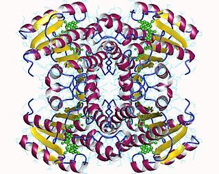
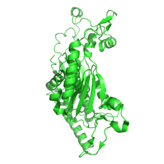
The primary structure of prostaglandin F synthase consists of 323 amino acid residues.[5] The secondary structure consists of 17 α-helices which contain 130 residues and 18 β-strands which contain 55 residues as well as many random coils. The tertiary structure is a single subunit.[2]
The active site of the enzyme is referred to as an (α/β)8 barrel because it consists of 8 α-helices and 8 β-strands. More specifically, the eight α-helices surround the eight β-strands which form the cylindrical core of the active site.[2] In addition, the active site of the enzyme contains also three random coils which help to connect the helices and strands together.[6] The size of the active site of the enzyme is large enough not only to bind NADPH cofactor but also to bind the substrates PGD2 or PGH2.[3]
Reaction
Reduction of PGD2 and PGH2.
In order for the PGFS enzyme to catalyze the reduction of the substrates PGH2 or PGD2, the cofactor NADPH must be present in the active site. This cofactor is present deep within the cavity of the enzyme and forms a hydrogen bond with it, whereas the substrate is located closer to the mouth of the cavity which limits its interaction with PGFS. The rate determining step of the catalysis is the binding of NADPH cofactor in the active site of the enzyme. This is because the binding of NADPH occurs before the binding of the substrate. NADPH is an important cofactor because it is involved in the hydride transfer which is necessary for the reduction to take place.[3]
More specifically, in order for the hydride transfer to occur, the substrate (PGD2) has to bind to the active site of the enzyme PGFS. The substrate binds to the active site through hydrogen bonding between the carbonyl group of PGD2 and the hydroxyl group of tyrosine (Y55) as well as one of the imidazole nitrogen of histidine (H117). The hydride shift from NADPH reduces the carbonyl group of PGD2 and forms a new sp3 hydroxyl group (9α,11β–PGF2).[3]
The protonation of the carbonyl oxygen is facilitated at low pH when histidine is used and at high pH when tyrosine is used for hydrogen bonding with the substrate. On the one hand, histidine is an ideal proton donor at low pH because of its pKa value (6.00), which means that it is protonated at a pH below 6.00. On the other hand, tyrosine is an ideal proton donor at higher pH because of its pKa value (10.1). The type of amino acid that is used for protonation depends on the substrate. For example, reduction of PGD2 in the human body occurs at a pH range of 6-9, which makes histidine an ideal proton donor.[3]
The hydride that is transferred to the carbonyl oxygen of PGD2 causes weakening of the hydrogen bond between the substrate and the enzyme. This has as a result the cleavage of the product (9α,11β–PGF2) from the active site of the enzyme.[3]
Use
In general, prostaglandins are molecules that are used for inflammation, muscle contraction and blood clotting.[3] Prostaglandin F synthase (PGFS) is very important enzyme because it catalyzes the formation of 9α,11β–PGF2 and PGF2α which are critical for the contraction of bronchial, vascular and arterial smooth muscle.[2]
Also, this enzyme can be used in cancer research. Recent studies have shown that there is a correlation between high levels of PGFS in gastrointestinal tumors and the effectiveness of non-steroidal anti-inflammatory drugs (NSAID). The inhibition of PGFS by NSAID could turn out to be a very important medicinal field in the development of anti-cancer medication.[6]
Inhibition
Chemical structure of bimatoprost
Prostaglandin F synthase can be inhibited not only by NSAIDs such as indometacin and suprofen but also by a molecule known as bimatoprost (BMP). BMP, an analogue of PGD2 is an ocular hypotensive agent that binds to the active site of the PGFS enzyme. This means that it inhibits the action of PGFS to catalyze the conversion of PGD2 to 9α,11β–PGF2 and PGH2 to PGF2α because it inhibits the substrate to bind to the active site of the enzyme.[6]
Related Research Articles
Prostaglandins (PG) are a group of physiologically active lipid compounds called eicosanoids that have diverse hormone-like effects in animals. Prostaglandins have been found in almost every tissue in humans and other animals. They are derived enzymatically from the fatty acid arachidonic acid. Every prostaglandin contains 20 carbon atoms, including a 5-carbon ring. They are a subclass of eicosanoids and of the prostanoid class of fatty acid derivatives.
In biochemistry, flavin adenine dinucleotide (FAD) is a redox-active coenzyme associated with various proteins, which is involved with several enzymatic reactions in metabolism. A flavoprotein is a protein that contains a flavin group, which may be in the form of FAD or flavin mononucleotide (FMN). Many flavoproteins are known: components of the succinate dehydrogenase complex, α-ketoglutarate dehydrogenase, and a component of the pyruvate dehydrogenase complex.
In molecular biology, prostanoids are active lipid mediators that regulate inflammatory response. Prostanoids are a subclass of eicosanoids consisting of the prostaglandins, the thromboxanes, and the prostacyclins. Prostanoids are seen to target NSAIDS which allow for therapeutic potential. Prostanoids are present within areas of the body such as the gastrointestinal tract, urinary tract, respiratory and cardiovascular systems, reproductive tract and vascular system. Prostanoids can even be seen with aid to the water and ion transportation within cells.
In enzymology, aldose reductase is a cytosolic NADPH-dependent oxidoreductase that catalyzes the reduction of a variety of aldehydes and carbonyls, including monosaccharides. It is primarily known for catalyzing the reduction of glucose to sorbitol, the first step in polyol pathway of glucose metabolism.
Prostaglandin D2 (or PGD2) is a prostaglandin that binds to the receptor PTGDR (DP1), as well as CRTH2 (DP2). It is a major prostaglandin produced by mast cells – recruits Th2 cells, eosinophils, and basophils. In mammalian organs, large amounts of PGD2 are found only in the brain and in mast cells. It is critical to development of allergic diseases such as asthma. Research carried out in 1989 found PGD2 is the primary mediator of vasodilation (the "niacin flush") after ingestion of niacin (nicotinic acid).
In enzymology, a carbonyl reductase (NADPH) (EC 1.1.1.184) is an enzyme that catalyzes the chemical reaction
In enzymology, a GDP-L-fucose synthase (EC 1.1.1.271) is an enzyme that catalyzes the chemical reaction
In enzymology, a prostaglandin-E2 9-reductase (EC 1.1.1.189) is an enzyme that catalyzes the chemical reaction
In enzymology, a 15-hydroxyprostaglandin-D dehydrogenase (NADP+) (EC 1.1.1.196) is an enzyme that catalyzes the chemical reaction
In enzymology, a ferredoxin-NADP+ reductase (EC 1.18.1.2) abbreviated FNR, is an enzyme that catalyzes the chemical reaction
[Methionine synthase] reductase, or Methionine synthase reductase, encoded by the gene MTRR, is an enzyme that is responsible for the reduction of methionine synthase inside human body. This enzyme is crucial for maintaining the one carbon metabolism, specifically the folate cycle. The enzyme employs one coenzyme, flavoprotein.
In enzymology, a glutamate synthase (NADH) (EC 1.4.1.14) is an enzyme that catalyzes the chemical reaction
In enzymology, a glutamate synthase (NADPH) (EC 1.4.1.13) is an enzyme that catalyzes the chemical reaction
In enzymology, a prostaglandin-D synthase is an enzyme that catalyzes the chemical reaction
The enzyme chorismate synthase catalyzes the chemical reaction
Fatty-acyl-CoA Synthase, or more commonly known as yeast fatty acid synthase, is an enzyme complex responsible for fatty acid biosynthesis, and is of Type I Fatty Acid Synthesis (FAS). Yeast fatty acid synthase plays a pivotal role in fatty acid synthesis. It is a 2.6 MDa barrel shaped complex and is composed of two, unique multi-functional subunits: alpha and beta. Together, the alpha and beta units are arranged in an α6β6 structure. The catalytic activities of this enzyme complex involves a coordination system of enzymatic reactions between the alpha and beta subunits. The enzyme complex therefore consists of six functional centers for fatty acid synthesis.
Prostaglandin F2α, pharmaceutically termed dinoprost is a naturally occurring prostaglandin used in medicine to induce labor and as an abortifacient. Prostaglandins are lipids throughout the entire body that have a hormone-like function. In pregnancy, PGF2 is medically used to sustain contracture and provoke myometrial ischemia to accelerate labor and prevent significant blood loss in labor. Additionally, PGF2 has been linked to being naturally involved in the process of labor. It has been seen that there are higher levels of PGF2 in maternal fluid during labor when compared to at term. This signifies that there is likely a biological use and significance to the production and secretion of PGF2 in labor. Prostaglandin is also used to treat uterine infections in domestic animals.
Aldo-keto reductase family 1 member C3 (AKR1C3), also known as 17β-hydroxysteroid dehydrogenase type 5 or 3α-hydroxysteroid dehydrogenase type 2 (3α-HSD2) is a steroidogenic enzyme that in humans is encoded by the AKR1C3 gene.
The aldo-keto reductase family is a family of proteins that are subdivided into 16 categories; these include a number of related monomeric NADPH-dependent oxidoreductases, such as aldehyde reductase, aldose reductase, prostaglandin F synthase, xylose reductase, rho crystallin, and many others.
3-Deoxy-D-arabinoheptulosonate 7-phosphate (DAHP) synthase is the first enzyme in a series of metabolic reactions known as the shikimate pathway, which is responsible for the biosynthesis of the amino acids phenylalanine, tyrosine, and tryptophan. Since it is the first enzyme in the shikimate pathway, it controls the amount of carbon entering the pathway. Enzyme inhibition is the primary method of regulating the amount of carbon entering the pathway. Forms of this enzyme differ between organisms, but can be considered DAHP synthase based upon the reaction that is catalyzed by this enzyme.
1 2 3 4 5 6 7 Komoto J, Yamada T, Watanabe K, Takusagawa F (March 2004). "Crystal structure of human prostaglandin F synthase (AKR1C3)". Biochemistry. 43 (8): 2188–98. doi:10.1021/bi036046x. PMID14979715.
↑ Yoshikawa K, Takei S, Hasegawa-Ishii S, Chiba Y, Furukawa A, Kawamura N, etal. (January 2011). "Preferential localization of prostamide/prostaglandin F synthase in myelin sheaths of the central nervous system". Brain Research. 1367: 22–32. doi:10.1016/j.brainres.2010.10.019. PMID20950588. S2CID43094318.
1 2 3 Komoto J, Yamada T, Watanabe K, Woodward DF, Takusagawa F (February 2006). "Prostaglandin F2alpha formation from prostaglandin H2 by prostaglandin F synthase (PGFS): crystal structure of PGFS containing bimatoprost". Biochemistry. 45 (7): 1987–96. doi:10.1021/bi051861t. PMID16475787.
Further reading
Reingold DF, Kawasaki A, Needleman P (May 1981). "A novel prostaglandin 11-keto reductase found in rabbit liver". Biochimica et Biophysica Acta (BBA) - Enzymology. 659 (1): 179–88. doi:10.1016/0005-2744(81)90282-5. PMID7248318.
Watanabe K, Shimizu T, Hayaishi O (1981). "Enzymatic conversion of prostaglandin-D2 to prostaglandin-F2α in the rat lung". Biochem. Int. 2: 603–610.
Wong PY (May 1981). "Purification and partial characterization of prostaglandin D2 11-keto reductase in rabbit liver". Biochimica et Biophysica Acta (BBA) - Enzymology. 659 (1): 169–78. doi:10.1016/0005-2744(81)90281-3. PMID7248317.
This page is based on this Wikipedia article Text is available under the CC BY-SA 4.0 license; additional terms may apply. Images, videos and audio are available under their respective licenses.