Cytochrome

Last updated
Cytochrome c with heme c. Cytochrome c.png
Cytochrome c with heme c.

Cytochromes are redox-active proteins containing a heme, with a central iron (Fe) atom at its core, as a cofactor. They are involved in the electron transport chain and redox catalysis. They are classified according to the type of heme and its mode of binding. Four varieties are recognized by the International Union of Biochemistry and Molecular Biology (IUBMB), cytochromes a, cytochromes b, cytochromes c and cytochrome d. [1]

Contents

Cytochrome function is linked to the reversible redox change from ferrous (Fe(II)) to the ferric (Fe(III)) oxidation state of the iron found in the heme core. [2] In addition to the classification by the IUBMB into four cytochrome classes, several additional classifications such as cytochrome o [3] and cytochrome P450 can be found in biochemical literature.

History

Cytochromes were initially described in 1884 by Charles Alexander MacMunn as respiratory pigments (myohematin or histohematin). [4] In the 1920s, Keilin rediscovered these respiratory pigments and named them the cytochromes, or “cellular pigments”. [5] He classified these heme proteins on the basis of the position of their lowest energy absorption band in their reduced state, as cytochromes a (605 nm), b (≈565 nm), and c (550 nm). The ultra-violet (UV) to visible spectroscopic signatures of hemes are still used to identify heme type from the reduced bis-pyridine-ligated state, i.e., the pyridine hemochrome method. Within each class, cytochrome a, b, or c, early cytochromes are numbered consecutively, e.g. cyt c, cyt c1, and cyt c2, with more recent examples designated by their reduced state R-band maximum, e.g. cyt c559. [6]

Structure and function

The heme group is a highly conjugated ring system (which allows its electrons to be very mobile) surrounding an iron ion. The iron in cytochromes usually exists in a ferrous (Fe2+) and a ferric (Fe3+) state with a ferroxo (Fe4+) state found in catalytic intermediates. [1] Cytochromes are, thus, capable of performing electron transfer reactions and catalysis by reduction or oxidation of their heme iron. The cellular location of cytochromes depends on their function. They can be found as globular proteins and membrane proteins.

In the process of oxidative phosphorylation, a globular cytochrome cc protein is involved in the electron transfer from the membrane-bound complex III to complex IV. Complex III itself is composed of several subunits, one of which is a b-type cytochrome while another one is a c-type cytochrome. Both domains are involved in electron transfer within the complex. Complex IV contains a cytochrome a/a3-domain that transfers electrons and catalyzes the reaction of oxygen to water. Photosystem II, the first protein complex in the light-dependent reactions of oxygenic photosynthesis, contains a cytochrome b subunit. Cyclooxygenase 2, an enzyme involved in inflammation, is a cytochrome b protein.

In the early 1960s, a linear evolution of cytochromes was suggested by Emanuel Margoliash [7] that led to the molecular clock hypothesis. The apparently constant evolution rate of cytochromes can be a helpful tool in trying to determine when various organisms may have diverged from a common ancestor. [8]

Types

Several kinds of cytochrome exist and can be distinguished by spectroscopy, exact structure of the heme group, inhibitor sensitivity, and reduction potential. [9]

Four types of cytochromes are distinguished by their prosthetic groups:

TypeProsthetic group
Cytochrome a heme A
Cytochrome b heme B
Cytochrome c heme C (covalently bound heme b) [10]
Cytochrome d heme D (Heme B with γ-spirolactone) [11]

There is no "cytochrome e," but cytochrome f, found in the cytochrome b6f complex of plants is a c-type cytochrome. [12]

In mitochondria and chloroplasts, these cytochromes are often combined in electron transport and related metabolic pathways: [13]

CytochromesCombination
a and a3 Cytochrome c oxidase ("Complex IV") with electrons delivered to complex by soluble cytochrome c (hence the name)
b and c1 Coenzyme Q - cytochrome c reductase ("Complex III")
b6 and f Plastoquinol—plastocyanin reductase

A distinct family of cytochromes is the cytochrome P450 family, so named for the characteristic Soret peak formed by absorbance of light at wavelengths near 450 nm when the heme iron is reduced (with sodium dithionite) and complexed to carbon monoxide. These enzymes are primarily involved in steroidogenesis and detoxification. [14] [9]

Related Research Articles

<span class="mw-page-title-main">Oxidative phosphorylation</span> Metabolic pathway

Oxidative phosphorylation or electron transport-linked phosphorylation or terminal oxidation is the metabolic pathway in which cells use enzymes to oxidize nutrients, thereby releasing chemical energy in order to produce adenosine triphosphate (ATP). In eukaryotes, this takes place inside mitochondria. Almost all aerobic organisms carry out oxidative phosphorylation. This pathway is so pervasive because it releases more energy than alternative fermentation processes such as anaerobic glycolysis.

An electron transport chain (ETC) is a series of protein complexes and other molecules that transfer electrons from electron donors to electron acceptors via redox reactions (both reduction and oxidation occurring simultaneously) and couples this electron transfer with the transfer of protons (H+ ions) across a membrane. Many of the enzymes in the electron transport chain are embedded within the membrane.

<span class="mw-page-title-main">Cytochrome c</span> Protein-coding gene in the species Homo sapiens

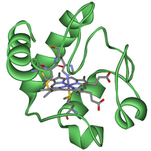
The cytochrome complex, or cyt c, is a small hemeprotein found loosely associated with the inner membrane of the mitochondrion where it plays a critical role in cellular respiration. It transfers electrons between Complexes III and IV. Cytochrome c is highly water-soluble, unlike other cytochromes. It is capable of undergoing oxidation and reduction as its iron atom converts between the ferrous and ferric forms, but does not bind oxygen. It also plays a major role in cell apoptosis. In humans, cytochrome c is encoded by the CYCS gene.

<span class="mw-page-title-main">Hemoprotein</span> Protein containing a heme prosthetic group

A hemeprotein, or heme protein, is a protein that contains a heme prosthetic group. They are a very large class of metalloproteins. The heme group confers functionality, which can include oxygen carrying, oxygen reduction, electron transfer, and other processes. Heme is bound to the protein either covalently or noncovalently or both.

<span class="mw-page-title-main">Coenzyme Q – cytochrome c reductase</span> Class of enzymes

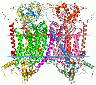
The coenzyme Q : cytochrome c – oxidoreductase, sometimes called the cytochrome bc1 complex, and at other times complex III, is the third complex in the electron transport chain, playing a critical role in biochemical generation of ATP. Complex III is a multisubunit transmembrane protein encoded by both the mitochondrial and the nuclear genomes. Complex III is present in the mitochondria of all animals and all aerobic eukaryotes and the inner membranes of most eubacteria. Mutations in Complex III cause exercise intolerance as well as multisystem disorders. The bc1 complex contains 11 subunits, 3 respiratory subunits, 2 core proteins and 6 low-molecular weight proteins.

<span class="mw-page-title-main">Heme</span> Chemical coordination complex of an iron ion chelated to a porphyrin

Heme, or haem, is a precursor to hemoglobin, which is necessary to bind oxygen in the bloodstream. Heme is biosynthesized in both the bone marrow and the liver.

Cytochrome C1 is a protein encoded by the CYC1 gene. Cytochrome is a heme-containing subunit of the cytochrome b-c1 complex, which accepts electrons from Rieske protein and transfers electrons to cytochrome c in the mitochondrial respiratory chain. It is formed in the cytosol and targeted to the mitochondrial intermembrane space. Cytochrome c1 belongs to the cytochrome c family of proteins.

<span class="mw-page-title-main">Flavin adenine dinucleotide</span> Redox-active coenzyme

In biochemistry, flavin adenine dinucleotide (FAD) is a redox-active coenzyme associated with various proteins, which is involved with several enzymatic reactions in metabolism. A flavoprotein is a protein that contains a flavin group, which may be in the form of FAD or flavin mononucleotide (FMN). Many flavoproteins are known: components of the succinate dehydrogenase complex, α-ketoglutarate dehydrogenase, and a component of the pyruvate dehydrogenase complex.

Cytochrome b<sub>6</sub>f complex Enzyme

The cytochrome b6f complex (plastoquinol/plastocyanin reductase or plastoquinol/plastocyanin oxidoreductase; EC 7.1.1.6) is an enzyme found in the thylakoid membrane in chloroplasts of plants, cyanobacteria, and green algae, that catalyzes the transfer of electrons from plastoquinol to plastocyanin:

<span class="mw-page-title-main">Rieske protein</span> Protein family with an iron–sulfur center transferring electrons

Rieske proteins are iron–sulfur protein (ISP) components of cytochrome bc1 complexes and cytochrome b6f complexes and are responsible for electron transfer in some biological systems. John S. Rieske and co-workers first discovered the protein and in 1964 isolated an acetylated form of the bovine mitochondrial protein. In 1979 Trumpower's lab isolated the "oxidation factor" from bovine mitochondria and showed it was a reconstitutively-active form of the Rieske iron-sulfur protein
It is a unique [2Fe-2S] cluster in that one of the two Fe atoms is coordinated by two histidine residues rather than two cysteine residues. They have since been found in plants, animals, and bacteria with widely ranging electron reduction potentials from -150 to +400 mV.

<span class="mw-page-title-main">Cytochrome b</span> Membrane protein involved in the electron transport chain

Cytochrome b within both molecular and cell biology, is a protein found in the membranes of aerobic cells. In eukaryotic mitochondria and in aerobic prokaryotes, cytochrome b is a component of respiratory chain complex III — also known as the bc1 complex or ubiquinol-cytochrome c reductase. In plant chloroplasts and cyanobacteria, there is an homologous protein, cytochrome b6, a component of the plastoquinone-plastocyanin reductase, also known as the b6f complex. These complexes are involved in electron transport, the pumping of protons to create a proton-motive force (PMF). This proton gradient is used for the generation of ATP. These complexes play a vital role in cells.

Iron-binding proteins are carrier proteins and metalloproteins that are important in iron metabolism and the immune response. Iron is required for life.

<span class="mw-page-title-main">Heme C</span> Chemical compound

Heme C is an important kind of heme.

<span class="mw-page-title-main">Heme A</span> Chemical compound

Heme A is a heme, a coordination complex consisting of a macrocyclic ligand called a porphyrin, chelating an iron atom. Heme A is a biomolecule and is produced naturally by many organisms. Heme A, often appears a dichroic green/red when in solution, is a structural relative of heme B, a component of hemoglobin, the red pigment in blood.

In enzymology, a cellobiose dehydrogenase (acceptor) (EC 1.1.99.18) is an enzyme that catalyzes the chemical reaction

<span class="mw-page-title-main">Cytochrome b559</span> Family of protein complexes

Cytochrome b559 is an important component of Photosystem II (PSII) is a multisubunit protein-pigment complex containing polypeptides both intrinsic and extrinsic to the photosynthetic membrane. Within the core of the complex, the chlorophyll and beta-carotene pigments are mainly bound to the antenna proteins CP43 (PsbC) and CP47 (PsbB), which pass the excitation energy on to chlorophylls in the reaction centre proteins D1 and D2 that bind all the redox-active cofactors involved in the energy conversion process. The PSII oxygen-evolving complex (OEC) provides electrons to re-reduce the PSII reaction center, and oxidizes 2 water molecules to recover its reduced initial state. It consists of OEE1 (PsbO), OEE2 (PsbP) and OEE3 (PsbQ). The remaining subunits in PSII are of low molecular weight, and are involved in PSII assembly, stabilisation, dimerization, and photoprotection.

<span class="mw-page-title-main">Cytochrome c family</span> Protein family

Cytochromes c cytochromes, or heme-containing proteins, that have heme C covalently attached to the peptide backbone via one or two thioether bonds. These bonds are in most cases part of a specific Cys-X-X-Cys-His (CXXCH) binding motif, where X denotes a miscellaneous amino acid. Two thioether bonds of cysteine residues bind to the vinyl sidechains of heme, and the histidine residue coordinates one axial binding site of the heme iron. Less common binding motifs can include a single thioether linkage, a lysine or a methionine instead of the axial histidine or a CXnCH binding motif with n>2. The second axial site of the iron can be coordinated by amino acids of the protein, substrate molecules or water. Cytochromes c possess a wide range of properties and function as electron transfer proteins or catalyse chemical reactions involving redox processes. A prominent member of this family is mitochondrial cytochrome c.

<span class="mw-page-title-main">Light-dependent reactions</span> Photosynthetic reactions

Light-dependent reactions refers to certain photochemical reactions that are involved in photosynthesis, the main process by which plants acquire energy. There are two light dependent reactions, the first occurs at photosystem II (PSII) and the second occurs at photosystem I (PSI).

<span class="mw-page-title-main">Cytochrome d</span>

Cytochrome d, previously known as cytochrome a2, is a name for all cytochromes that contain heme D as a cofactor. Two unrelated classes of cytochrome d are known: Cytochrome bd, an enzyme that generates a charge across the membrane so that protons will move, and cytochrome cd1, a nitrite reductase.

In the field of enzymology, murburn is a term coined by Kelath Murali Manoj that explains the catalytic mechanism of certain redox-active proteins. The term describes the equilibrium among molecules, unbound ions and radicals, signifying a process of "mild unrestricted redox catalysis".

References

  1. 1 2 "Nomenclature Committee of the International Union of Biochemistry (NC-IUB). Nomenclature of electron-transfer proteins. Recommendations 1989". Journal of Biological Chemistry. 267 (1): 665–677. 1992-01-05. doi: 10.1016/S0021-9258(18)48544-4 . ISSN   0021-9258. PMID   1309757.
  2. L., Lehninger, Albert (2000). Lehninger Principles of Biochemistry (3rd ed.). New York: Worth Publishers. ISBN   978-1572591530. OCLC   42619569.{{cite book}}: CS1 maint: multiple names: authors list (link)
  3. Puustinen, A.; Wikström, M. (1991-07-15). "The heme groups of cytochrome o from Escherichia coli". Proceedings of the National Academy of Sciences. 88 (14): 6122–6126. Bibcode:1991PNAS...88.6122P. doi: 10.1073/pnas.88.14.6122 . ISSN   0027-8424. PMC   52034 . PMID   2068092.
  4. Mac Munn, C. A. (1886). "Researches on Myohaematin and the Histohaematins". Philosophical Transactions of the Royal Society of London. 177: 267–298. doi:10.1098/rstl.1886.0007. JSTOR   109482. S2CID   110335335.
  5. Keilin, D. (1925-08-01). "On cytochrome, a respiratory pigment, common to animals, yeast, and higher plants". Proc. R. Soc. Lond. B. 98 (690): 312–339. Bibcode:1925RSPSB..98..312K. doi: 10.1098/rspb.1925.0039 . ISSN   0950-1193.
  6. Reedy, C. J.; Gibney, B. R. (February 2004). "Heme protein assemblies". Chem Rev. 104 (2): 617–49. doi:10.1021/cr0206115. PMID   14871137.
  7. Margoliash, E. (1963). "Primary Structure and Evolution of Cytochrome C". Proceedings of the National Academy of Sciences of the United States of America. 50 (4): 672–679. Bibcode:1963PNAS...50..672M. doi: 10.1073/pnas.50.4.672 . ISSN   0027-8424. PMC   221244 . PMID   14077496.
  8. Kumar, Sudhir (2005). "Molecular clocks: four decades of evolution". Nature Reviews. Genetics. 6 (8): 654–662. doi:10.1038/nrg1659. ISSN   1471-0056. PMID   16136655. S2CID   14261833.
  9. 1 2 "Investigation of biological oxidation, oxidative phosphorylation and ATP synthesis. Inhibitor and Uncouplers of oxidative phosphorylation". Archived from the original on 2020-06-28. Retrieved 2020-02-02.
  10. Cytochrome+c+Group at the U.S. National Library of Medicine Medical Subject Headings (MeSH).
  11. Murshudov, G.; Grebenko, A.; Barynin, V.; Dauter, Z.; Wilson, K.; Vainshtein, B.; Melik-Adamyan, W.; Bravo, J.; Ferrán, J.; Ferrer, J. C.; Switala, J.; Loewen, P. C.; Fita, I. (1996). "Structure of the heme d of Penicillium vitale and Escherichia coli catalases". The Journal of Biological Chemistry. 271 (15): 8863–8868. doi: 10.1074/jbc.271.15.8863 . PMID   8621527.
  12. Bendall, Derek S. (2004). "The Unfinished Story of Cytochrome f". Photosynthesis Research. 80 (1–3): 265–276. doi:10.1023/b:pres.0000030454.23940.f9. ISSN   0166-8595. PMID   16328825. S2CID   16716904.
  13. Doidge, Norman (2015). The brain's way of healing : remarkable discoveries and recoveries from the frontiers of neuroplasticity. Penguin Group. p. 173. ISBN   978-0-698-19143-3.
  14. Miller, Walter L.; Gucev, Zoran S. (2014), "Disorders in the Initial Steps in Steroidogenesis", Genetic Steroid Disorders, Elsevier, pp. 145–164, doi:10.1016/b978-0-12-416006-4.00011-9, ISBN   9780124160064