Cytochrome d

Last updated
Ubiquinol oxidase (electrogenic, proton-motive force generating; Cytochrome bd)
Cytochrome bd CydABX predicted 26335199.png
Predicted structure of E. coli Cytochrome bd-1
Identifiers
EC no. 7.1.1.7
Databases
IntEnz IntEnz view
BRENDA BRENDA entry
ExPASy NiceZyme view
KEGG KEGG entry
MetaCyc metabolic pathway
PRIAM profile
PDB structures RCSB PDB PDBe PDBsum

Cytochrome d, previously known as cytochrome a2, is a name for all cytochromes (electron-transporting heme proteins) that contain heme D as a cofactor. Two unrelated classes of cytochrome d are known: Cytochrome bd, an enzyme that generates a charge across the membrane so that protons will move, [1] and cytochrome cd1 (NirS; SCOP b.70.2 ), a nitrite reductase. [2]

Contents

Cytochrome bd is found in plenty of aerobic bacteria, especially when it has grown with a limited oxygen supply. Compared to other terminal oxidases, it is notable for its high oxygen affinity and resistance to cyanide poisoning. It has a group of very similar relatives that do not use heme D, known as cyanide insensitive oxidases (CIOs). [3]

Function

Structure of Heme D. Heme d.svg
Structure of Heme D.

Cytochrome d is, as other proteins of its family, a membrane-bound hemeprotein, but unlike cytochromes a and b, cytochrome D has a heme D instead of a heme A or heme B group. [4]

Cytochrome d is part of the cytochrome bd terminal oxidase which catalyse the two electron oxidation of ubiquinol. This process is an oxidative phosphorylation that oxidizes the ubiquinol-8 to ubiquinone. The chemical reaction followed by this process is:

Ubiquinol-8 + O2 → Ubiquinone-8 + H2O [5]

By a similar reaction, it also catalyses the reduction of oxygen to water, which involves 4 electrons.

As a terminal oxidase, the reaction generates a proton motive force:

2 ubiquinol[inner membrane] + O2 + 4 H+[cytoplasm] → 2 ubiquinone[inner membrane] + 2 H2O + 4 H+[periplasm]

Some members of the family may accept or prefer other electron-transporting quinols such as menaquinol or plastoquinol in lieu of ubiquinol. [3]

Structure

Cytochrome bd (OPM family 805) is a tri-heme oxidase as it is compound by cytochromes b558, b595 and d. Its main function is the reduction of O2 to H2O. It is thought that it uses a di-heme active site, which is formed by the hemes of cytochromes b595 and d. These two cytochromes are considered high-spin complexes, what is directly related to the electrons' spin. While other respiratory terminal oxidases which catalyze that same reaction have a heme-copper active site and use a proton pump, cytochrome bd has an active site with iron instead of copper and need no proton pump as they can produce a proton-motion force themselves. [6] They are embedded in the bacterial cytoplasmic bilayer and serve as terminal oxidases in the respiratory chain. [7]

The oxidases tend to have two or three subunits. Subunits 1 (InterPro :  IPR003317 ) and 2 (InterPro :  IPR002585 ) are predicted to have pseudo-symmetry, and are sufficient to bind the two heme b molecules. [8] Some proteobacterial assemblies require a third subunit (InterPro :  IPR012994 ) to bind heme d; others do not. [9]

The high-resolution structure heterotrimeric Cytochromes bd from Geobacillus species has been determined ( PDB: 5IR6, 5DOQ ). The third subunit does not share sequence homology with the third subunit proteobacteria, but does come into the assemblies at a similar position. [10]

Occurrence

Escherichia coli

E. coli possess two sets of Cytochrome bd. [7] The bd-I complex (CydABX) is a heterotrimer, while the bd-II complex (AppCB) is a heterodimer. There is an AppX gene that may correspond to a subunit 3 for AppCB. [9]

The ability of bd-II to generate a proton motive force is a matter of recent debate, putting it under the nonelectrogenic Ubiquinol oxidase (H+-transporting) in some categorizations. [11]

Azotobacter vinelandii

Azotobacter vinelandii is a nitrogen-fixing bacteria which is known by its high respiratory rate among aerobic organisms. Some physiological studies postulate that cytochrome d functions as a terminal oxidase in the membranes of this organism, taking part in the electron transport system. The studies characterized the different genes in the two subunits ( Q09049 , C1DEL1 ; third subunit C1DEL0 ). A very extensive homology with CydAB of the E. coli was found in these studies. [12]

Spectra

Generally, in protein complexes, cytochrome D gives an absorption band of approximately 636 nm or 638 nm, depending on the cytochrome d form. If it is oxidized, the band has a length of 636 nm, and a 638 nm length if it is reduced. It is commonly associated to certain prosthetic groups when found in multiple subunit complexes. Detecting cytochrome d as Fe(II) pyridine alkaline hemachrome is very difficult because the stability under these conditions is limited. If cytochrome d is pulled out of the protein complex (as heme D) and placed in ether containing from 1 to 5 % of HCl, it gives a different absorption band (603 nm, in the oxidized form). [2]

Related Research Articles

Cytochrome Redox-active proteins containing a heme with a Fe atom as a cofactor

Cytochromes are redox-active proteins containing a heme, with a central Fe atom at its core, as a cofactor. They are involved in electron transport chain and redox catalysis. They are classified according to the type of heme and its mode of binding. Four varieties are recognized by the International Union of Biochemistry and Molecular Biology (IUBMB), cytochromes a, cytochromes b, cytochromes c and cytochrome d. Cytochrome function is linked to the reversible redox change from ferrous to the ferric oxidation state of the iron found in the heme core. In addition to the classification by the IUBMB into four cytochrome classes, several additional classifications such as cytochrome o and cytochrome P450 can be found in biochemical literature.

Oxidative phosphorylation The phosphorylation of ADP to ATP that accompanies the oxidation of a metabolite through the operation of the respiratory chain. Oxidation of compounds establishes a proton gradient across the membrane, providing the energy for ATP synthesis.

Oxidative phosphorylation or electron transport-linked phosphorylation or terminal oxidation is the metabolic pathway in which cells use enzymes to oxidize nutrients, thereby releasing the chemical energy stored within the nutrients in order to produce adenosine triphosphate (ATP). In eukaryotes, this takes place inside mitochondria. Almost all aerobic organisms carry out oxidative phosphorylation. This pathway is so pervasive because it releases more energy than alternative fermentation processes such as anaerobic glycolysis.

Electron transport chain Process in which a series of electron carriers operate together to transfer electrons from donors to any of several different terminal electron acceptors to generate a transmembrane electrochemical gradient.

The electron transport chain (ETC; respiratory chain) is a series of protein complexes that transfer electrons from electron donors to electron acceptors via redox reactions (both reduction and oxidation occurring simultaneously) and couples this electron transfer with the transfer of protons (H+ ions) across a membrane. The electron transport chain is built up of peptides, enzymes, and other molecules.

Cytochrome c oxidase

The enzyme cytochrome c oxidase or Complex IV, EC 1.9.3.1, is a large transmembrane protein complex found in bacteria, archaea, and the mitochondria of eukaryotes.

Respiratory complex I

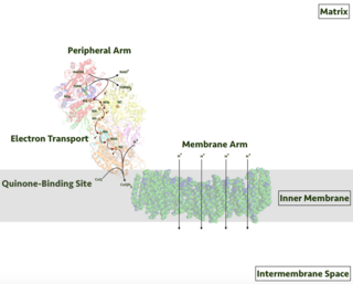
Respiratory complex I, EC 7.1.1.2 is the first large protein complex of the respiratory chains of many organisms from bacteria to humans. It catalyzes the transfer of electrons from NADH to coenzyme Q10 (CoQ10) and translocates protons across the inner mitochondrial membrane in eukaryotes or the plasma membrane of bacteria.

Coenzyme Q – cytochrome c reductase Class of enzymes

The coenzyme Q : cytochrome c – oxidoreductase, sometimes called the cytochrome bc1 complex, and at other times complex III, is the third complex in the electron transport chain, playing a critical role in biochemical generation of ATP. Complex III is a multisubunit transmembrane protein encoded by both the mitochondrial and the nuclear genomes. Complex III is present in the mitochondria of all animals and all aerobic eukaryotes and the inner membranes of most eubacteria. Mutations in Complex III cause exercise intolerance as well as multisystem disorders. The bc1 complex contains 11 subunits, 3 respiratory subunits, 2 core proteins and 6 low-molecular weight proteins.

Succinate dehydrogenase

Succinate dehydrogenase (SDH) or succinate-coenzyme Q reductase (SQR) or respiratory complex II is an enzyme complex, found in many bacterial cells and in the inner mitochondrial membrane of eukaryotes. It is the only enzyme that participates in both the citric acid cycle and the electron transport chain. Histochemical analysis showing high succinate dehydrogenase in muscle demonstrates high mitochondrial content and high oxidative potential.

Rieske protein

Rieske proteins are iron–sulfur protein (ISP) components of cytochrome bc1 complexes and cytochrome b6f complexes and are responsible for electron transfer in some biological systems. John S. Rieske and co-workers first discovered the protein and in 1964 isolated an acetylated form of the bovine mitochondrial protein. In 1979 Trumpower's lab isolated the "oxidation factor" from bovine mitochondria and showed it was a reconstitutively-active form of the Rieske iron-sulfur protein
It is a unique [2Fe-2S] cluster in that one of the two Fe atoms is coordinated by two histidine residues rather than two cysteine residues. They have since been found in plants, animals, and bacteria with widely ranging electron reduction potentials from -150 to +400 mV.

Q cycle

The Q cycle describes a series of reactions that describe how the sequential oxidation and reduction of the lipophilic electron carrier, Coenzyme Q10 (CoQ10), between the ubiquinol and ubiquinone forms, can result in the net movement of protons across a lipid bilayer.

Heme A Chemical compound

Heme A is a heme, a coordination complex consisting of a macrocyclic ligand called a porphyrin, chelating an iron atom. Heme A is a biomolecule and is produced naturally by many organisms. Heme A, often appears a dichroic green/red when in solution, is a structural relative of heme B, a component of hemoglobin, the red pigment in blood.

Ubiquinol oxidases are enzymes in the bacterial electron transport chain that oxidise ubiquinol into ubiquinone and reduce oxygen to water. These enzymes are one set of the many alternative terminal oxidases in the branched prokaryotic electron transport chain. The overall structure of the E. coli ubiquinol oxidase is similar to that of the mammalian Cytochrome c oxidase, with the addition of a polar ubiquinol-binding site embedded in the membrane.

Cytochrome c oxidase subunit I Enzyme of the respiratory chain encoded by the mitochondrial genome

Cytochrome c oxidase I (COX1) also known as mitochondrially encoded cytochrome c oxidase I (MT-CO1) is a protein that in humans is encoded by the MT-CO1 gene. In other eukaryotes, the gene is called COX1, CO1, or COI. Cytochrome c oxidase I is the main subunit of the cytochrome c oxidase complex. Mutations in MT-CO1 have been associated with Leber's hereditary optic neuropathy (LHON), acquired idiopathic sideroblastic anemia, Complex IV deficiency, colorectal cancer, sensorineural deafness, and recurrent myoglobinuria.

Cytochrome c oxidase subunit III Enzyme of the respiratory chain encoded by the mitochondrial genome

Cytochrome c oxidase subunit III (COX3) is an enzyme that in humans is encoded by the MT-CO3 gene. It is one of main transmembrane subunits of cytochrome c oxidase. Cytochrome c oxidase subunit III is also one of the three mitochondrial DNA (mtDNA) encoded subunits of respiratory complex IV. Variants of MT-CO3 have been associated with isolated myopathy, severe encephalomyopathy, Leber hereditary optic neuropathy, mitochondrial complex IV deficiency, and recurrent myoglobinuria.

COX5B

Cytochrome c oxidase subunit 5B, mitochondrial is an enzyme in humans that is a subunit of the cytochrome c oxidase complex, also known as Complex IV, the last enzyme in the mitochondrial electron transport chain. In humans, cytochrome c oxidase subunit 5B is encoded by the COX5B gene.

UQCRB

Ubiquinol-cytochrome c reductase binding protein, also known as UQCRB, Complex III subunit 7, QP-C, or Ubiquinol-cytochrome c reductase complex 14 kDa protein is a protein which in humans is encoded by the UQCRB gene. This gene encodes a subunit of the ubiquinol-cytochrome c oxidoreductase complex, which consists of one mitochondrial-encoded and 10 nuclear-encoded subunits. Mutations in this gene are associated with mitochondrial complex III deficiency. Alternatively spliced transcript variants have been found for this gene. Related pseudogenes have been identified on chromosomes 1, 5 and X.

Fumarate reductase (quinol)

Fumarate reductase (quinol) is an enzyme with systematic name succinate:quinone oxidoreductase. This enzyme catalyzes the following chemical reaction:

Pyruvate dehydrogenase (quinone) (EC 1.2.5.1, pyruvate dehydrogenase, pyruvic dehydrogenase, pyruvic (cytochrome b1) dehydrogenase, pyruvate:ubiquinone-8-oxidoreductase, pyruvate oxidase (ambiguous)) is an enzyme with systematic name pyruvate:ubiquinone oxidoreductase. This enzyme catalyses the following chemical reaction

Ubiquinol oxidase (H+-transporting) (EC 7.1.1.3, cytochrome bb3 oxidase, cytochrome bo oxidase, cytochrome bd-I oxidase) is an enzyme with systematic name ubiquinol:O2 oxidoreductase (H+-transporting). This enzyme catalyses the following chemical reaction

COX8A

Cytochrome c oxidase subunit 8A (COX8A) is a protein that in humans is encoded by the COX8A gene. Cytochrome c oxidase 8A is a subunit of the cytochrome c oxidase complex, also known as Complex IV. Mutations in the COX8A gene have been associated with complex IV deficiency with Leigh syndrome and epilepsy.

NDUFA4

NDUFA4, mitochondrial complex associated is a protein that in humans is encoded by the NDUFA4 gene. The NDUFA3 protein is a subunit of NADH dehydrogenase (ubiquinone), which is located in the mitochondrial inner membrane and is the largest of the five complexes of the electron transport chain. Mutations in the NDUFA4 gene are associated with Leigh's syndrome.

References

  1. EC 7.1.1.7
  2. 1 2 "Nomenclature Committee of the International Union of Biochemistry (NC-IUB). Nomenclature of electron-transfer proteins. Recommendations 1989" (PDF). European Journal of Biochemistry. 200 (3): 599–611. September 1991. doi: 10.1111/j.1432-1033.1991.tb16223.x . PMID   1655423.
  3. 1 2 Borisov VB, Gennis RB, Hemp J, Verkhovsky MI (November 2011). "The cytochrome bd respiratory oxygen reductases". Biochimica et Biophysica Acta (BBA) - Bioenergetics. 1807 (11): 1398–413. doi:10.1016/j.bbabio.2011.06.016. PMC   3171616 . PMID   21756872.
  4. Belevich I, Borisov VB, Konstantinov AA, Verkhovsky MI (August 2005). "Oxygenated complex of cytochrome bd from Escherichia coli: stability and photolability". FEBS Letters. 579 (21): 4567–70. doi:10.1016/j.febslet.2005.07.011. PMID   16087180. S2CID   36465802.
  5. Cytochrome d ubiquinol oxidase subunit 1
  6. Borisov VB, Verkhovsky MI (January 2013). "Accommodation of CO in the di-heme active site of cytochrome bd terminal oxidase from Escherichia coli". Journal of Inorganic Biochemistry. 118: 65–7. doi:10.1016/j.jinorgbio.2012.09.016. PMID   23123340.
  7. 1 2 Michael J. Miller, Robert B. Gennis. The Cytochrome d Complex Is a Coupling Site in the Aerobic Respiratory Chain of Escherichia coli. The Journal of Biological Chemistry Vol.260 No.26 (1985)
  8. Ovchinnikov S, Kinch L, Park H, Liao Y, Pei J, Kim DE, Kamisetty H, Grishin NV, Baker D (September 2015). "Large-scale determination of previously unsolved protein structures using evolutionary information". eLife. 4: e09248. doi:10.7554/eLife.09248. PMC   4602095 . PMID   26335199.
  9. 1 2 Escherichia coli K-12 substr. MG1655 Transporter: cytochrome bd-I terminal oxidase
  10. Safarian S, Rajendran C, Müller H, Preu J, Langer JD, Ovchinnikov S, Hirose T, Kusumoto T, Sakamoto J, Michel H (April 2016). "Structure of a bd oxidase indicates similar mechanisms for membrane-integrated oxygen reductases". Science. 352 (6285): 583–6. Bibcode:2016Sci...352..583S. doi:10.1126/science.aaf2477. PMC   5515584 . PMID   27126043.
  11. Borisov VB, Murali R, Verkhovskaya ML, Bloch DA, Han H, Gennis RB, Verkhovsky MI (October 2011). "Aerobic respiratory chain of Escherichia coli is not allowed to work in fully uncoupled mode". Proceedings of the National Academy of Sciences of the United States of America. 108 (42): 17320–4. Bibcode:2011PNAS..10817320B. doi: 10.1073/pnas.1108217108 . PMC   3198357 . PMID   21987791.
  12. Jones CW, Redfearn ER. The cytochrome system of Azotobacter vinelandii. Biochim Biophys Acta. 1967 Sep 6;143(2):340–353