Female reproductive system

Last updated
Female reproductive system (human)
Female reproductive system lateral 1.png
A pictorial illustration of the female reproductive system
Details
Identifiers
Latin systema genitale femininum
MeSH D005836
TA98 A09.1.00.001
TA2 3469
FMA 45663
Anatomical terminology
1. Vulva: 2. Labia majora; 3. Labia minora; 4. Vestibule; 5. Clitoris: (with 6. Glans and 7. Body). 8. Bulb of vestibule
9. Vagina: 10. Hymen; 11. Lumen; 12. Wall; 13. Fornix (lateral)
14. Uterus: Parts: 15. Cervix; 16. Body and 17. Fundus. 18. Orifices: external and internal; 19. Cervical canal; 20. Uterine cavity; Layers: 21. Endometrium; 22. Myometrium and 23. Perimetrium
24. Fallopian tube: 25. Isthmus; 26. Ampulla; 27. Infundibulum; 28. Fimbriae (with 29. Fimbria ovarica)
30. Ovary
31. Visceral pelvic peritoneum: 32. Broad ligament (with 33. Mesosalpinx; 34. Mesovarium and 35. Mesometrium)
Ligaments: 36. Round; 37. Ovarian; 38. Suspensory of ovary
Blood vessels: 39. Ovarian artery and vein; 40. Uterine artery and veins; 41. Vaginal artery and veins
Other: 42. Ureter; 43. Pelvic floor (Levator ani); 44. Femoral head; 45. Hip bone; 46. Internal iliac vessels (anterior branches); 47. External iliac vessels; 48. Abdominal cavity Scheme female reproductive system-number-full-cropped.svg
1. Vulva : 2.  Labia majora; 3.  Labia minora; 4.  Vestibule; 5.  Clitoris : (with 6. Glans and 7. Body). 8.  Bulb of vestibule
9. Vagina : 10.  Hymen; 11. Lumen; 12. Wall; 13.  Fornix (lateral)
14. Uterus : Parts: 15.  Cervix; 16.  Body and 17.  Fundus. 18. Orifices: external and internal; 19.  Cervical canal; 20.  Uterine cavity; Layers: 21.  Endometrium; 22.  Myometrium and 23.  Perimetrium
24. Fallopian tube : 25.  Isthmus; 26.  Ampulla; 27.  Infundibulum; 28.  Fimbriae (with 29. Fimbria ovarica)
30. Ovary
31. Visceral pelvic peritoneum : 32.  Broad ligament (with 33.  Mesosalpinx; 34.  Mesovarium and 35.  Mesometrium)
Ligaments: 36.  Round; 37.  Ovarian; 38.  Suspensory of ovary
Blood vessels: 39.  Ovarian artery and vein; 40.  Uterine artery and veins; 41.  Vaginal artery and veins
Other: 42.  Ureter; 43.  Pelvic floor (Levator ani); 44.  Femoral head; 45.  Hip bone; 46.  Internal iliac vessels (anterior branches); 47.  External iliac vessels; 48.  Abdominal cavity

The female reproductive system is made up of the internal and external sex organs that function in the reproduction of new offspring. The human female reproductive system is immature at birth and develops to maturity at puberty to be able to produce gametes, and to carry a fetus to full term. The internal sex organs are the vagina, uterus, fallopian tubes, and ovaries. The female reproductive tract includes the vagina, uterus, and fallopian tubes and is prone to infections. [1] The vagina allows for sexual intercourse and childbirth, and is connected to the uterus at the cervix. The uterus or womb accommodates the embryo, which develops into the fetus. The uterus also produces secretions, which help the transit of sperm to the fallopian tubes, where sperm fertilize ova (egg cells) produced by the ovaries. The external sex organs are also known as the genitals and these are the organs of the vulva including the labia, clitoris, and vaginal opening. [2]

Contents

During the menstrual cycle, the ovaries release an ovum, which transits through the fallopian tube into the uterus. If an egg cell meets with sperm on its way to the uterus, a single sperm cell can enter and merge with it, fertilizing it into a zygote.

Fertilization usually occurs in the fallopian tubes and marks the beginning of embryogenesis. The zygote will then divide over enough generations of cells to form a blastocyst, which implants itself in the wall of the uterus. This begins the period of gestation and the embryo will continue to develop until full-term. When the fetus has developed enough to survive outside the uterus, the cervix dilates and contractions of the uterus propel the newborn through the birth canal (the vagina).

The corresponding equivalent among males is the male reproductive system.

External genitalia

External female genitalia; a human vulva Michaela.JPG
External female genitalia; a human vulva

Vulva

The vulva is of all of the external parts and tissues and includes the following: [3]

Internal genitalia

Sagittal MRI showing an extreme anteversion of the uterus and dislocation of the cervix uteri. Female pelvis with extreme anteversion of the uterus.png
Sagittal MRI showing an extreme anteversion of the uterus and dislocation of the cervix uteri.
Illustration depicting a healthy female reproductive system (sagittal view) Blausen 0400 FemaleReproSystem 02.png
Illustration depicting a healthy female reproductive system (sagittal view)
Frontal view as scheme of reproductive organs Scheme female reproductive system-en.svg
Frontal view as scheme of reproductive organs

Vagina

The vagina is a fibromuscular (made up of fibrous and muscular tissue) canal leading from the outside of the body to the cervix of the uterus. It is also referred to as the birth canal in the context of pregnancy. The vagina accommodates a penis during sexual intercourse. Semen containing spermatozoa is ejaculated from the penis at orgasm, into the vagina potentially enabling fertilization of the egg cell (ovum) to take place.

Cervix

The cervix is the neck of the uterus, the lower, narrow portion where it joins with the upper part of the vagina. It is cylindrical or conical in shape and protrudes through the upper anterior vaginal wall. Approximately half its length is visible, the remainder lies above the vagina beyond view. The vagina has a thick layer outside and it is the opening where the fetus emerges during delivery.

Uterus

The uterus or womb is the major female reproductive organ. The uterus provides mechanical protection, nutritional support, and waste removal for the developing embryo (weeks 1 to 8) and fetus (from week 9 until the delivery). In addition, contractions in the muscular wall of the uterus are important in pushing out the fetus at the time of birth.

The uterus contains three suspensory ligaments that help stabilize the position of the uterus and limits its range of movement. The uterosacral ligaments keep the body from moving inferiorly and anteriorly. The round ligaments restrict posterior movement of the uterus. The cardinal ligaments also prevent the inferior movement of the uterus.

The uterus is a pear-shaped muscular organ. Its major function is to accept a fertilized ovum, which becomes implanted into the endometrium, and derives nourishment from blood vessels, which develop exclusively for this purpose. The fertilized ovum becomes an embryo, develops into a fetus and gestates until childbirth. If the egg does not embed in the wall of the uterus, the female begins menstruation. [4]

Fallopian tubes

The fallopian tubes are two tubes leading from the ovaries into the uterus. On maturity of an ovum, the follicle and the ovary's wall rupture, allowing the ovum to escape and enter the fallopian tube. There it travels toward the uterus, pushed along by movements of cilia on the inner lining of the tubes. This trip takes hours or days. If the ovum is fertilized while in the fallopian tube, then it normally implants in the endometrium when it reaches the uterus, which signals the beginning of pregnancy. [5]

Ovaries

The ovaries are small, paired gonads located near the lateral walls of the pelvic cavity. These organs are responsible for the production of the egg cells (ova) and the secretion of hormones. The process by which the egg cell (ovum) is released is called ovulation. The speed of ovulation is periodic and impacts directly to the length of a menstrual cycle.

After ovulation, the egg cell is travels through the fallopian tube toward the uterus. If fertilization is going to occur, it often happens in the fallopian tube; the fertilized egg can then proceed to try and implant on the uterus's lining. [6] [7] During fertilization the egg cell plays a role; it releases certain molecules that are essential to guiding the sperm and allows the surface of the egg to attach to the sperm's surface. The egg can then absorb the sperm and fertilization can then begin. [8]

Function

The female reproductive system functions to produce offspring.

In the absence of fertilization, the ovum will eventually traverse the entire reproductive tract from the fallopian tube until exiting the vagina through menstruation.

The reproductive tract can be used for various transluminal procedures such as fertiloscopy, intrauterine insemination, and transluminal sterilization.

Development

Chromosome characteristics determine the genetic sex of a fetus at conception. This is specifically based on the 23rd pair of chromosomes that is inherited. Since the mother's egg contains an X chromosome and the father's sperm contains either an X or Y chromosome, it is the male who determines the fetus' sex. If the fetus inherits the X chromosome from the father, the fetus will be a female. In this case, testosterone is not made and the Wolffian duct will degrade thus, the Müllerian duct will develop into female sex organs. The clitoris is the remnants of the Wolffian duct. On the other hand, if the fetus inherits the Y chromosome from the father, the fetus will be a male. The presence of testosterone will stimulate the Wolffian duct, which will bring about the development of the male sex organs and the Müllerian duct will degrade. [9]

Clinical significance

Vaginitis

Vaginitis is inflammation of the vagina and largely caused by an infection. It is the most common gynaecological condition presented. [10] It is difficult to determine any one organism most responsible for vaginitis because it varies from range of age, sexual activity, and method of microbial identification. Vaginitis is not necessarily caused by a sexually transmitted infection as there are many infectious agents that make use of the close proximity to mucous membranes and secretions. Vaginitis is usually diagnosed based on the presence of vaginal discharge, which can have a certain color, odor, or quality. [11]

Bacterial vaginosis

This is a vaginal infection in women. It differs from vaginitis in that there is no inflammation. Bacterial vaginosis is polymicrobial, consisting of many bacteria species. The diagnosis for bacterial vaginosis is made if three of the following four criteria are present: (1) Homogenous, thin discharge, (2) a pH of 4.5 in the vagina, (3) epithelial cells in the vagina with bacteria attached to them, or (4) a fishy odor. It has been associated with an increased risk of other genital tract infections such as endometritis. [11]

Yeast infection

This is a common cause of vaginal irritation and according to the Centers for Disease Control and Prevention at least 75% of adult women have experienced one at least once in their lifetime. Yeast infections are caused by an overgrowth of fungus in the vagina known as Candida. Yeast infections are usually caused by an imbalance of the pH in the vagina, which is usually acidic. Other factors such as pregnancy, diabetes, weakened immune systems, tight fitting clothing, or douching can also be a cause. Symptoms of yeast infections include itching, burning, irritation, and a white cottage-cheese-like discharge from the vagina. Women have also reported that they experience painful intercourse and urination as well. Taking a sample of the vaginal secretions and placing them under a microscope for evidence of yeast can diagnose a yeast infection. Treatment varies from creams that can be applied in or around the vaginal area to oral tablets that stop the growth of fungus. [11]

Genital mutilation

There are many practices of mutilating female genitalia in different cultures. The most common two types of genital mutilation practiced are clitoridectomy, the circumcision of the clitoris and the excision of the clitoral prepuce. They can all involve a range of adverse health consequences such as bleeding, irreparable tissue damage, and sepsis, which can sometimes prove fatal.

Genital surgery

Genitoplasty refers to surgery that is carried out to repair damaged sex organs particularly following cancer and its treatment. There are also elective surgical procedures, which change the appearance of the external genitals.

Birth control

There are many types of birth control available to females. Birth control can be hormonal or physical in nature. Oral contraception can assist with management of various medical conditions, such as menorrhagia. [12] However, oral contraceptives can have a variety of side effects, including depression. [13]

Reproductive rights

The International Federation of Gynaecology and Obstetrics was founded in 1954 to promote the well-being of women particularly in raising the standards of gynaecological practice and care. As of 2010, there were 124 countries involved.

Reproductive rights are legal rights related to reproduction and reproductive health. Women have the right to control matters involving their sexuality including their sexual and reproductive health. Violation of these rights include forced pregnancy, forced sterilization, forced abortion and genital mutilation. Female genital mutilation is the complete or partial removal of a female's external genitals.

History

It is claimed in the Hippocratic writings that both males and females contribute their seed to conception; otherwise, children would not resemble either or both of their parents. Four-hundred years later, Galen "identified" the source of 'female semen' as the ovaries in female reproductive organs. [14]

See also

Related Research Articles

<span class="mw-page-title-main">Sex organ</span> Biological part involved in sexual reproduction

A sex organ, also known as a reproductive organ, is a part of an organism that is involved in sexual reproduction. Sex organs constitute the primary sex characteristics of an organism. Sex organs are responsible for producing and transporting gametes, as well as facilitating fertilization and supporting the development and birth of offspring. Sex organs are found in many species of animals and plants, with their features varying depending on the species.

<span class="mw-page-title-main">Uterus</span> Female sex organ in mammals

The uterus or womb is the organ in the reproductive system of most female mammals, including humans, that accommodates the embryonic and fetal development of one or more embryos until birth. The uterus is a hormone-responsive sex organ that contains glands in its lining that secrete uterine milk for embryonic nourishment.

<span class="mw-page-title-main">Vagina</span> Part of the female reproductive tract

In mammals and other animals, the vagina is the elastic, muscular reproductive organ of the female genital tract. In humans, it extends from the vestibule to the cervix. The outer vaginal opening is normally partly covered by a thin layer of mucosal tissue called the hymen. At the deep end, the cervix bulges into the vagina. The vagina allows for copulation and birth. It also channels menstrual flow, which occurs in humans and closely related primates as part of the menstrual cycle.

<span class="mw-page-title-main">Paramesonephric duct</span> Paired ducts in the embryo in the primitive urogenital structures

The paramesonephric ducts are paired ducts of the embryo in the female reproductive system that run down the lateral sides of the genital ridge and terminate at the sinus tubercle in the primitive urogenital sinus. In the female, they will develop to form the fallopian tubes, uterus, cervix, and the upper one-third of the vagina.

Insemination is the introduction of sperm into a female's reproductive system for the purpose of impregnating, also called fertilizing, the female for sexual reproduction. The sperm is introduced into the uterus of a mammal or the oviduct of an oviparous (egg-laying) animal. In mammals, insemination normally occurs during sexual intercourse or copulation, but insemination can take place in other ways, such as by artificial insemination.

The development of the urinary system begins during prenatal development, and relates to the development of the urogenital system – both the organs of the urinary system and the sex organs of the reproductive system. The development continues as a part of sexual differentiation.

<span class="mw-page-title-main">Human reproductive system</span> Organs involved in reproduction

The human reproductive system includes the male reproductive system which functions to produce and deposit sperm; and the female reproductive system which functions to produce egg cells, and to protect and nourish the fetus until birth. Humans have a high level of sexual differentiation. In addition to differences in nearly every reproductive organ, there are numerous differences in typical secondary sex characteristics.

Vaginal atresia is a condition in which the vagina is abnormally closed or absent. The main causes can either be complete vaginal hypoplasia, or a vaginal obstruction, often caused by an imperforate hymen or, less commonly, a transverse vaginal septum. It results in uterovaginal outflow tract obstruction. This condition does not usually occur by itself within an individual, but coupled with other developmental disorders within the female. The disorders that are usually coupled with a female who has vaginal atresia are Mayer-Rokitansky-Küster-Hauser syndrome, Bardet-Biedl syndrome, or Fraser syndrome. One out of every 5,000 women have this abnormality.

<span class="mw-page-title-main">Human fertilization</span> Union of a human egg and sperm

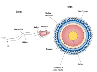
Human fertilization is the union of an egg and sperm, occurring primarily in the ampulla of the fallopian tube. The result of this union leads to the production of a fertilized egg called a zygote, initiating embryonic development. Scientists discovered the dynamics of human fertilization in the 19th century.

<span class="mw-page-title-main">Human reproduction</span> Procreative biological processes of humanity

Human reproduction is sexual reproduction that results in human fertilization to produce a human offspring. It typically involves sexual intercourse between a sexually mature human male and female. During sexual intercourse, the interaction between the male and female reproductive systems results in fertilization of the ovum by the sperm to form a zygote. While normal cells contain 46 chromosomes, gamete cells only contain 23 single chromosomes, and it is when these two cells merge into one zygote cell that genetic recombination occurs and the new zygote contains 23 chromosomes from each parent, giving it 46 chromosomes. The zygote then undergoes a defined development process that is known as human embryogenesis, and this starts the typical 9-month gestation period that is followed by childbirth. The fertilization of the ovum may be achieved by artificial insemination methods, which do not involve sexual intercourse. Assisted reproductive technology also exists.

A reproductive system disease is any disease of the human reproductive system.

<span class="mw-page-title-main">Sexual differentiation in humans</span> Process of development of sex differences in humans

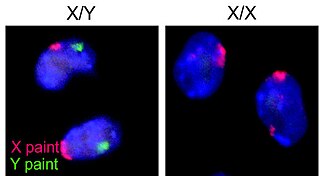
Sexual differentiation in humans is the process of development of sex differences in humans. It is defined as the development of phenotypic structures consequent to the action of hormones produced following gonadal determination. Sexual differentiation includes development of different genitalia and the internal genital tracts and body hair plays a role in sex identification.

<span class="mw-page-title-main">Fallopian tube obstruction</span> Medical condition

Fallopian tube obstruction, also known as fallopian tube occlusion, is a major cause of female infertility. Blocked fallopian tubes are unable to let the ovum and the sperm converge, thus making fertilization impossible.

The development of the reproductive system is the part of embryonic growth that results in the sex organs and contributes to sexual differentiation. Due to its large overlap with development of the urinary system, the two systems are typically described together as the genitourinary system.

A vaginal disease is a pathological condition that affects part or all of the vagina.

The reproductive system of an organism, also known as the genital system, is the biological system made up of all the anatomical organs involved in sexual reproduction. Many non-living substances such as fluids, hormones, and pheromones are also important accessories to the reproductive system. Unlike most organ systems, the sexes of differentiated species often have significant differences. These differences allow for a combination of genetic material between two individuals, which allows for the possibility of greater genetic fitness of the offspring.

<span class="mw-page-title-main">Fallopian tube</span> Tubes in the human female reproductive system

The fallopian tubes, also known as uterine tubes, oviducts or salpinges, are paired tubes in the human female body that stretch from the ovaries to the uterus. The fallopian tubes are part of the female reproductive system. In other vertebrates, they are only called oviducts.

<span class="mw-page-title-main">Fertility testing</span>

Fertility testing is the process by which fertility is assessed, both generally and also to find the "fertile window" in the menstrual cycle. General health affects fertility, and STI testing is an important related field.

<span class="mw-page-title-main">Mammalian reproduction</span> Most mammals are viviparous, giving birth to live young

Most mammals are viviparous, giving birth to live young. However, the five species of monotreme, the platypuses and the echidnas, lay eggs. The monotremes have a sex determination system different from that of most other mammals. In particular, the sex chromosomes of a platypus are more like those of a chicken than those of a therian mammal.

References

  1. Scoullar, Michelle J. L.; Boeuf, Philippe; Peach, Elizabeth (2021). "Mycoplasma genitalium and Other Reproductive Tract Infections in Pregnant Women, Papua New Guinea, 2015–2017 - Volume 27, Number 3—March 2021 - Emerging Infectious Diseases journal - CDC". Emerging Infectious Diseases. 27 (3): 894–904. doi:10.3201/eid2703.201783. PMC   7920647 . PMID   33622474. Archived from the original on 9 October 2022. Retrieved 9 October 2022.
  2. Ellis, Harold; Mahadevan, Vishy (2013). Clinical anatomy: applied anatomy for students and junior doctors (13th ed.). Chichester, West Sussex, UK: Wiley-Blackwell. ISBN   978-1-118-37376-7.
  3. "Female Reproductive System: Structure & Function". Cleveland Clinic. Retrieved 2023-12-19.
  4. "Genital Tract | SEER Training". training.seer.cancer.gov. Retrieved 2023-12-19.
  5. "Fallopian Tubes: Location, Anatomy, Function & Conditions". Cleveland Clinic. Retrieved 2023-12-19.
  6. "Female Reproductive System: Structure & Function". Cleveland Clinic. Retrieved 2023-12-19.
  7. "Ovaries: Anatomy, Function, Hormones & Conditions". Cleveland Clinic. Retrieved 2023-12-19.
  8. Alberts, Bruce; Johnson, Alexander; Lewis, Julian; Raff, Martin; Roberts, Keith; Walter, Peter (2002), "Fertilization", Molecular Biology of the Cell. 4th edition, Garland Science, retrieved 2023-12-19
  9. "Details of genital development". Archived from the original on February 25, 2020. Retrieved August 6, 2010.
  10. Egan ME, Lipsky MS (2000). "Diagnosis of Vaginitis". American Family Physician. 62 (5): 1095–104. PMID   10997533. Archived from the original on 6 June 2011. Retrieved 7 July 2020.
  11. 1 2 3 Zaino, Richard J.; Robboy, Stanley J.; Bentley, Rex; Kurman, Robert J. (2011). "Diseases of the Vagina". Blaustein's Pathology of the Female Genital Tract. pp. 105–154. doi:10.1007/978-1-4419-0489-8_3. ISBN   978-1-4419-0488-1.
  12. Iyer, V; Farquhar, C; Jepson, R (2000). Iyer, Vadeihi (ed.). "Oral contraceptive pills for heavy menstrual bleeding". Cochrane Database Syst Rev (2): CD000154. doi:10.1002/14651858.CD000154. PMID   10796696.
  13. de Wit, AE; Booij, SH; Giltay, EJ; Joffe, H; Schoevers, RA; Oldehinkel, AJ (2020). "Association of Use of Oral Contraceptives With Depressive Symptoms Among Adolescents and Young Women". JAMA Psychiatry. 77 (1): 52–59. doi:10.1001/jamapsychiatry.2019.2838. PMC   6777223 . PMID   31577333.
  14. Anwar, Etin. "The Transmission of Generative Self and Women's Contribution to Conception." Gender and Self in Islam. London: Routledge, 2006. 75. Print.