Complications of pregnancy are health problems that are related to, or arise during pregnancy. Complications that occur primarily during childbirth are termed obstetric labor complications, and problems that occur primarily after childbirth are termed puerperal disorders. While some complications improve or are fully resolved after pregnancy, some may lead to lasting effects, morbidity, or in the most severe cases, maternal or fetal mortality.[1][2][3]
Common complications of pregnancy include anemia, gestational diabetes, infections, gestational hypertension and pre-eclampsia.[4][5] Presence of these types of complications can have implications on monitoring lab work, imaging, and medical management during pregnancy.[4]
Severe complications of pregnancy, childbirth, and the puerperium are present in 1.6% of mothers in the US,[6] and in 1.5% of mothers in Canada.[7] In the immediate postpartum period (puerperium), 87% to 94% of women report at least one health problem.[8][9] Long-term health problems (persisting after six months postpartum) are reported by 31% of women.[10]
Complications of pregnancy can sometimes arise from abnormally severe presentations of symptoms and discomforts of pregnancy, which usually do not significantly interfere with activities of daily living or pose any significant threat to the health of the birthing person or fetus. For example, morning sickness is a fairly common mild symptom of pregnancy that generally resolves in the second trimester, but hyperemesis gravidarum is a severe form of this symptom that sometimes requires medical intervention to prevent electrolyte imbalances from severe vomiting.
Maternal problems
The following problems originate in the mother, however, they may have serious consequences for the fetus as well.
Gestational diabetes
Gestational diabetes is when a woman, without a previous diagnosis of diabetes, develops high blood sugar levels during pregnancy.[13][14] There are many non-modifiable and modifiable risk factors that lead to the devopment of this complication. Non-modifiable risk factors include a family history of diabetes, advanced maternal age, and ethnicity. Modifiable risk factors include maternal obesity.[14] There is an elevated demand for insulin during pregnancy which leads to increased insulin production from pancreaticbeta cells. The elevated demand is a result of increased maternal calorie intake and weight gain, and increased production of prolactin and growth hormone. Gestational diabetes increases risk for further maternal and fetal complications such as development of pre-eclampsia, need for cesarean delivery, preterm delivery, polyhydramnios, macrosomia, shoulder dystocia, fetal hypoglycemia, hyperbilirubinemia, and admission into the neonatal intensive care unit. The increased risk is correlated with the how well the gestational diabetes is controlled during pregnancy with poor control associated with worsened outcomes. A multidisciplinary approach is used to treat gestational diabetes and involves monitoring of blood-glucose levels, nutritional and dietary modifications, lifestyle changes such as increasing physical activity, maternal weight management, and medication such as insulin.[14]
Hyperemesis gravidarum
Hyperemesis gravidarum is the presence of severe and persistent vomiting, causing dehydration and weight loss. It is similar although more severe than the common morning sickness.[15][16] It is estimated to affect 0.3–3.6% of pregnant women and is the greatest contributor to hospitalizations under 20 weeks of gestation. Most often, nausea and vomiting symptoms during pregnancy resolve in the first trimester, however, some continue to experience symptoms. Hyperemesis gravidarum is diagnosed by the following criteria: greater than 3 vomiting episodes per day, ketonuria, and weight loss of more than 3kg or 5% of body weight. There are several non-modifiable and modifiable risk factors that predispose women to development of this condition such as female fetus, psychiatric illness history, high or low BMI pre-pregnancy, young age, African American or Asian ethnicity, type I diabetes, multiple pregnancies, and history of pregnancy affected by hyperemesis gravidarum. There are currently no known mechanisms for the cause of this condition. This complication can cause nutritional deficiency, low pregnancy weight gain, dehydration, and vitamin, electrolyte, and acid-based disturbances in the mother. It has been shown to cause low birth weight, small gestational age, preterm birth, and poor APGAR scores in the infant. Treatments for this condition focus on preventing harm to the fetus while improving symptoms and commonly include fluid replacement and consumption of small, frequent, bland meals. First-line treatments include ginger and acupuncture. Second-line treatments include vitamin B6 +/- doxylamine, antihistamines, dopamine antagonists, and serotonin antagonists. Third-line treatments include corticosteroids, transdermal clonidine, and gabapentin. Treatments chosen are dependent on severity of symptoms and response to therapies.[17]
Pelvic girdle pain
Pelvic girdle pain (PGP) disorder is pain in the area between the posterior iliac crest and gluteal fold beginning peri or postpartum caused by instability and limitation of mobility. It is associated with pubic symphysis pain and sometimes radiation of pain down the hips and thighs. For most pregnant individuals, PGP resolves within three months following delivery, but for some it can last for years, resulting in a reduced tolerance for weight bearing activities. PGP affects around 45% of individuals during pregnancy: 25% report serious pain and 8% are severely disabled.[18][19] Risk factors for complication development include multiparity, increased BMI, physically strenuous work, smoking, distress, history of back and pelvic trauma, and previous history of pelvic and lower back pain. This syndrome results from a growing uterus during pregnancy that causes increased stress on the lumbar and pelvic regions of the mother, thereby, resulting in postural changes and reduced lumbopelvic muscle strength leading to pelvic instability and pain. It is unclear whether specific hormones in pregnancy are associated with complication development. PGP can result in poor quality of life, predisposition to chronic pain syndrome, extended leave from work, and psychosocial distress. Many treatment options are available based on symptom severity. Non-invasive treatment options include activity modification, pelvic support garments, analgesia with or without short periods of bed rest, and physiotherapy to increase strength of gluteal and adductor muscles reducing stress on the lumbar spine. Invasive surgical management is considered a last-line treatment if all other treatment modalities have failed and symptoms are severe.[19]
Potential severe hypertensive states of pregnancy are mainly:
Pre-eclampsia – gestational hypertension, proteinuria (>300mg), and edema. Severe pre-eclampsia involves a BP over 160/110 (with additional signs). It affects 5–8% of pregnancies.[20]
Eclampsia – seizures in a pre-eclamptic patient, affect around 1.4% of pregnancies.[21]
Gestational hypertension can develop after 20 weeks but has no other symptoms, and later rights itself, but it can develop into pre-eclampsia.[22]
Acute fatty liver of pregnancy is sometimes included in the pre-eclamptic spectrum. It occurs in approximately one in 7,000 to one in 15,000 pregnancies.[24][25]
Women who have chronic hypertension before their pregnancy are at increased risk of complications such as premature birth, low birthweight or stillbirth.[26] Women who have high blood pressure and had complications in their pregnancy have three times the risk of developing cardiovascular disease compared to women with normal blood pressure who had no complications in pregnancy. Monitoring pregnant women's blood pressure can help prevent both complications and future cardiovascular diseases.[27][28]
Venous thromboembolism
Venous thromboembolism, consisting of deep vein thrombosis and pulmonary embolism, is a major risk factor for postpartum morbidity and mortality, especially in highly developed countries. A combination of pregnancy-exacerbated hypercoagulability and additional risk factors such as obesity and thrombophilias makes pregnant women vulnerable to thrombotic events[29] T.he prophylactic measures that include the usage of low molecular weight heparin, in fact, can significantly reduce risks associated with surgery, particularly in high-risk patients. Awareness among healthcare givers and prompt response in early identification and management of venous thromboembolism during pregnancy and the postpartum period are both crucial for prompt response. Deep vein thrombosis, a form of venous thromboembolism, has an incidence of 0.5 to 7 per 1,000 pregnancies, and is the second most common cause of maternal death in developed countries after bleeding.[30]
Treatment: Prophylactic treatment, e.g. with low molecular weight heparin may be indicated when there are additional risk factors for deep vein thrombosis.[30]
Anemia is a globally recognized complication of pregnancy worldwide and is a condition with a low hemoglobin amount in one of the trimesters. Such physiological modifications are more pronounced among individuals who suffer from undernutrition as well as chronic diseases associated with hemoglobin rehoming, like sickle cell anemia. Prevention of anemia during pregnancy is complicated, and is often treated by a team effort of dietary supplementation, iron therapy, and continuous assessment of mother and fetal indices in a multidisciplinary approach[31]. As an additional measure, emphasis is placed on the astute determination of the respective triggering points, and the application of optimal prenatal care to better maternal and fetal outcome.
Levels of hemoglobin are lower in the third trimesters. According to the United Nations (UN) estimates, approximately half of pregnant individuals develop anemia worldwide. Anemia prevalences during pregnancy differed from 18% in developed countries to 75% in South Asia; culminating to a global rate of 38% of pregnancies world wide.[32][33][34]
Treatment varies due to the severity of the anaemia, and can be used by increasing iron containing foods, oral iron tablets or by the use of parenteral iron.[13]
Pregnancy is a critical period for the expectant mom to experience additional dangers associated with infections. Moreover, a mother and baby's health is exposed to danger when she is in this condition. The prenatal physiology complexity and immunity modulation inherently increase the risk of influenza, hepatitis E, and cytomegalovirus transmission[35]. Avoidance actions like vaccines and strict infectious control protocols can be given priority in the policies aimed at limiting the risk of transmission among high-risk populations. In addition, it is early diagnosis and management of maternal infections are among the main methods to flatline vertical transmission and fetal aberrations.
Peripartum cardiomyopathy is a heart failure caused by a decrease in left ventricular ejection fraction (LVEF) to <45% which occurs towards the end of pregnancy or a few months postpartum. Symptoms include shortness of breath in various positions and/or with exertion, fatigue, pedal edema, and chest tightness. Risk factors associated with the development of this complication include maternal age over 30 years, multi gestational pregnancy, family history of cardiomyopathy, previous diagnosis of cardiomyopathy, pre-eclampsia, hypertension, and African ancestry. The pathogenesis of peripartum cardiomyopathy is not yet known, however, it is suggested that multifactorial potential causes could include autoimmune processes, viral myocarditis, nutritional deficiencies, and maximal cardiovascular changes during which increase cardiac preload. Peripartum cardiomyopathy can lead to many complications such as cardiopulmonary arrest, pulmonary edema, thromboembolisms, brain injury, and death. Treatment of this condition is very similar to treatment of non-gravid heart failure patients, however, safety of the fetus must be prioritized. For example, for anticoagulation due to increased risk for thromboembolism, low molecular weight heparin which is safe for use during pregnancy is used instead of warfarin which crosses the placenta.[39]
Hypothyroidism (commonly caused by Hashimoto's disease) is an autoimmune disease that affects the thyroid by causing low thyroid hormone levels. Symptoms of hypothyroidism can include low energy, cold intolerance, muscle cramps, constipation, and memory and concentration problems.[40] It is diagnosed by the presence of elevated levels of thyroid stimulation hormone or TSH. Patients with elevated TSH and decreased levels of free thyroxine or T4 are considered to have overt hypothyroidism. While those with elevated TSH and normal levels of free T4 are considered to have subclinical hypothyroidism.[41] Risk factors for developing hypothyroidism during pregnancy include iodine deficiency, history of thyroid disease, visible goiter, hypothyroidism symptoms, family history of thyroid disease, history of type 1 diabetes or autoimmune conditions, and history of infertility or fetal loss. Various hormones during pregnancy affect the thyroid and increase thyroid hormone demand. For example, during pregnancy, there is increased urinary iodine excretion as well as increased thyroxine binding globulin and thyroid hormone degradation which all increase thyroid hormone demands.[42] This condition can have a profound effect during pregnancy on the mother and fetus. The infant may be seriously affected and have a variety of birth defects. Complications in the mother and fetus can include pre-eclampsia, anemia, miscarriage, low birth weight, still birth, congestive heart failure, impaired neurointellectual development, and if severe, congenital iodine deficiency syndrome.[40][42] This complication is treated by iodine supplementation, levothyroxine which is a form of thyroid hormone replacement, and close monitoring of thyroid function.[42]
Fetal and placental problems
The following problems occur in the fetus or placenta, but may have serious consequences on the mother as well.
Ectopic pregnancy
Ectopic pregnancy is implantation of the embryo outside the uterus. This form of complicated pregnancy, which is a non-implication of a normally fertilized egg at any spot other than the uterus, involves operation failure, which can cause life-threatening conditions. However, the underlying reasons for this are not exactly known. This phenomenon is often accompanied by PID (pelvic inflammatory disease) or salpingectomy (surgery).
Caused by: Unknown, but risk factors include smoking, advanced maternal age, and prior surgery or trauma to the fallopian tubes.
Risk factors include untreated pelvic inflammatory disease, likely due to fallopian tube scarring.[43]
Treatment: In most cases, keyhole surgery must be carried out to remove the fetus, along with the fallopian tube. If the pregnancy is very early, it may resolve on its own, or it can be treated with methotrexate, an abortifacient.[44]
Miscarriage
Miscarriage is the loss of a pregnancy prior to 20 weeks.[45][46] In the UK, miscarriage is defined as the loss of a pregnancy during the first 23 weeks.[47] Comprehensive support, consists of the consultation of the genomics as well as the provision of the medical or surgical operations required. The psychological relevance of family members, relatives, and friends to the bereaved ones is also a necessity. The most effective tools that can be used to minimize the psychological implications of the mourners include autopsy and bereavement counseling.
Approximately 80% of pregnancy loss occurs in the first trimester, with a decrease in risk after 12 weeks gestation. Some variables, such as the mother’s being older or chromosomal abnormalities, possess a higher likelihood of causing multiple miscarriages[48]. Spontaneous abortions can be further categorized into complete, inevitable, missed, and threatened abortions:[citation needed]
Complete: Vaginal bleeding occurs followed by the complete passing of conception products through the cervix.
Inevitable: Vaginal bleeding occurs; the cervical os is closed indicating that conception products will pass in the near future.
Missed: Vaginal bleeding occurs and some products of conception may have passed through the cervix; the cervical os is closed and ultrasound shows a nonviable fetus and remaining products of conception.
Threatened: Vaginal bleeding occurs; the cervical os is closed and ultrasound shows a viable fetus.
Stillbirth
Stillbirth is defined as fetal loss or death after 20 weeks gestation. Early stillbirth is between 20 and 27 weeks gestation, while late stillbirth is between 28 and 36 weeks gestation. A term stillbirth is when the fetus dies 37 weeks and above.[49] This phenomenon can go beyond grief and can lead to worries about strange maternal feelings or postpartum treatment regarding complications of childbirth[50]. Such parents would require more than empathy; generally, adequate medical programs should be considered for parents having such unbearable grief. Along with psychiatric help, counseling, and peer support, which should be useful in the process of assisting parents who have lost their children.
Epidemiology: There are over 2 million stillbirths a year and there are about 6 stillbirths per 1000 births (0.6%)[51]
Clinical presentation: Fetal behavioral changes like decreased movements or a loss in fetal sensation may indicate stillbirth, but the presentation can vary greatly.
Risk factors: Maternal weight, age, and smoking, as well as pre-existing maternal diabetes or hypertension[49]
Treatment: If fetal passing occurs before labor, treatment options include induced labor or cesarean section. Otherwise, stillbirths can pass with natural birth.
Placental abruption
Placental abruption defined as the separation of the placenta from the uterus prior to delivery, is a major cause of third trimester vaginal bleeding and complicates about 1% of pregnancies.[13][52] Symptomatic presentations are variable: Some women can entirely ignore the symptoms, while others have mild bleeding or abdominal discomfort and pain. Hence, though symptom severity variance and precipitous placental separation are not relevant, they can still cause the diagnosis and clinical management to be complicated.
Several contributors may result in placental abruption. This includes: pre-existing maternal factors (e.g., smoking, hypertension, advanced age)[53], as well as pregnancy-related factors such as multiple pregnancies or the presence of in-utero infections. Identifying risk factors beforehand in order to take steps and make quick reactions to minimize the likelihood of unfavorable outcomes for the mother or the fetus is essential. The therapy techniques of placental rupture are based on the fetal gestation age and the status of both the mother and the baby. Instant delivery should be medically warranted for full-term babies (36 weeks or more) and in case of distress. Milder cases with immature embryos being monitored closely, any necessary intervention is done in time after careful observation.
The implementation of preventive measures, which include pre-conception counseling to deal with the modifiable risk factors, can significantly contribute to the reduction of incidents of placental abruption. Knowing the long-term impacts on the mother and the baby after giving birth is essential. Continuous research and evidence-based approaches help in providing management that works. Collaboration between healthcare providers and patients is the core of the outcomes of placenta abruption.
Clinical Presentation: Varies widely from asymptomatic to vaginal bleeding and abdominal pain.
Treatment: Immediate delivery if the fetus is mature (36 weeks or older), or if a younger fetus or the mother is in distress. In less severe cases with immature fetuses, the situation may be monitored in hospital, with treatment if necessary.
Placenta previa
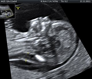
Placenta previa is a condition that occurs when the placenta fully or partially covers the cervix.[13] Placenta previa can be further categorized into complete previa, partial previa, marginal previa, and low-lying placenta, depending on the degree to which the placenta covers the internal cervical os. Placenta previa is primarily diagnosed by ultrasound, either during a routine examination or following an episode of abnormal vaginal bleeding, often in the second trimester of pregnancy. Most diagnosis of placenta previa occurs during the second-trimester.[citation needed]
Treatments are adapted according to their severity and the mother's state of health, from strict monitoring to cesarean section.
Placenta accreta is an abnormal adherence of the placenta to the uterine wall.[55] Specifically, placenta accreta involves abnormal adherence of the placental trophoblast to the uterine myometrium.[56]
Placenta accreta risk factors include placenta previa, abnormally elevated second-trimester AFP and free β-hCG levels, and advanced gestational parent age, specifically over the age of 35.[57][58]Furthermore, prior cesarean delivery is one of the most common risk factors for placenta accreta, due to the presence of a uterine scar leading to abnormal decidualization of the placenta.[59]
Due to abnormal adherence of the placenta to the uterine wall, cesarean delivery is often indicated, as well as cesarean hysterectomy.[56]
Multiple births may become monochorionic, sharing the same chorion, with resultant risk of twin-to-twin transfusion syndrome. Monochorionic multiples may even become monoamniotic, sharing the same amniotic sac, resulting in risk of umbilical cord compression and entanglement. In very rare cases, there may be conjoined twins, possibly impairing function of internal organs.[citation needed] Control of multiple pregnancies, such as special prenatal care and birth plans, can help in the control of placenta accreta[60]. Moreover, early detection and response to the health problems arising from multiple pregnancies can help both the expectant parents and medical care providers deal with this particular aspect of reproductive health consciously.
Since the embryo and fetus have little or no immune function, they depend on the immune function of their mother. Several pathogens can cross the placenta and cause (perinatal) infection. Often microorganisms that produce minor illness in the mother are very dangerous for the developing embryo or fetus. This can result in spontaneous abortion or major developmental disorders. For many infections, the baby is more at risk at particular stages of pregnancy. Problems related to perinatal infection are not always directly noticeable.[citation needed]
The term TORCH complex refers to a set of several different infections that may be caused by transplacental infection:
T - Toxoplasmosis
O - other infections (i.e. Parvovirus B19, Coxsackievirus, Chickenpox, Chlamydia, HIV, HTLV, syphilis, Zika)
R - Rubella
C - Cytomegalovirus
H - HSV
Babies can also become infected by their mother during birth. During birth, babies are exposed to maternal blood and body fluids without the placental barrier intervening and to the maternal genital tract[61]. Because of this, blood-borne microorganisms (hepatitis B, HIV), organisms associated with sexually transmitted disease (e.g., gonorrhoea and chlamydia), and normal fauna of the genito-urinary tract (e.g., Candida) are among those commonly seen in infection of newborns. Furthermore, vaccination, commitment to safe birth practices, and prenatal screening and treatment of infections are also strategic measures that can help reduce the risk of newborn infections.
General risk factors
Factors increasing the risk (to either the pregnant individual, the fetus/es, or both) of pregnancy complications beyond the normal level of risk may be present in the pregnant individual's medical profile either before they become pregnant or during the pregnancy.[62] These pre-existing factors may related to the individual's genetics, physical or mental health, their environment and social issues, or a combination of those.[63]
Older parents: As they age, both mothers and fathers are at an increased risk for complications in the fetus and during pregnancy and childbirth. Complications for those 45 or older include increased risk of primary Caesarean delivery (i.e. C-section).[65]
Height: Pregnancy in individuals whose height is less than 1.5 meters (5 feet) correlates with a higher incidence of preterm birth and underweight babies. Also, these individuals are more likely to have a small pelvis, which can result in such complications during childbirth as shoulder dystocia.[63]
Risks arising from previous pregnancies: Complications experienced during a previous pregnancy are more likely to recur.[66][67]
Multiple pregnancies: Individuals who have had greater than five previous pregnancies face increased risks of rapid labor and excessive bleeding after delivery.
Prenatal methamphetamine exposure: Can cause premature birth and congenital abnormalities.[72] Other investigations have revealed short-term neonatal outcomes to include small deficits in infant neurobehavioral function and growth restriction when compared to control infants.[73] Also, prenatal methamphetamine use is believed to have long-term effects in terms of brain development, which may last for many years.[72]
Cannabis: Possibly associated with adverse effects on the child later in life.
Social and socioeconomic factors: Generally speaking, unmarried individuals and those in lower socioeconomic groups experience an increased level of risk in pregnancy, due at least in part to lack of access to appropriate prenatal care.[63][74]
Unintended pregnancy: Unintended pregnancies preclude preconception care and delays prenatal care. They preclude other preventive care, may disrupt life plans and on average have worse health and psychological outcomes for the mother and, if birth occurs, the child.[75][76]
An elevated level of stress during pregnancy leads to notorious pregnancy outcomes, including preterm birth, low birth weight, and mental health problems for the mother.
Prolonged effects of chronic stressors such as discrimination, intimate partner violence, housing issues, and poverty lead to widespread maternal health issues and adverse pregnancy outcomes. [citation needed]
Cultural norms, convictions, and traditions connected to pregnancy and childbirth lead people to establish perceptions, habits, and treatment-seeking. Cultural determinants affect the assessment of prenatal care utilization, childbirth practice, dietary habits and reproductive health beliefs, which are direct outcomes of pregnancy and health situations.[citation needed]
High-risk pregnancy
Some disorders and conditions can mean that pregnancy is considered high-risk (about 6-8% of pregnancies in the USA) and in extreme cases may be contraindicated. High-risk pregnancies are the main focus of doctors specialising in maternal-fetal medicine. Serious pre-existing disorders which can reduce a woman's physical ability to survive pregnancy include a range of congenital defects (that is, conditions with which the woman herself was born, for example, those of the heart or reproductive organs, some of which are listed above) and diseases acquired at any time during the woman's life.
Absolute and relative incidence of venous thromboembolism (VTE) during pregnancy and the postpartum period
Absolute incidence of first VTE per 10,000 person–years during pregnancy and the postpartum period
Swedish data A
Swedish data B
English data
Danish data
Time period
N
Rate (95% CI)
N
Rate (95% CI)
N
Rate (95% CI)
N
Rate (95% CI)
Outside pregnancy
1105
4.2 (4.0–4.4)
1015
3.8 (?)
1480
3.2 (3.0–3.3)
2895
3.6 (3.4–3.7)
Antepartum
995
20.5 (19.2–21.8)
690
14.2 (13.2–15.3)
156
9.9 (8.5–11.6)
491
10.7 (9.7–11.6)
Trimester 1
207
13.6 (11.8–15.5)
172
11.3 (9.7–13.1)
23
4.6 (3.1–7.0)
61
4.1 (3.2–5.2)
Trimester 2
275
17.4 (15.4–19.6)
178
11.2 (9.7–13.0)
30
5.8 (4.1–8.3)
75
5.7 (4.6–7.2)
Trimester 3
513
29.2 (26.8–31.9)
340
19.4 (17.4–21.6)
103
18.2 (15.0–22.1)
355
19.7 (17.7–21.9)
Around delivery
115
154.6 (128.8–185.6)
79
106.1 (85.1–132.3)
34
142.8 (102.0–199.8)
–
Postpartum
649
42.3 (39.2–45.7)
509
33.1 (30.4–36.1)
135
27.4 (23.1–32.4)
218
17.5 (15.3–20.0)
Early postpartum
584
75.4 (69.6–81.8)
460
59.3 (54.1–65.0)
177
46.8 (39.1–56.1)
199
30.4 (26.4–35.0)
Late postpartum
65
8.5 (7.0–10.9)
49
6.4 (4.9–8.5)
18
7.3 (4.6–11.6)
319
3.2 (1.9–5.0)
Incidence rate ratios (IRRs) of first VTE during pregnancy and the postpartum period
Swedish data A
Swedish data B
English data
Danish data
Time period
IRR* (95% CI)
IRR* (95% CI)
IRR (95% CI)†
IRR (95% CI)†
Outside pregnancy
Reference (i.e., 1.00)
Antepartum
5.08 (4.66–5.54)
3.80 (3.44–4.19)
3.10 (2.63–3.66)
2.95 (2.68–3.25)
Trimester 1
3.42 (2.95–3.98)
3.04 (2.58–3.56)
1.46 (0.96–2.20)
1.12 (0.86–1.45)
Trimester 2
4.31 (3.78–4.93)
3.01 (2.56–3.53)
1.82 (1.27–2.62)
1.58 (1.24–1.99)
Trimester 3
7.14 (6.43–7.94)
5.12 (4.53–5.80)
5.69 (4.66–6.95)
5.48 (4.89–6.12)
Around delivery
37.5 (30.9–44.45)
27.97 (22.24–35.17)
44.5 (31.68–62.54)
–
Postpartum
10.21 (9.27–11.25)
8.72 (7.83–9.70)
8.54 (7.16–10.19)
4.85 (4.21–5.57)
Early postpartum
19.27 (16.53–20.21)
15.62 (14.00–17.45)
14.61 (12.10–17.67)
8.44 (7.27–9.75)
Late postpartum
2.06 (1.60–2.64)
1.69 (1.26–2.25)
2.29 (1.44–3.65)
0.89 (0.53–1.39)
Notes: Swedish data A = Using any code for VTE regardless of confirmation. Swedish data B = Using only algorithm-confirmed VTE. Early postpartum = First 6 weeks after delivery. Late postpartum = More than 6 weeks after delivery. * = Adjusted for age and calendar year. † = Unadjusted ratio calculated based on the data provided. Source:[78]
List of complications (complete)
Obstetric complications are those complications that develop during pregnancy. A woman may develop an infection, syndrome or complication that is not unique to pregnancy and that may have existed before pregnancy. Pregnancy often is complicated by preexisting and concurrent conditions. Though these pre-existing and concurrent conditions may have great impact on pregnancy, they are not included in the following list.
Stillbirth is typically defined as fetal death at or after 20 or 28 weeks of pregnancy, depending on the source. It results in a baby born without signs of life. A stillbirth can often result in the feeling of guilt or grief in the mother. The term is in contrast to miscarriage, which is an early pregnancy loss, and sudden infant death syndrome, where the baby dies a short time after being born alive.
Eclampsia is the onset of seizures (convulsions) in a woman with pre-eclampsia. Pre-eclampsia is a hypertensive disorder of pregnancy that presents with three main features: new onset of high blood pressure, large amounts of protein in the urine or other organ dysfunction, and edema. If left untreated, pre-eclampsia can result in long-term consequences for the mother, namely increased risk of cardiovascular diseases and associated complications. In more severe cases, it may be fatal for both the mother and the fetus.
Pre-eclampsia is a multi-system disorder specific to pregnancy, characterized by the onset of high blood pressure and often a significant amount of protein in the urine. When it arises, the condition begins after 20 weeks of pregnancy. In severe cases of the disease there may be red blood cell breakdown, a low blood platelet count, impaired liver function, kidney dysfunction, swelling, shortness of breath due to fluid in the lungs, or visual disturbances. Pre-eclampsia increases the risk of undesirable as well as lethal outcomes for both the mother and the fetus including preterm labor. If left untreated, it may result in seizures at which point it is known as eclampsia.
Placenta praevia is when the placenta attaches inside the uterus but in a position near or over the cervical opening. Symptoms include vaginal bleeding in the second half of pregnancy. The bleeding is bright red and tends not to be associated with pain. Complications may include placenta accreta, dangerously low blood pressure, or bleeding after delivery. Complications for the baby may include fetal growth restriction.
Gestational hypertension or pregnancy-induced hypertension (PIH) is the development of new hypertension in a pregnant woman after 20 weeks' gestation without the presence of protein in the urine or other signs of pre-eclampsia. Gestational hypertension is defined as having a blood pressure greater than 140/90 on two occasions at least 6 hours apart.
Placental abruption is when the placenta separates early from the uterus, in other words separates before childbirth. It occurs most commonly around 25 weeks of pregnancy. Symptoms may include vaginal bleeding, lower abdominal pain, and dangerously low blood pressure. Complications for the mother can include disseminated intravascular coagulopathy and kidney failure. Complications for the baby can include fetal distress, low birthweight, preterm delivery, and stillbirth.
Pregnancy is the time during which one or more offspring develops (gestates) inside a woman's uterus (womb). A multiple pregnancy involves more than one offspring, such as with twins.
Intrauterine hypoxia occurs when the fetus is deprived of an adequate supply of oxygen. It may be due to a variety of reasons such as prolapse or occlusion of the umbilical cord, placental infarction, maternal diabetes and maternal smoking. Intrauterine growth restriction may cause or be the result of hypoxia. Intrauterine hypoxia can cause cellular damage that occurs within the central nervous system. This results in an increased mortality rate, including an increased risk of sudden infant death syndrome (SIDS). Oxygen deprivation in the fetus and neonate have been implicated as either a primary or as a contributing risk factor in numerous neurological and neuropsychiatric disorders such as epilepsy, attention deficit hyperactivity disorder, eating disorders and cerebral palsy.
Velamentous cord insertion is a complication of pregnancy where the umbilical cord is inserted in the fetal membranes. It is a major cause of antepartum hemorrhage that leads to loss of fetal blood and associated with high perinatal mortality. In normal pregnancies, the umbilical cord inserts into the middle of the placental mass and is completely encased by the amniotic sac. The vessels are hence normally protected by Wharton's jelly, which prevents rupture during pregnancy and labor. In velamentous cord insertion, the vessels of the umbilical cord are improperly inserted in the chorioamniotic membrane, and hence the vessels traverse between the amnion and the chorion towards the placenta. Without Wharton's jelly protecting the vessels, the exposed vessels are susceptible to compression and rupture.
A placental disease is any disease, disorder, or pathology of the placenta.
Circumvallate placenta is a rare condition affecting about 1-2% of pregnancies, in which the amnion and chorion fetal membranes essentially "double back" on the fetal side around the edges of the placenta. After delivery, a circumvallate placenta has a thick ring of membranes on its fetal surface. Circumvallate placenta is a placental morphological abnormality associated with increased fetal morbidity and mortality due to the restricted availability of nutrients and oxygen to the developing fetus.
Birth injury refers to damage or injury to the child before, during, or just after the birthing process. "Birth trauma" refers specifically to mechanical damage sustained during delivery.
Thyroid disease in pregnancy can affect the health of the mother as well as the child before and after delivery. Thyroid disorders are prevalent in women of child-bearing age and for this reason commonly present as a pre-existing disease in pregnancy, or after childbirth. Uncorrected thyroid dysfunction in pregnancy has adverse effects on fetal and maternal well-being. The deleterious effects of thyroid dysfunction can also extend beyond pregnancy and delivery to affect neurointellectual development in the early life of the child. Due to an increase in thyroxine binding globulin, an increase in placental type 3 deioidinase and the placental transfer of maternal thyroxine to the fetus, the demand for thyroid hormones is increased during pregnancy. The necessary increase in thyroid hormone production is facilitated by high human chorionic gonadotropin (hCG) concentrations, which bind the TSH receptor and stimulate the maternal thyroid to increase maternal thyroid hormone concentrations by roughly 50%. If the necessary increase in thyroid function cannot be met, this may cause a previously unnoticed (mild) thyroid disorder to worsen and become evident as gestational thyroid disease. Currently, there is not enough evidence to suggest that screening for thyroid dysfunction is beneficial, especially since treatment thyroid hormone supplementation may come with a risk of overtreatment. After women give birth, about 5% develop postpartum thyroiditis which can occur up to nine months afterwards. This is characterized by a short period of hyperthyroidism followed by a period of hypothyroidism; 20–40% remain permanently hypothyroid.
A high-risk pregnancy is a pregnancy where the mother or the fetus has an increased risk of adverse outcomes compared to uncomplicated pregnancies. No concrete guidelines currently exist for distinguishing “high-risk” pregnancies from “low-risk” pregnancies; however, there are certain studied conditions that have been shown to put the mother or fetus at a higher risk of poor outcomes. These conditions can be classified into three main categories: health problems in the mother that occur before she becomes pregnant, health problems in the mother that occur during pregnancy, and certain health conditions with the fetus.
A pre-existing disease in pregnancy is a disease that is not directly caused by the pregnancy, in contrast to various complications of pregnancy, but which may become worse or be a potential risk to the pregnancy. A major component of this risk can result from necessary use of drugs in pregnancy to manage the disease.
Obstetric medicine, similar to maternal medicine, is a sub-specialty of general internal medicine and obstetrics that specializes in process of prevention, diagnosing, and treating medical disorders in with pregnant humans. It is closely related to the specialty of maternal-fetal medicine, although obstetric medicine does not directly care for the fetus. The practice of obstetric medicine, or previously known as "obstetric intervention," primarily consisted of the extraction of the baby during instances of duress, such as obstructed labor or if the baby was positioned in breech.
Hypertensive disease of pregnancy, also known as maternal hypertensive disorder, is a group of high blood pressure disorders that include preeclampsia, preeclampsia superimposed on chronic hypertension, gestational hypertension, and chronic hypertension.
Fetal programming, also known as prenatal programming, is the theory that environmental cues experienced during fetal development play a seminal role in determining health trajectories across the lifespan.
The Pregnancy Outcome Prediction (POP) Study is a prospective cohort study of 4,512 women who have never given birth, recruited at the Rosie Hospital between January 2008 and July 2012.
↑ Glazener CM, Abdalla M, Stroud P, Naji S, Templeton A, Russell IT (April 1995). "Postnatal maternal morbidity: extent, causes, prevention and treatment". British Journal of Obstetrics and Gynaecology. 102 (4): 282–287. doi:10.1111/j.1471-0528.1995.tb09132.x. PMID7612509. S2CID38872754.
↑ Thompson JF, Roberts CL, Currie M, Ellwood DA (June 2002). "Prevalence and persistence of health problems after childbirth: associations with parity and method of birth". Birth. 29 (2): 83–94. doi:10.1046/j.1523-536X.2002.00167.x. PMID12051189.
↑ Borders N (2006). "After the afterbirth: a critical review of postpartum health relative to method of delivery". Journal of Midwifery & Women's Health. 51 (4): 242–248. doi:10.1016/j.jmwh.2005.10.014. PMID16814217.
↑ Goodwin TM (September 2008). "Hyperemesis gravidarum". Obstetrics and Gynecology Clinics of North America. 35 (3): 401–17, viii. doi:10.1016/j.ogc.2008.04.002. PMID18760227.
↑ Villar J, Say L, Gulmezoglu AM, Meraldi M, Lindheimer MD, Betran AP, Piaggio G (2003). "Eclampsia and pre-eclampsia: a health problem for 2000 years.". In Critchly H, MacLean A, Poston L, Walker J (eds.). Pre-eclampsia. London: RCOG Press. pp.189–207.
↑ Abalos E, Cuesta C, Grosso AL, Chou D, Say L (September 2013). "Global and regional estimates of preeclampsia and eclampsia: a systematic review". European Journal of Obstetrics, Gynecology, and Reproductive Biology. 170 (1): 1–7. doi:10.1016/j.ejogrb.2013.05.005. PMID23746796.
1 2 3 Venös tromboembolism (VTE) – Guidelines for treatment in C counties. Bengt Wahlström, Emergency department, Uppsala Academic Hospital. January 2008
↑ Wang S, An L, Cochran SD (2002). "Women". In Detels R, McEwen J, Beaglehole R, Tanaka H (eds.). Oxford Textbook of Public Health (4thed.). Oxford University Press. pp.1587–601.
↑ Wortman AC, Alexander JM (March 2013). "Placenta accreta, increta, and percreta". Obstetrics and Gynecology Clinics of North America. 40 (1): 137–154. doi:10.1016/j.ogc.2012.12.002. PMID23466142.
↑ Koniak-Griffin D, Turner-Pluta C (September 2001). "Health risks and psychosocial outcomes of early childbearing: a review of the literature". The Journal of Perinatal & Neonatal Nursing. 15 (2): 1–17. doi:10.1097/00005237-200109000-00002. PMID12095025. S2CID42701860.
↑ Williams PM, Fletcher S (September 2010). "Health effects of prenatal radiation exposure". American Family Physician. 82 (5): 488–493. PMID20822083. S2CID22400308.
↑ Kirk E, Bottomley C, Bourne T (1 March 2014). "Diagnosing ectopic pregnancy and current concepts in the management of pregnancy of unknown location". Human Reproduction Update. 20 (2): 250–261. doi:10.1093/humupd/dmt047. PMID24101604.
1 2 3 4 5 6 7 8 "ICD-10 Version:2016". International Statistical Classification of Diseases and Related Health Problems 10th Revision. Retrieved 16 May 2017.
Further reading
Leveno K (2013). Williams manual of pregnancy complications. New York: McGraw-Hill Medical. ISBN978-0071765626.
This page is based on this Wikipedia article Text is available under the CC BY-SA 4.0 license; additional terms may apply. Images, videos and audio are available under their respective licenses.