Molar pregnancy

Last updated
Molar pregnancy
Other namesHydatid mole, hydatidiform mole
Hydatidiform mole (1) complete type.jpg
Histopathologic image of hydatidiform mole (complete type). H&E stain.
Specialty Obstetrics

A molar pregnancy, also known as a hydatidiform mole, is an abnormal form of pregnancy in which a non-viable fertilized egg implants in the uterus. It falls under the category of gestational trophoblastic diseases. [1] During a molar pregnancy, the uterus contains a growing mass characterized by swollen chorionic villi, resembling clusters of grapes. [2] The occurrence of a molar pregnancy can be attributed to the fertilized egg lacking an original maternal nucleus. As a result, the products of conception may or may not contain fetal tissue. These molar pregnancies are categorized into two types: partial moles and complete moles, where the term 'mole' simply denotes a clump of growing tissue or a ‘growth

Contents

A complete mole is caused by either a single sperm (90% of the time) or two sperm (10% of the time) combining with an egg that has lost its DNA. In the former case, the sperm reduplicates, leading to the formation of a "complete" 46-chromosome set. [3] Typically, the genotype is 46,XX (diploid) due to subsequent mitosis of the fertilizing sperm, but it can also be 46,XY (diploid). [3] However, 46,YY (diploid) is not observed. On the other hand, a partial mole occurs when a normal egg is fertilized by one or two sperm, which then reduplicates itself, resulting in genotypes of 69,XXY (triploid) or 92,XXXY (tetraploid). [3]

Complete moles carry a 2–4% risk, in Western countries, of developing into choriocarcinoma and a higher risk of 10–15% in Eastern countries, with an additional 15% risk of becoming an invasive mole. In contrast, incomplete moles can become invasive as well but are not associated with choriocarcinoma. [3] Notably, complete hydatidiform moles account for 50% of all cases of choriocarcinoma.

Molar pregnancies are relatively rare complications of pregnancy, occurring in approximately 1 in 1,000 pregnancies in the United States, while in Asia, the rates are considerably higher, reaching up to 1 in 100 pregnancies in countries like Indonesia. [4]

Signs and symptoms

Vesicular mole Vesicular mole.jpg
Vesicular mole

Molar pregnancies usually present with painless vaginal bleeding in the fourth to fifth months of pregnancy. [5] The uterus may be larger than expected, or the ovaries may be enlarged. There may also be more vomiting than would be expected (hyperemesis). Sometimes there is an increase in blood pressure along with protein in the urine. Blood tests will show very high levels of human chorionic gonadotropin (hCG). [6]

Cause

The cause of this condition is not completely understood. Potential risk factors may include defects in the egg, abnormalities within the uterus, or nutritional deficiencies. Women under 20 or over 40 years of age have a higher risk. Other risk factors include diets low in protein, folic acid, and carotene. [7] The diploid set of sperm-only DNA means that all chromosomes have sperm-patterned methylation suppression of genes. This leads to overgrowth of the syncytiotrophoblast whereas dual egg-patterned methylation leads to a devotion of resources to the embryo, with an underdeveloped syncytiotrophoblast. This is considered to be the result of evolutionary competition, with male genes driving for high investment into the fetus versus female genes driving for resource restriction to maximise the number of children. [8]

Pathophysiology

A hydatidiform mole is a pregnancy/conceptus in which the placenta contains grapelike vesicles (small sacs) that are usually visible to the naked eye. The vesicles arise by distention of the chorionic villi by fluid. When inspected under the microscope, hyperplasia of the trophoblastic tissue is noted. If left untreated, a hydatidiform mole will almost always end as a spontaneous abortion (miscarriage).

Based on morphology, hydatidiform moles can be divided into two types: in complete moles, all the chorionic villi are vesicular, and no sign of embryonic or fetal development is present. In partial moles, some villi are vesicular, whereas others appear more normal, and embryonic/fetal development may be seen but the fetus is always malformed and is never viable.

Uterus with complete hydatidiform mole Complete Hydatidiform Mole (39810634804).jpg
Uterus with complete hydatidiform mole

In rare cases, a hydatidiform mole co-exists in the uterus with a normal, viable fetus. These cases are due to twinning. The uterus contains the products of two conceptions: one with an abnormal placenta and no viable fetus (the mole), and one with a normal placenta and a viable fetus. Under careful surveillance, it is often possible for the woman to give birth to the normal child and to be cured of the mole. [9]

Parental origin

In most complete moles, all nuclear genes are inherited from the father only (androgenesis). In approximately 80% of these androgenetic moles, the most probable mechanism is that an egg with an empty nucleus or no nucleus is fertilized by a single sperm, followed by a duplication of all chromosomes/genes (a process called endoreduplication). In approximately 20% of complete moles, the most probable mechanism is that an empty egg is fertilized by two sperm. In both cases, the moles are diploid (i.e. there are two copies of every chromosome). In all these cases, the mitochondrial genes are inherited from the mother, as usual.

Most partial moles are triploid (three chromosome sets). The nucleus contains one maternal set of genes and two paternal sets. The mechanism is usually the reduplication of the paternal haploid set from a single sperm, but may also be the consequence of dispermic (two sperm) fertilization of the egg. [10]

In rare cases, hydatidiform moles are tetraploid (four chromosome sets) or have other chromosome abnormalities.

A small percentage of hydatidiform moles have biparental diploid genomes, as in normal living persons; they have two sets of chromosomes, one inherited from each biological parent. Some of these moles occur in women who carry mutations in the gene NLRP7 , predisposing them towards molar pregnancy. These rare variants of hydatidiform mole may be complete or partial. [11] [12] [13]

Diagnosis

Transvaginal ultrasonography showing a molar pregnancy Molar pregnancy.jpg
Transvaginal ultrasonography showing a molar pregnancy
Molar pregnancy in ultrasound Molar pregnancy 0001.jpg
Molar pregnancy in ultrasound
Hydatidiform mole on CT, sagittal view Blasenmole Computertomographie sagittal.jpg
Hydatidiform mole on CT, sagittal view
Hydatidiform mole on CT, axial view Blasenmole Computertomographie axial.jpg
Hydatidiform mole on CT, axial view

The diagnosis is strongly suggested by ultrasound (sonogram), but definitive diagnosis requires histopathological examination. On ultrasound, the mole resembles a bunch of grapes ("cluster of grapes" or "honeycombed uterus" or "snow-storm"). [14] There is increased trophoblast proliferation and enlarging of the chorionic villi, and angiogenesis in the trophoblasts is impaired. [15]

Sometimes symptoms of hyperthyroidism are seen, due to the extremely high levels of hCG, which can mimic the effects of thyroid-stimulating hormone. [15]

Complete MolePartial Mole
Karyotype [16] 46,XX or 46,XY69,XXY or 69, XXX
hCG↑↑↑↑
Uterine Size[ clarification needed ]
Fetal PartsNoYes
Components [16] 2 sperm + empty egg

or Empty egg + 1 sperm that duplicates

2 sperm + 1 egg

or 1 sperm + 1 egg that duplicates

Risk of Choriocarcinoma [17] 15%0.5%

Treatment

Hydatidiform moles should be treated by evacuating the uterus by uterine suction or by surgical curettage as soon as possible after diagnosis, in order to avoid the risks of choriocarcinoma. [18] Patients are followed up until their serum human chorionic gonadotrophin (hCG) level has fallen to an undetectable level. Invasive or metastatic moles (cancer) may require chemotherapy and often respond well to methotrexate. As they contain paternal antigens, the response to treatment is nearly 100%. Patients are advised not to conceive for half a year after hCG levels have normalized. The chances of having another molar pregnancy are approximately 1%.

Management is more complicated when the mole occurs together with one or more normal fetuses.

In some women, the growth can develop into gestational trophoblastic neoplasia. For women who have complete hydatidiform mole and are at high risk of this progression, evidence suggests giving prophylactic chemotherapy (known as P-chem) may reduce the risk of this happening. [19] However P-chem may also increase toxic side effects, so more research is needed to explore its effects. [19]

Anesthesia

The uterine curettage is generally done under the effect of anesthesia, preferably spinal anesthesia in hemodynamically stable patients. The advantages of spinal anesthesia over general anesthesia include ease of technique, favorable effects on the pulmonary system, safety in patients with hyperthyroidism and non-tocolytic pharmacological properties. Additionally, by maintaining patient's consciousness one can diagnose the complications like uterine perforation, cardiopulmonary distress and thyroid storm at an earlier stage than when the patient is sedated or is under general anesthesia. [20]

Prognosis

More than 80% of hydatidiform moles are benign. The outcome after treatment is usually excellent. Close follow-up is essential to ensure that treatment has been successful. [21] Highly effective means of contraception are recommended to avoid pregnancy for at least 6 to 12 months. Women who have had a prior partial or complete mole have a slightly increased risk of a second hydatidiform mole in a subsequent pregnancy, meaning a future pregnancy will require an earlier ultrasound scan. [21]

In 10 to 15% of cases, hydatidiform moles may develop into invasive moles. This condition is named persistent trophoblastic disease (PTD). The moles may intrude so far into the uterine wall that hemorrhage or other complications develop. It is for this reason that a post-operative full abdominal and chest X-ray will often be requested.

In 2 to 3% of cases, hydatidiform moles may develop into choriocarcinoma, which is a malignant, rapidly growing, and metastatic (spreading) form of cancer. Despite these factors which normally indicate a poor prognosis, the rate of cure after treatment with chemotherapy is high.

Over 90% of women with malignant, non-spreading cancer are able to survive and retain their ability to conceive and bear children. In those with metastatic (spreading) cancer, remission remains at 75 to 85%, although their childbearing ability is usually lost.

Epidemiology

Hydatidiform moles are a rare complication of pregnancy, occurring once in every 1,000 pregnancies in the US, with much higher rates in Asia (e.g. up to one in 100 pregnancies in Indonesia). [4]

Etymology

The etymology is derived from hydatisia (Greek "a drop of water"), referring to the watery contents of the cysts, and mole (from Latin mola = millstone/false conception). [22] The term, however, comes from the similar appearance of the cyst to a hydatid cyst in an echinococcosis. [23]

Related Research Articles

<span class="mw-page-title-main">Amniocentesis</span> Sampling of amniotic fluid done mainly to detect fetal chromosomal abnormalities

Amniocentesis is a medical procedure used primarily in the prenatal diagnosis of genetic conditions. It has other uses such as in the assessment of infection and fetal lung maturity. Prenatal diagnostic testing, which includes amniocentesis, is necessary to conclusively diagnose the majority of genetic disorders, with amniocentesis being the gold-standard procedure after 15 weeks' gestation.

<span class="mw-page-title-main">Human chorionic gonadotropin</span> Hormone

Human chorionic gonadotropin (hCG) is a hormone for the maternal recognition of pregnancy produced by trophoblast cells that are surrounding a growing embryo, which eventually forms the placenta after implantation. The presence of hCG is detected in some pregnancy tests. Some cancerous tumors produce this hormone; therefore, elevated levels measured when the patient is not pregnant may lead to a cancer diagnosis and, if high enough, paraneoplastic syndromes, however, it is not known whether this production is a contributing cause, or an effect of carcinogenesis. The pituitary analog of hCG, known as luteinizing hormone (LH), is produced in the pituitary gland of males and females of all ages.

<span class="mw-page-title-main">Chorion</span> Outermost fetal membrane around the embryo in amniotes

The chorion is the outermost fetal membrane around the embryo in mammals, birds and reptiles (amniotes). It develops from an outer fold on the surface of the yolk sac, which lies outside the zona pellucida, known as the vitelline membrane in other animals. In insects, it is developed by the follicle cells while the egg is in the ovary. Some mollusks also have chorions as part of their eggs. For example, fragile octopus eggs have only a chorion as their envelope.

<span class="mw-page-title-main">Blastocyst</span> Structure formed around day 5 of mammalian embryonic development

The blastocyst is a structure formed in the early embryonic development of mammals. It possesses an inner cell mass (ICM) also known as the embryoblast which subsequently forms the embryo, and an outer layer of trophoblast cells called the trophectoderm. This layer surrounds the inner cell mass and a fluid-filled cavity known as the blastocoel. In the late blastocyst, the trophectoderm is known as the trophoblast. The trophoblast gives rise to the chorion and amnion, the two fetal membranes that surround the embryo. The placenta derives from the embryonic chorion and the underlying uterine tissue of the mother.

<span class="mw-page-title-main">Female reproductive system</span> Reproductive system of human females

The female reproductive system is made up of the internal and external sex organs that function in the reproduction of new offspring. The human female reproductive system is immature at birth and develops to maturity at puberty to be able to produce gametes, and to carry a fetus to full term. The internal sex organs are the vagina, uterus, fallopian tubes, and ovaries. The female reproductive tract includes the vagina, uterus, and fallopian tubes and is prone to infections. The vagina allows for sexual intercourse and childbirth, and is connected to the uterus at the cervix. The uterus or womb accommodates the embryo, which develops into the fetus. The uterus also produces secretions, which help the transit of sperm to the fallopian tubes, where sperm fertilize ova produced by the ovaries. The external sex organs are also known as the genitals and these are the organs of the vulva including the labia, clitoris, and vaginal opening.

<span class="mw-page-title-main">Trophoblast</span> Early embryonic structure that gives rise to the placenta

The trophoblast is the outer layer of cells of the blastocyst. Trophoblasts are present four days after fertilization in humans. They provide nutrients to the embryo and develop into a large part of the placenta. They form during the first stage of pregnancy and are the first cells to differentiate from the fertilized egg to become extraembryonic structures that do not directly contribute to the embryo. After blastulation, the trophoblast is contiguous with the ectoderm of the embryo and is referred to as the trophectoderm. After the first differentiation, the cells in the human embryo lose their totipotency because they can no longer form a trophoblast. They become pluripotent stem cells.

<span class="mw-page-title-main">Choriocarcinoma</span> Malignant, trophoblastic cancer, usually of the placenta

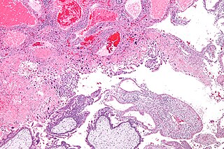
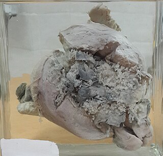
Choriocarcinoma is a malignant, trophoblastic cancer, usually of the placenta. It is characterized by early hematogenous spread to the lungs. It belongs to the malignant end of the spectrum in gestational trophoblastic disease (GTD). It is also classified as a germ cell tumor and may arise in the testis or ovary.

<span class="mw-page-title-main">Gestational trophoblastic disease</span> Pregnancy-related tumours

Gestational trophoblastic disease (GTD) is a term used for a group of pregnancy-related tumours. These tumours are rare, and they appear when cells in the womb start to proliferate uncontrollably. The cells that form gestational trophoblastic tumours are called trophoblasts and come from tissue that grows to form the placenta during pregnancy.

Gestational choriocarcinoma is a form of gestational trophoblastic neoplasia, which is a type of gestational trophoblastic disease (GTD), that can occur during pregnancy. It is a rare disease where the trophoblast, a layer of cells surrounding the blastocyst, undergoes abnormal developments, leading to trophoblastic tumors. The choriocarcinoma can metastasize to other organs, including the lungs, kidney, and liver. The amount and degree of choriocarcinoma spread to other parts of the body can vary greatly from person to person.

<span class="mw-page-title-main">Human reproductive system</span> Organs involved in reproduction

The human reproductive system includes the male reproductive system which functions to produce and deposit sperm; and the female reproductive system which functions to produce egg cells, and to protect and nourish the fetus until birth. Humans have a high level of sexual differentiation. In addition to differences in nearly every reproductive organ, there are numerous differences in typical secondary sex characteristics.

Dilation and evacuation (D&E) or dilatation and evacuation is the dilation of the cervix and surgical evacuation of the uterus after the first trimester of pregnancy. It is a method of abortion as well as a common procedure used after miscarriage to remove all pregnancy tissue.

Prenatal development includes the development of the embryo and of the fetus during a viviparous animal's gestation. Prenatal development starts with fertilization, in the germinal stage of embryonic development, and continues in fetal development until birth.

<span class="mw-page-title-main">Trophoblastic neoplasm</span> Medical condition

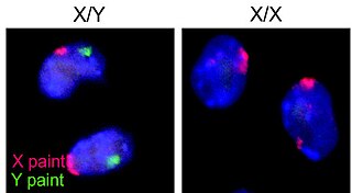
Gestational trophoblastic neoplasia (GTN) is group of rare diseases related to pregnancy and included in gestational trophoblastic disease (GTD) in which abnormal trophoblast cells grow in the uterus. GTN can be classified into benign and malignant lesions. Benign lesions include placental site nodule and hydatidiform moles while malignant lesions have four subtypes including invasive mole, gestational choriocarcinoma, placental site trophoblastic tumor (PSTT) and epithelioid trophoblastic tumor (ETT). The choriocarcinoma has 2 significant subtypes including gestational and non-gestational and they are differentiated by their different biological feature and prognosis. Signs and symptoms of GTN will appear vary from person to person and depending upon the type of the disease. They may include uterine bleeding not related to menstruation, pain or pressure in pelvis, large uterus and high blood pressure during pregnancy. The cause of this disease is unknown but the identification of the tumor based on total beta-human chorionic gonadotropin (β-hCG) in the serum.

<span class="mw-page-title-main">Human reproduction</span> Procreative biological processes of humanity

Human reproduction is sexual reproduction that results in human fertilization to produce a human offspring. It typically involves sexual intercourse between a sexually mature human male and female. During sexual intercourse, the interaction between the male and female reproductive systems results in fertilization of the ovum by the sperm to form a zygote. While normal cells contain 46 chromosomes, gamete cells only contain 23 single chromosomes, and it is when these two cells merge into one zygote cell that genetic recombination occurs and the new zygote contains 23 chromosomes from each parent, giving it 46 chromosomes. The zygote then undergoes a defined development process that is known as human embryogenesis, and this starts the typical 9-month gestation period that is followed by childbirth. The fertilization of the ovum may be achieved by artificial insemination methods, which do not involve sexual intercourse. Assisted reproductive technology also exists.

<span class="mw-page-title-main">Cytotrophoblast</span> Layer of an embryo

"Cytotrophoblast" is the name given to both the inner layer of the trophoblast or the cells that live there. It is interior to the syncytiotrophoblast and external to the wall of the blastocyst in a developing embryo.

<span class="mw-page-title-main">Triploid syndrome</span> Chromosomal disorder in which there are three copies of every chromosome

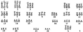
Triploid syndrome, also called triploidy, is a chromosomal disorder in which a fetus has three copies of every chromosome instead of the normal two. If this occurs in only some cells, it is called mosaic triploidy and is less severe.

<span class="mw-page-title-main">Invasive hydatidiform mole</span> Medical condition

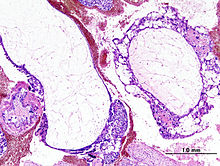
Invasive hydatidiform mole is a type of neoplasia that grows into the muscular wall of the uterus. It is formed after conception. It may spread to other parts of the body, such as the vagina, vulva, and lung.

<span class="mw-page-title-main">Placental disease</span> Medical condition

A placental disease is any disease, disorder, or pathology of the placenta.

<span class="mw-page-title-main">Placental site trophoblastic tumor</span> Medical condition

Placental site trophoblastic tumor is a form of gestational trophoblastic disease, which is thought to arise from intermediate trophoblast.

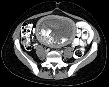
Theca lutein cyst is a type of bilateral functional ovarian cyst filled with clear, straw-colored fluid. These cysts result from exaggerated physiological stimulation due to elevated levels of beta-human chorionic gonadotropin (beta-hCG) or hypersensitivity to beta-hCG. On ultrasound and MRI, theca lutein cysts appear in multiples on ovaries that are enlarged.

References

  1. "About molar pregnancy | Gestational trophoblastic disease (GTD) | Cancer Research UK". www.cancerresearchuk.org. Retrieved 18 June 2022.
  2. "hydatidiform mole". Merriam Webster . Retrieved May 7, 2012.
  3. 1 2 3 4 Kumar V, ed. (2010). Pathologic Basis of Disease (8th ed.). Saunders Elsevier. pp. 1057–1058. ISBN   978-1-4377-0792-2.
  4. 1 2 Di Cintio E, Parazzini F, Rosa C, Chatenoud L, Benzi G (November 1997). "The epidemiology of gestational trophoblastic disease". General & Diagnostic Pathology. 143 (2–3): 103–8. PMID   9443567.
  5. Cotran RS, Kumar V, Fausto N, Nelso F, Robbins SL, Abbas AK (2005). Robbins and Cotran pathologic basis of disease (7th ed.). St. Louis, Mo: Elsevier Saunders. p. 1110. ISBN   0-7216-0187-1.
  6. Ganong WF, McPhee SJ, Lingappa VR (2005). Pathophysiology of Disease: An Introduction to Clinical Medicine (Lange). McGraw-Hill Medical. p. 639. ISBN   0-07-144159-X.
  7. MedlinePlus Encyclopedia : Hydatidiform mole
  8. Paoloni-Giacobino A (May 2007). "Epigenetics in reproductive medicine". Pediatric Research. 61 (5 Pt 2): 51R–57R. doi: 10.1203/pdr.0b013e318039d978 . PMID   17413849.
  9. Sebire NJ, Foskett M, Paradinas FJ, Fisher RA, Francis RJ, Short D, et al. (June 2002). "Outcome of twin pregnancies with complete hydatidiform mole and healthy co-twin". Lancet. 359 (9324): 2165–6. doi:10.1016/S0140-6736(02)09085-2. PMID   12090984. S2CID   33752037.
  10. Monga A, ed. (2006). Gynaecology by Ten Teachers (18th ed.). Hodder Arnold. pp.  99–101. ISBN   0-340-81662-7.
  11. Lawler SD, Fisher RA, Dent J (May 1991). "A prospective genetic study of complete and partial hydatidiform moles". American Journal of Obstetrics and Gynecology. 164 (5 Pt 1): 1270–7. doi:10.1016/0002-9378(91)90698-q. PMID   1674641.
  12. Wallace DC, Surti U, Adams CW, Szulman AE (1982). "Complete moles have paternal chromosomes but maternal mitochondrial DNA". Human Genetics. 61 (2): 145–7. doi:10.1007/BF00274205. PMID   6290372. S2CID   1417706.
  13. Slim R, Mehio A (January 2007). "The genetics of hydatidiform moles: new lights on an ancient disease". Clinical Genetics. 71 (1): 25–34. doi:10.1111/j.1399-0004.2006.00697.x. PMID   17204043. S2CID   32421323. Review.
  14. Woo JS, Hsu C, Fung LL, Ma HK (May 1983). "Partial hydatidiform mole: ultrasonographic features". The Australian & New Zealand Journal of Obstetrics & Gynaecology. 23 (2): 103–7. doi:10.1111/j.1479-828X.1983.tb00174.x. PMID   6578773. S2CID   6572375.
  15. 1 2 "The 6 questions that pregnant women should be ask to the doctor". HealthGuru. 15 March 2019.
  16. 1 2 Ghassemzadeh, Sassan; Farci, Fabiola; Kang, Michael (2023), "Hydatidiform Mole", StatPearls, Treasure Island (FL): StatPearls Publishing, PMID   29083593 , retrieved 2024-01-16
  17. Duffy L, Zhang L, Sheath K, Love DR, George AM (December 2015). "The Diagnosis of Choriocarcinoma in Molar Pregnancies: A Revised Approach in Clinical Testing". Journal of Clinical Medicine Research. 7 (12): 961–966. doi: 10.14740/jocmr2236w . PMC   4625817 . PMID   26566410.
  18. Cotran RS, Kumar V, Fausto N, Nelso F, Robbins SL, Abbas AK (2005). Robbins and Cotran pathologic basis of disease (7th ed.). St. Louis, Mo: Elsevier Saunders. p. 1112. ISBN   0-7216-0187-1.
  19. 1 2 Wang Q, Fu J, Hu L, Fang F, Xie L, Chen H, et al. (September 2017). "Prophylactic chemotherapy for hydatidiform mole to prevent gestational trophoblastic neoplasia". The Cochrane Database of Systematic Reviews. 9 (12): CD007289. doi:10.1002/14651858.cd007289.pub3. PMC   6483742 . PMID   28892119.
  20. Biyani G, Bhatia P (November 2011). "Mortality in hydatidiform mole: Should we blame thyroid?". Indian Journal of Anaesthesia. 55 (6): 628–9. doi: 10.4103/0019-5049.90629 . PMC   3249877 . PMID   22223914.
  21. 1 2 Cavaliere A, Ermito S, Dinatale A, Pedata R (January 2009). "Management of molar pregnancy". Journal of Prenatal Medicine. 3 (1): 15–7. PMC   3279094 . PMID   22439034.
  22. Entries HYDATID n. (a.) and MOLE, n.6 in the Oxford English Dictionary online. (http://dictionary.oed.com/ subscription required.)
  23. "Hydatidiform".