Antepartum bleeding

Last updated
Antepartum bleeding
Other namesAntepartum haemorrhage (APH), prepartum haemorrhage
Specialty Obstetrics   OOjs UI icon edit-ltr-progressive.svg

Antepartum bleeding, also known as antepartum haemorrhage (APH) or prepartum hemorrhage, is genital bleeding during pregnancy after the 28th week of pregnancy up to delivery. [1] [2]

Contents

It can be associated with reduced fetal birth weight. [3] Use of aspirin before 16 weeks of pregnancy to prevent pre-eclampsia also appears effective at preventing antepartum bleeding. [4]

In regard to treatment, it should be considered a medical emergency (regardless of whether there is pain), as if it is left untreated it can lead to death of the mother or baby.

Classification

The total amount of blood loss and signs of circulatory shock due to blood determine the severity of the antepartum haemorrhaging. There are 4 degrees of antepartum haemorrhaging: [5]

StageAmount of Blood Loss
SpottingStains, streaking, or spotting of blood
Minor HaemorrhageLess than 50mL
Major Haemorrhage50-1000mL without signs of circulatory shock
Massive HaemorrhageGreater than 1000mL with or without signs of circulatory shock

Causes

Placenta praevia

Placenta praevia refers to when the placenta of a growing foetus is attached abnormally low within the uterus. Intermittent antepartum haemorrhaging occurs in 72% of women living with placenta praevia. [6] The severity of a patient's placenta praevia depends on the location of placental attachment;

TypeLocation of Placental Attachment
Type 1Lower segment of uterus, no attachment to the cervix
Type 2Touching but not covering the internal orifice of the cervix
Type 3Partially covering the internal orifice of the cervix
Type 4Completely covering the internal orifice of the cervix

Types 1 and 2 are classified as minor placental praevia as these typically result in minor antepartum haemorrhaging. Types 3 and 4 are referred to as major placental praevia due to the risk of heavy haemorrhaging in the case of a rupture due to the location of placental attachment. [7] During the third trimester of pregnancy, thinning of the lower uterine segment or contractions caused by cervical dilation can increase the amount of stress placed on the placental attachment to the uterine wall. In patients with placenta praevia, these stresses can cause detachment of the placenta from the uterine wall causing haemorrhaging. To prevent further haemorrhaging patients with major placental praevia are recommended to have a caesarean delivery. [8]

Abnormal placentation

During pregnancy the layer of endometrium that attaches directly to developing blastocyst becomes the maternal portion of the placenta, also known as the decidua basalis. [9] In the absence of a decidua basalis, trophoblast cells on the developing blastocyst form an abnormally deep attachment to the uterine wall, this is known as abnormal placentation. Abnormal placentation can categorised into 3 types, depending on the depth of infiltration of the chorionic villi into the uterine wall: [10]

TypeDepth of Chorionic Villus
Placenta Accreta Attached to outermost portion of the myometrium
Placenta IncretaInvading the myometrium
Placenta PercretaInvading past the myometrium into the perimetrium

In placenta percreta, the chorionic villi have grow entirely through the myometrium and invade into the perimetrium. Placenta percreta results in the most intense haemorrhaging that can be expected caused by abnormal placentation. In the event of placental detachment from the uterine wall, the depth of chorionic villi attachment dictates the amount of haemorrhaging that can be expected. [11] The chance of abnormal placentation increases in subsequent pregnancies, if there is scar tissue present from previous pregnancies. For example, previously undergoing a caesarean or placenta previa increases the likelihood of abnormal placentation, therefore increasing the chances of antepartum haemorrhaging. [12]

Placental abruption

Placental abruption occurs when the placenta detaches from the endometrium. Detachment causes antepartum haemorrhaging at the location of abruption. Depending on the site of detachment, haemorrhaging may or may not be apparent. If abruption occurs behind the placenta where blood cannot escape through the cervix, blood will pool and form a retroplacental clot. Only when the site of detachment occurs on the side facing the cervical opening can the total amount of haemorrhaging be measured by vaginal bleeding. Using vaginal bleeding as a measurement of the severity of the placental abruption is therefore ineffective. The scale of haemorrhaging depends on the degree to which the placenta has separated from the uterine wall. In the case of partial placental separation, haemorrhaging can be minor. However, in the case of total placental separation haemorrhaging will be major and emergency delivery will typically be the course of action. [13] Placental abruption causes blood loss from the mother and loss of oxygen and nutrients to the placenta occasionally leading to preterm labour. [14] Other causes of placental abruption can be abdominal trauma or sudden decompression of amniotic fluid, however it is not uncommon for the cause of placental abruption to be unknown. [15]

Vasa praevia

Vasa praevia is the presence of unprotected foetal blood vessels running along the placenta and over the internal cervical opening. Vasa praevia is a very rare, presenting only 4:10,000 cases from the largest study of the condition. [16] Risks of antepartum bleeding due to vasa praevia greatly increase during the third trimester of pregnancy during cervical dilation or placenta praevia. Vessel rupture is very likely in the event of a membranous rupture as foetal blood vessels aren't protected by the umbilical cord of the placenta. In the event of foetal vessel rupture, antepartum haemorrhaging occurs however blood is lost from the foetal blood supply. If the foetus is developed enough caesarean sections are often recommended. [17]

Abnormal placental shape

A circumvallate placenta refers to when the foetal membrane wraps twice around, over the foetal side around the edge of the placenta. This is to compensate for an undersized chorionic plate resulting in a decreased nutritional supply to the foetus. Thickening of the placental edge due to a circumvallate placenta can lead to placental abruption, causing antepartum haemorrhaging. [18]

A bilobed placenta has a cleavage in the middle dividing it into two lobes with membranous vessels branching in between. The exposed connecting membranous vessels present risk of rupturing due to limited protection from thrombosis and trauma. Bilobing of the placenta can be caused by placental implantation occurring over areas of uterine fibroid scarring, previous surgery, decreased blood supply or implantation occurring over the internal cervical orifice. Chances of vasa previa and placental abruption increase in the presence of a bilobed placenta due to decreased surface area for attachment to the uterine wall and the exposure of membranous vessels. [19]

When a placenta has multiple lobes which are distal and not of equal size this is referred to as a succenturiate placenta. Distal lobes are connected by a placental artery and vein extending from the main placenta, which tends to be centrally located and is the largest in mass. [20] At the time of contraction or delivery the connecting placental arteries and veins may rupture resulting in significant haemorrhaging. Incidences of vasa previa and haemorrhaging in the presence of a succenturiate placenta are highly increased. [21]

Minor causes

Cervical ectropion

There are 2 types of epithelial cells present within the cervical canal. In the endocervix the epithelia is columnar glandular which transitions into stratified squamous towards the ectocervix and external cervical orifice. During cervical ectropion the epithelial transitional zone (also called the squamo-columnar junction) migrates from the endocervical canal towards the ectocervix exposing some columnar glandular cells on the external cervical orifice. Unlike stratified squamous epithelial cells, glandular cells are columnar and not used to external stresses such as abrasion. The translocation of these cells causes bleeding and mucus secretion. Cervical ectropion can be attributed to rises in oestrogen levels during foetal development. Antepartum haemorrhage caused by cervical ectropion can be expected and is typically harmless. [22]

Vaginal infection

The presence of severe vaginal infections at the time of pregnancy may cause minor antepartum haemorrhaging. For example, the presence of chlamydia, thrush, cervicitis or other infections are all irritants to the vaginal and cervical lining, causing bleeding from those surfaces where the infection is severe. [23]

Cervical canal and distal genital tract

Most cases of Antepartum haemorrhaging originate from within the cervical canal or vagina. The amount of bleeding in these areas are typically limited to spotting or minor antepartum haemorrhaging. Cervical ectropion, dysplasia, polyps or cervical carcinoma may cause lesions in the cervix leading to minor haemorrhaging or spotting. Abrasion or slight trauma caused by intercourse, clinical examinations and pap smear may also cause spotting from the cervix. Vaginal bleeding from atrophy, vaginitis, and ulcers also attribute to minor haemorrhaging. Similarly, varicosities, tumours or inflammation in the vulva can cause minor antepartum haemorrhaging. Non genital tract bleeding caused by haematuria or haemorrhoids can often be mistaken for antepartum haemorrhaging and are typically harmless. [24]

Endometriosis

Estimates of the frequency of endometriosis in women range between 6-15% across several sources. In a 30-year reproductive and pregnancy outcome study of 14,000+ women, those with endometriosis had a significantly increased risk of antepartum haemorrhage during pregnancy. [25]

Diagnosis

Differential diagnosis

See also

Related Research Articles

Obstetrics is the field of study concentrated on pregnancy, childbirth and the postpartum period. As a medical specialty, obstetrics is combined with gynecology under the discipline known as obstetrics and gynecology (OB/GYN), which is a surgical field.

<span class="mw-page-title-main">Placenta praevia</span> Medical condition

Placenta praevia is when the placenta attaches inside the uterus but in a position near or over the cervical opening. Symptoms include vaginal bleeding in the second half of pregnancy. The bleeding is bright red and tends not to be associated with pain. Complications may include placenta accreta, dangerously low blood pressure, or bleeding after delivery. Complications for the baby may include fetal growth restriction.

<span class="mw-page-title-main">Placental abruption</span> Medical condition

Placental abruption is when the placenta separates early from the uterus, in other words separates before childbirth. It occurs most commonly around 25 weeks of pregnancy. Symptoms may include vaginal bleeding, lower abdominal pain, and dangerously low blood pressure. Complications for the mother can include disseminated intravascular coagulopathy and kidney failure. Complications for the baby can include fetal distress, low birthweight, preterm delivery, and stillbirth.

<span class="mw-page-title-main">Vaginal bleeding</span> Medical condition

Vaginal bleeding is any expulsion of blood from the vagina. This bleeding may originate from the uterus, vaginal wall, or cervix. Generally, it is either part of a normal menstrual cycle or is caused by hormonal or other problems of the reproductive system, such as abnormal uterine bleeding.

Bloody show or show is the passage of a small amount of blood or blood-tinged mucus through the vagina near the end of pregnancy. It is caused by thinning and dilation of the cervix, leading to detachment of the cervical mucus plug that seals the cervix during pregnancy and tearing of small cervical blood vessels, and is one of the signs that labor may be imminent. The bloody show may be expelled from the vagina in pieces or altogether and often appears as a jelly-like piece of mucus stained with blood. Although the bloody show may be alarming at first, it is not a concern of patient health after 37 weeks gestation.

Obstetrical bleeding is bleeding in pregnancy that occurs before, during, or after childbirth. Bleeding before childbirth is that which occurs after 24 weeks of pregnancy. Bleeding may be vaginal or less commonly into the abdominal cavity. Bleeding which occurs before 24 weeks is known as early pregnancy bleeding.

<span class="mw-page-title-main">Complications of pregnancy</span> Medical condition

Complications of pregnancy are health problems that are related to, or arise during pregnancy. Complications that occur primarily during childbirth are termed obstetric labor complications, and problems that occur primarily after childbirth are termed puerperal disorders. While some complications improve or are fully resolved after pregnancy, some may lead to lasting effects, morbidity, or in the most severe cases, maternal or fetal mortality.

<span class="mw-page-title-main">Placenta accreta spectrum</span> Medical condition

Placenta accreta occurs when all or part of the placenta attaches abnormally to the myometrium. Three grades of abnormal placental attachment are defined according to the depth of attachment and invasion into the muscular layers of the uterus:

  1. Accreta – chorionic villi attached to the myometrium, rather than being restricted within the decidua basalis.
  2. Increta – chorionic villi invaded into the myometrium.
  3. Percreta – chorionic villi invaded through the perimetrium.
<span class="mw-page-title-main">Uterine atony</span> Loss of tone in the uterine musculature

Uterine atony is the failure of the uterus to contract adequately following delivery. Contraction of the uterine muscles during labor compresses the blood vessels and slows flow, which helps prevent hemorrhage and facilitates coagulation. Therefore, a lack of uterine muscle contraction can lead to an acute hemorrhage, as the vasculature is not being sufficiently compressed. Uterine atony is the most common cause of postpartum hemorrhage, which is an emergency and potential cause of fatality. Across the globe, postpartum hemorrhage is among the top five causes of maternal death. Recognition of the warning signs of uterine atony in the setting of extensive postpartum bleeding should initiate interventions aimed at regaining stable uterine contraction.

<span class="mw-page-title-main">Postpartum bleeding</span> Loss of blood following childbirth

Postpartum bleeding or postpartum hemorrhage (PPH) is often defined as the loss of more than 500 ml or 1,000 ml of blood following childbirth. Some have added the requirement that there also be signs or symptoms of low blood volume for the condition to exist. Signs and symptoms may initially include: an increased heart rate, feeling faint upon standing, and an increased breathing rate. As more blood is lost, the patient may feel cold, blood pressure may drop, and they may become restless or unconscious. The condition can occur up to six weeks following delivery.

<span class="mw-page-title-main">Vasa praevia</span> Condition in which fetal blood vessels cross or run near the internal opening of the uterus

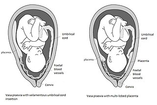
Vasa praevia is a condition in which fetal blood vessels cross or run near the internal opening of the uterus. These vessels are at risk of rupture when the supporting membranes rupture, as they are unsupported by the umbilical cord or placental tissue.

<span class="mw-page-title-main">Vaginal delivery</span> Delivery through the vagina

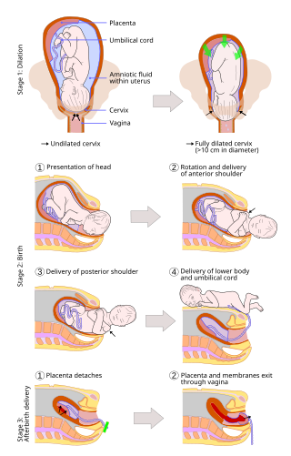
A vaginal delivery is the birth of offspring in mammals through the vagina. It is the most common method of childbirth worldwide. It is considered the preferred method of delivery, with lower morbidity and mortality than caesarean sections (C-sections).

<span class="mw-page-title-main">Velamentous cord insertion</span> Velamentous placenta

Velamentous cord insertion is a complication of pregnancy where the umbilical cord is inserted in the fetal membranes. It is a major cause of antepartum hemorrhage that leads to loss of fetal blood and associated with high perinatal mortality. In normal pregnancies, the umbilical cord inserts into the middle of the placental mass and is completely encased by the amniotic sac. The vessels are hence normally protected by Wharton's jelly, which prevents rupture during pregnancy and labor. In velamentous cord insertion, the vessels of the umbilical cord are improperly inserted in the chorioamniotic membrane, and hence the vessels traverse between the amnion and the chorion towards the placenta. Without Wharton's jelly protecting the vessels, the exposed vessels are susceptible to compression and rupture.

<span class="mw-page-title-main">Placental disease</span> Medical condition

A placental disease is any disease, disorder, or pathology of the placenta.

<span class="mw-page-title-main">Chorionic hematoma</span> Medical condition

Chorionic hematoma is the pooling of blood (hematoma) between the chorion, a membrane surrounding the embryo, and the uterine wall. It occurs in about 3.1% of all pregnancies, it is the most common sonographic abnormality and the most common cause of first trimester bleeding.

The following outline is provided as an overview of and topical guide to obstetrics:

<span class="mw-page-title-main">Circumvallate placenta</span> Medical condition

Circumvallate placenta is a rare condition affecting about 1-2% of pregnancies, in which the amnion and chorion fetal membranes essentially "double back" on the fetal side around the edges of the placenta. After delivery, a circumvallate placenta has a thick ring of membranes on its fetal surface. Circumvallate placenta is a placental morphological abnormality associated with increased fetal morbidity and mortality due to the restricted availability of nutrients and oxygen to the developing fetus.

Uterine Tachysystole is a condition of excessively frequent uterine contractions during pregnancy. It is most often seen in induced or augmented labor, though it can also occur during spontaneous labor, and this may result in fetal hypoxia and acidosis. This may have serious effects on both the mother and the fetus including hemorrhaging and death. There are still major gaps in understanding treatment as well as clinical outcomes of this condition. Uterine tachysystole is defined as more than 5 contractions in 10 minutes, averaged over a 30-minute period.

Early pregnancy bleeding refers to vaginal bleeding before 14 weeks of gestational age. If the bleeding is significant, hemorrhagic shock may occur. Concern for shock is increased in those who have loss of consciousness, chest pain, shortness of breath, or shoulder pain.

The anomaly scan, also sometimes called the anatomy scan, 20-week ultrasound, or level 2 ultrasound, evaluates anatomic structures of the fetus, placenta, and maternal pelvic organs. This scan is an important and common component of routine prenatal care. The function of the ultrasound is to measure the fetus so that growth abnormalities can be recognized quickly later in pregnancy, to assess for congenital malformations and multiple pregnancies, and to plan method of delivery.

References

  1. patient.info » PatientPlus » Antepartum Haemorrhage
  2. The Royal Women's Hospital > antepartum haemorrhage Archived 2010-01-08 at the Wayback Machine Retrieved on Jan 13, 2009
  3. Lam CM, Wong SF, Chow KM, Ho LC (2000). "Women with placenta praevia and antepartum haemorrhage have a worse outcome than those who do not bleed before delivery". Journal of Obstetrics and Gynaecology. 20 (1): 27–31. doi:10.1080/01443610063417. PMID   15512459. S2CID   218855273.
  4. Roberge, S; Bujold, E; Nicolaides, KH (May 2018). "Meta-analysis on the effect of aspirin use for prevention of preeclampsia on placental abruption and antepartum hemorrhage" (PDF). American Journal of Obstetrics and Gynecology. 218 (5): 483–489. doi: 10.1016/j.ajog.2017.12.238 . PMID   29305829.
  5. Antepartum Haemorrhage. (2015). Perth, Western Australia: Department of Health Western Australia, pp.3-6.
  6. Love, C. and Wallace, E. (1996). Pregnancies complicated by placenta praevia: what is appropriate management?. BJOG: An International Journal of Obstetrics and Gynaecology, 103(9), pp.864-867.
  7. Royal College of Obstetricians & Gynaecologists. (2018). Placenta Praevia, Placenta Praevia Accreta and Vasa Praevia: Diagnosis and Management (Green-top Guideline No. 27). [online] Available at: https://www.rcog.org.uk/en/guidelines-research-services/guidelines/gtg27/ [Accessed 27 Apr. 2018].
  8. Better Health Channel. (2018). Placenta Previa. [online] Available at: https://www.betterhealth.vic.gov.au/health/healthyliving/placenta-previa [Accessed 27 Apr. 2018].
  9. Gest, T. (2018). Placenta and Extraembryonic Membranes. [online] The University of Michigan Medical School - Anatomy. Available at: https://www.med.umich.edu/lrc/coursepages/m1/embryology/embryo/06placenta.htm [Accessed 31 May 2018].
  10. Silver, R. (2015). Abnormal Placentation. Obstetrics & Gynecology, 126(3), pp.654-668.
  11. Smith, Z., Sehgal, S., Van Arsdalen, K. and Goldstein, I. (2014). Placenta Percreta With Invasion into the Urinary Bladder. Urology Case Reports, 2(1), pp.31-32.
  12. Nielsen, T., Hagberg, H. and Ljungblad, U. (1989). Placenta previa and Antepartum Hemorrhage after Previous Cesarean Section. Gynecologic and Obstetric Investigation, 27(2), pp.88-90.
  13. American Pregnancy Association. (2018). Placental Abruption: Risks, Causes, Symptoms and Treatment. [online] Available at: http://americanpregnancy.org/pregnancy-complications/placental-abruption/ [Accessed 27 Apr. 2018].
  14. Better Health Channel. (2014). Placental Abruption. [online] Available at: https://www.betterhealth.vic.gov.au/health/healthyliving/placental-abruption [Accessed 27 Apr. 2018].
  15. TIKKANEN, M. (2010). Placental abruption: epidemiology, risk factors and consequences. Acta Obstetricia et Gynecologica Scandinavica, 90(2), pp.140-149.
  16. Derbala, Y., Grochal, F. and Jeanty, P. (2007). Vasa Praevia. Journal of Prenatal Medicine.
  17. Ruiter, L., Kok, N., Limpens, J., Derks, J., de Graaf, I., Mol, B. and Pajkrt, E. (2015). Incidence of and risk indicators for vasa praevia: a systematic review. BJOG: An International Journal of Obstetrics & Gynaecology, 123(8), pp.1278-1287.
  18. Hermann, Z. (1963). Circumvallate Placenta, a Cause of Antepartum Bleeding, Premature Delivery, and Perinatal Mortality. Obstetrics & Gynecology, 22(6), pp.798-802.
  19. Rathburn, K. and Hildebrand, J. (2017). Placenta, Abnormalities.
  20. Antepartum haemorrhage or bleeding in the second half of pregnancy. (2013). p.14.
  21. Suzuki, S. and Igarashi, M. (2007). Clinical significance of pregnancies with succenturiate lobes of placenta. Archives of Gynecology and Obstetrics, 277(4), pp.299-301.
  22. Cervical ectropion (cervical erosion). (2015). Guy's and St Rhomas' NHS Foundation Trust, pp.1-2.
  23. Mater Patient Information. (2018). Antepartum haemorrhage - Mater Patient Information. [online] Available at: http://brochures.mater.org.au/brochures/mater-mothers-hospital/antepartum-haemorrhage [Accessed 27 Apr. 2018].
  24. Antepartum haemorrhage or bleeding in the second half of pregnancy. (2013). p.11.
  25. Saraswat, Lucky (2015). "ESHRE2015: Endometriosis associated with a greater risk of complications in pregnancy". endometriosis.org. European Society of Human Reproduction and Embryology. Retrieved 14 February 2024.