Chorioamnionitis

Last updated
Chorioamnionitis
Histopathology of chorioamnionitis.jpg
Micrograph showing acute chorioamnionitis, with neutrophils in the chorion. Also seen are fibrin thrombi, which indicate a severe fetal inflammatory response. [1] H&E stain.
Specialty Obstetrics and gynaecology   OOjs UI icon edit-ltr-progressive.svg

Chorioamnionitis, also known as intra-amniotic infection (IAI), is inflammation of the fetal membranes (amnion and chorion), usually due to bacterial infection. [1] In 2015, a National Institute of Child Health and Human Development Workshop expert panel recommended use of the term "triple I" to address the heterogeneity of this disorder. The term triple I refers to intrauterine infection or inflammation or both and is defined by strict diagnostic criteria, but this terminology has not been commonly adopted although the criteria are used. [2]

Contents

Chorioamnionitis results from an infection caused by bacteria ascending from the vagina into the uterus and is associated with premature or prolonged labor. [3] It triggers an inflammatory response to release various inflammatory signaling molecules, leading to increased prostaglandin and metalloproteinase release. These substances promote uterine contractions and cervical ripening, causations of premature birth. [4] The risk of developing chorioamnionitis increases with number of vaginal examinations performed in the final month of pregnancy, including labor. [5] [6] Tobacco and alcohol use also puts mothers at risk for chorioamnionitis development. [7]

Chorioamnionitis is caught early by looking at signs and symptoms such as fever, abdominal pain, or abnormal vaginal excretion. [8] Administration of antibiotics if the amniotic sac bursts prematurely can prevent chorioamnionitis occurrence. [9]

Signs and symptoms

The signs and symptoms of clinical chorioamnionitis include fever, leukocytosis (>15,000 cells/mm3), maternal (>100 bpm) [10] or fetal (>160 bpm) tachycardia, uterine tenderness and preterm rupture of membranes. [2]

Causes

Causes of chorioamnionitis stem from bacterial infection as well as obstetric and other related factors. [3] [7]

Microorganisms

Bacterial, viral, and even fungal infections can cause chorioamnionitis. Most commonly from Ureaplasma, Fusobacterium , and Streptococcus bacteria species. Less commonly, Gardnerella, Mycoplasma, and Bacteroides bacteria species. Sexually transmitted infections, chlamydia and gonorrhea, can cause development of the condition as well. [7] Studies are continuing to identify other microorganism classes and species as infection sources. [11]

Obstetric and other

Birthing-related events, lifestyle, and ethnic background have been linked to an increase in the risk of developing chorioamnionitis apart from bacterial causation. [11] Premature deliveries, ruptures of the amniotic sac membranes, prolonged labor, and primigravida childbirth are associated with this condition. [12] At term mothers who experience a combination of pre-labor membrane ruptures and multiple invasive vaginal examinations, prolonged labor, or have meconium appear in the amniotic fluid are at higher risk than at term mothers experiencing just one of those events. [11] In other studies, smoking, alcohol use and drug use are noted as risk factors. Those of African American ethnicity are noted to be at higher risk. [7] [12]

Anatomy

The chorion and amnion membranes are labelled in this depiction of a growing fetus in the uterus. FetalMembranes1L.jpg
The chorion and amnion membranes are labelled in this depiction of a growing fetus in the uterus.

The amniotic sac consists of two parts:

Diagnosis

Intermediate magnification micrograph of moderate chorioamnionitis. H&E stain. Chorioamnionitis - intermed mag.jpg
Intermediate magnification micrograph of moderate chorioamnionitis. H&E stain.
Acute choriodeciduitis, with neutrophils seen in the chorion and decidua. H&E stain. Histopathology of acute choriodeciduitis.jpg
Acute choriodeciduitis, with neutrophils seen in the chorion and decidua. H&E stain.

Pathologic

Chorioamnionitis is diagnosed from a histologic (tissue) examination of the fetal membranes. [12] Confirmed histologic chorioamnionitis without any clinical symptoms is termed subclinical chorioamnionitis and is more common than symptomatic clinical chorioamnionitis. [2]

Infiltration of the chorionic plate by neutrophils is diagnostic of (mild) chorioamnionitis. More severe chorioamnionitis involves subamniotic tissue and may have fetal membrane necrosis and/or abscess formation. [1]

Severe chorioamnionitis may be accompanied by vasculitis of the umbilical blood vessels due to the fetus' inflammatory cells. If very severe, funisitis, inflammation of the umbilical cord connective tissue, occurs. [12]

Suspected clinical diagnosis

The presence of fever between 38.0°C and 39.0°C alone is insufficient to indicate chorioamnionitis and is termed isolated maternal fever. Isolated maternal fever may not have an infectious cause and does not require antibiotic treatment. [2]

When intrapartum (during delivery) fever is higher than 39.0°C, suspected diagnosis of chorioamnionitis can be made. Alternatively, if intrapartum fever is between 38.0°C and 39.0°C, an additional risk factor must be present to make a presumptive diagnosis of chorioamnionitis. Additional risk factors include: [14]

Confirmed diagnosis

Diagnosis is typically not confirmed until after delivery. However, people with confirmed diagnosis and suspected diagnosis have the same post-delivery treatment regardless of diagnostic status. Diagnosis can be confirmed histologically or through amniotic fluid tests such as gram staining, glucose levels, or other culture results consistent with infection. [14]

Prevention

If the amniotic sac breaks early into pregnancy, the potential of introducing bacteria in the amniotic fluid can increase. Administering antibiotics maternally can potentially prevent chorioamnionitis and allow for a longer pregnancy. [9]  In addition, it has been shown that it is not necessary to deliver the fetus quickly after chorioamnionitis is diagnosed, so a C-section is not necessary unless maternal health concern is present. [12] However, research has found that beginning labor early at approximately 34 weeks can lessen the likelihood of fetal death, and reduce the potential for excessive infection within the mother. [12]

In addition, providers should interview people suspected to have chorioamnionitis about whether they are experiencing signs and symptoms at scheduled obstetrics visits during pregnancy, including whether the individual has experienced excretion vaginally, febrile, or abdominal pain. [8]

Treatment

The American College of Obstetricians and Gynecologists' Committee Opinion proposes the use of antibiotic treatment in intrapartum mothers with suspected or confirmed chorioamnionitis and maternal fever without an identifiable cause. [14]

Intrapartum antibiotic treatment consists of: [2]

However, there is not enough evidence to support the most efficient antimicrobial regimen. [16] Starting the treatment during the intrapartum period is more effective than starting it postpartum; it shortens the hospital stay for the mother and the neonate. [17] There is currently not enough evidence to dictate how long antibiotic therapy should last. Completion of treatment/cure is only considered after delivery. [2]

Supportive measures

Acetaminophen is often used for treating fevers and may be beneficial for fetal tachycardia. There can be increased likelihood for neonatal encephalopathy when mothers have intrapartum fever. [12]

Outcomes

Chorioamnionitis has possible associations with numerous neonatal conditions. Intrapartum (during labor) chorioamnionitis may be associated with neonatal pneumonia, meningitis, sepsis, and death. Long-term infant complications like bronchopulmonary dysplasia, cerebral palsy, and Wilson-Mikity syndrome have been associated to the bacterial infection. [14] Furthermore, histological chorioamnionitis may increase the likelihood of newborn necrotizing enterocolitis, where one or more sections of the bowel die. This occurs when the fetal gut barrier becomes compromised and is more susceptible to conditions like infection and sepsis. [18] In addition, chorioamnionitis can act as a risk factor for premature birth and periventricular leukomalacia. [19]

Complications

For mother and fetus, chorioamnionitis may lead to short-term and long-term issues when microbes move to different areas or trigger inflammatory responses due to infection. [12]

Maternal complications

Mothers with chorioamnionitis who undergo a C-section may be more likely to develop pelvic abscesses, septic pelvic thrombophlebitis, and infections at the surgical site. [11]

Fetal complications

Neonatal complications

In the long-term, infants may be more likely to experience cerebral palsy or neurodevelopmental disabilities. Disability development is related to the activation of the fetal inflammatory response syndrome (FIRS) when the fetus is exposed to infected amniotic fluid or other foreign entities. [4] [12] This systemic response results in neutrophil and cytokine release that can impair the fetal brain and other vital organs. [4] [9] Compared to infants with clinical chorioamnionitis, it appears cerebral palsy may occur at a higher rate for those with histologic chorioamnionitis. However, more research needs to be done to examine this association. [22] There is also concern about the impact of FIRS on infant immunity as this is a critical time for growth and development. For instance, it may be linked to chronic inflammatory disorders, such as asthma. [23]

Epidemiology

Chorioamnionitis is approximated to occur in about 4% [8] of births in the United States. However, many other factors can increase the risk of chorioamnionitis. For example, in births with premature rupture of membranes (PROM), between 40 and 70% involve chorioamnionitis. Furthermore, clinical chorioamnionitis is implicated in 12% of all cesarean deliveries. Some studies have shown that the risk of chorioamnionitis is higher in those of African American ethnicity, those with immunosuppression, and those who smoke, use alcohol, or abuse drugs. [12]

See also

Notes

  1. 1 2 3 4 Kim CJ, Romero R, Chaemsaithong P, Chaiyasit N, Yoon BH, Kim YM (October 2015). "Acute chorioamnionitis and funisitis: definition, pathologic features, and clinical significance". American Journal of Obstetrics and Gynecology. 213 (4 Suppl): S29–S52. doi:10.1016/j.ajog.2015.08.040. PMC   4774647 . PMID   26428501.
  2. 1 2 3 4 5 6 Tita AT (2019). Berghella V, Barss BA (eds.). "Intra-amniotic infection (clinical chorioamnionitis or triple I)". UpToDate.
  3. 1 2 Cheng YW, Delaney SS, Hopkins LM, Caughey AB (November 2009). "The association between the length of first stage of labor, mode of delivery, and perinatal outcomes in women undergoing induction of labor". American Journal of Obstetrics and Gynecology. 201 (5): 477.e1–477.e7. doi:10.1016/j.ajog.2009.05.024. PMID   19608153.
  4. 1 2 3 Galinsky R, Polglase GR, Hooper SB, Black MJ, Moss TJ (2013). "The consequences of chorioamnionitis: preterm birth and effects on development". Journal of Pregnancy. 2013: 412831. doi: 10.1155/2013/412831 . PMC   3606792 . PMID   23533760.
  5. Seaward PG, Hannah ME, Myhr TL, Farine D, Ohlsson A, Wang EE, et al. (November 1997). "International Multicentre Term Prelabor Rupture of Membranes Study: evaluation of predictors of clinical chorioamnionitis and postpartum fever in patients with prelabor rupture of membranes at term". American Journal of Obstetrics and Gynecology. 177 (5): 1024–1029. doi:10.1016/s0002-9378(97)70007-3. PMID   9396886.
  6. Gluck O, Mizrachi Y, Ganer Herman H, Bar J, Kovo M, Weiner E (April 2020). "The correlation between the number of vaginal examinations during active labor and febrile morbidity, a retrospective cohort study". BMC Pregnancy and Childbirth. 20 (1): 246. doi: 10.1186/s12884-020-02925-9 . PMC   7183634 . PMID   32334543.
  7. 1 2 3 4 Sweeney EL, Dando SJ, Kallapur SG, Knox CL (January 2017). "The Human Ureaplasma Species as Causative Agents of Chorioamnionitis". Clinical Microbiology Reviews. 30 (1): 349–379. doi:10.1128/CMR.00091-16. PMC   5217797 . PMID   27974410.
  8. 1 2 3 Fowler JR, Simon LV (October 2019). "Chorioamnionitis.". StatPearls [Internet]. StatPearls Publishing. PMID   30335284.
  9. 1 2 3 Ericson JE, Laughon MM (March 2015). "Chorioamnionitis: implications for the neonate". Clinics in Perinatology. 42 (1): 155–65, ix. doi:10.1016/j.clp.2014.10.011. PMC   4331454 . PMID   25678002.
  10. Sung JH, Choi SJ, Oh SY, Roh CR (March 2021). "Should the diagnostic criteria for suspected clinical chorioamnionitis be changed?". The Journal of Maternal-Fetal & Neonatal Medicine. 34 (5): 824–833. doi:10.1080/14767058.2019.1618822. PMID   31084245. S2CID   155091947.
  11. 1 2 3 4 Czikk MJ, McCarthy FP, Murphy KE (September 2011). "Chorioamnionitis: from pathogenesis to treatment". Clinical Microbiology and Infection. 17 (9): 1304–1311. doi: 10.1111/j.1469-0691.2011.03574.x . PMID   21672080.
  12. 1 2 3 4 5 6 7 8 9 10 11 Tita AT, Andrews WW (June 2010). "Diagnosis and management of clinical chorioamnionitis". Clinics in Perinatology. 37 (2): 339–354. doi:10.1016/j.clp.2010.02.003. PMC   3008318 . PMID   20569811.
  13. 1 2 Betts JG, Young KA, Wise JA, Johnson E, Poe B, Kruse DH, et al. (25 April 2013). "Embryonic Development". Anatomy and Physiology. Houston, Texas: OpenStax. Retrieved 29 July 2020.
  14. 1 2 3 4 "Committee Opinion No. 712 Summary: Intrapartum Management of Intraamniotic Infection". Obstetrics and Gynecology. 130 (2): 490–492. August 2017. doi:10.1097/AOG.0000000000002230. PMID   28742671.
  15. Zanella P, Bogana G, Ciullo R, Zambon A, Serena A, Albertin MA (June 2010). "[Chorioamnionitis in the delivery room]". Minerva Pediatrica. 62 (3 Suppl 1): 151–153. PMID   21090085.
  16. Hopkins L, Smaill F (2002). "Antibiotic regimens for management of intraamniotic infection". The Cochrane Database of Systematic Reviews. 2014 (3): CD003254. doi:10.1002/14651858.CD003254. PMC   6669261 . PMID   12137684.
  17. Chapman E, Reveiz L, Illanes E, Bonfill Cosp X (December 2014). Reveiz L (ed.). "Antibiotic regimens for management of intra-amniotic infection". The Cochrane Database of Systematic Reviews. 2014 (12): CD010976. doi:10.1002/14651858.CD010976.pub2. PMC   10562955 . PMID   25526426.
  18. Been JV, Lievense S, Zimmermann LJ, Kramer BW, Wolfs TG (February 2013). "Chorioamnionitis as a risk factor for necrotizing enterocolitis: a systematic review and meta-analysis". The Journal of Pediatrics. 162 (2): 236–42.e2. doi: 10.1016/j.jpeds.2012.07.012 . PMID   22920508.
  19. Wu YW, Colford JM (September 2000). "Chorioamnionitis as a risk factor for cerebral palsy: A meta-analysis". JAMA. 284 (11): 1417–1424. doi:10.1001/jama.284.11.1417. PMID   10989405.
  20. Casey BM, Cox SM (March 1997). "Chorioamnionitis and endometritis". Infectious Disease Clinics of North America. 11 (1): 203–222. doi:10.1016/S0891-5520(05)70349-4. PMID   9067792.
  21. Bersani I, Thomas W, Speer CP (April 2012). "Chorioamnionitis--the good or the evil for neonatal outcome?". The Journal of Maternal-Fetal & Neonatal Medicine. 25 (Suppl 1): 12–16. doi:10.3109/14767058.2012.663161. PMID   22309119. S2CID   11109172.
  22. Shi Z, Ma L, Luo K, Bajaj M, Chawla S, Natarajan G, et al. (June 2017). "Chorioamnionitis in the Development of Cerebral Palsy: A Meta-analysis and Systematic Review". Pediatrics. 139 (6): e20163781. doi:10.1542/peds.2016-3781. PMC   5470507 . PMID   28814548.
  23. Sabic D, Koenig JM (January 2020). "A perfect storm: fetal inflammation and the developing immune system". Pediatric Research. 87 (2): 319–326. doi:10.1038/s41390-019-0582-6. PMC   7875080 . PMID   31537013. S2CID   202702137.

Related Research Articles

<span class="mw-page-title-main">Amniocentesis</span> Sampling of amniotic fluid done mainly to detect fetal chromosomal abnormalities

Amniocentesis is a medical procedure used primarily in the prenatal diagnosis of genetic conditions. It has other uses such as in the assessment of infection and fetal lung maturity. Prenatal diagnostic testing, which includes amniocentesis, is necessary to conclusively diagnose the majority of genetic disorders, with amniocentesis being the gold-standard procedure after 15 weeks' gestation.

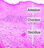
The amniotic sac, also called the bag of waters or the membranes, is the sac in which the embryo and later fetus develops in amniotes. It is a thin but tough transparent pair of membranes that hold a developing embryo until shortly before birth. The inner of these membranes, the amnion, encloses the amniotic cavity, containing the amniotic fluid and the embryo. The outer membrane, the chorion, contains the amnion and is part of the placenta. On the outer side, the amniotic sac is connected to the yolk sac, the allantois, and via the umbilical cord, the placenta.

Fetal distress, also known as non-reassuring fetal status, is a condition during pregnancy or labor in which the fetus shows signs of inadequate oxygenation. Due to its imprecision, the term "fetal distress" has fallen out of use in American obstetrics. The term "non-reassuring fetal status" has largely replaced it. It is characterized by changes in fetal movement, growth, heart rate, and presence of meconium stained fluid.

Oligohydramnios is a medical condition in pregnancy characterized by a deficiency of amniotic fluid, the fluid that surrounds the fetus in the abdomen, in the amniotic sac. It is typically diagnosed by ultrasound when the amniotic fluid index (AFI) measures less than 5 cm or when the single deepest pocket (SDP) of amniotic fluid measures less than 2 cm. Amniotic fluid is necessary to allow for normal fetal movement, lung development, and cushioning from uterine compression. Low amniotic fluid can be attributed to a maternal, fetal, placental or idiopathic cause and can result in poor fetal outcomes including death. The prognosis of the fetus is dependent on the etiology, gestational age at diagnosis, and the severity of the oligohydramnios.

<span class="mw-page-title-main">Amniotic fluid</span> Fluid surrounding a fetus within the amnion

The amniotic fluid is the protective liquid contained by the amniotic sac of a gravid amniote. This fluid serves as a cushion for the growing fetus, but also serves to facilitate the exchange of nutrients, water, and biochemical products between mother and fetus.

Rupture of membranes (ROM) or amniorrhexis is a term used during pregnancy to describe a rupture of the amniotic sac. Normally, it occurs spontaneously at full term either during or at the beginning of labor. Rupture of the membranes is known colloquially as "breaking (one's) water," especially when induced rather than spontaneous, or as one's "water breaking". A premature rupture of membranes (PROM) is a rupture of the amnion that occurs at full term and prior to the onset of labor. In cases of PROM, options include expectant management without intervention, or interventions such as oxytocin or other methods of labor induction, and both are usually accompanied by close monitoring of maternal and fetal health. Preterm premature rupture of membranes (PPROM) is when water breaks both before the onset of labor and before the pregnancy's 37 week gestation. In the United States, more than 120,000 pregnancies per year are affected by a premature rupture of membranes, which is the cause of about one third of preterm deliveries.

<span class="mw-page-title-main">Prelabor rupture of membranes</span> Medical condition

Prelabor rupture of membranes (PROM), previously known as premature rupture of membranes, is breakage of the amniotic sac before the onset of labor. Women usually experience a painless gush or a steady leakage of fluid from the vagina. Complications in the baby may include premature birth, cord compression, and infection. Complications in the mother may include placental abruption and postpartum endometritis.

<span class="mw-page-title-main">Cervical effacement</span>

Cervical effacement or cervical ripening refers to the thinning and shortening of the cervix. This process occurs during labor to prepare the cervix for dilation to allow the fetus to pass through the vagina. While this a normal, physiological process that occurs at the later end of pregnancy, it can also be induced through medications and procedures.

<span class="mw-page-title-main">Group B streptococcal infection</span> Medical condition

Group B streptococcal infection, also known as Group B streptococcal disease or just Group B strep infection, is the infectious disease caused by the bacterium Streptococcus agalactiae, which is the most common human pathogen belonging to the group B of the Lancefield classification of streptococci—hence the group B stretococcal (GBS) infection nomenclature. Infection with GBS can cause serious illness and sometimes death, especially in newborns, the elderly, and people with compromised immune systems. The most severe form of group B streptococcal disease is neonatal meningitis in infants, which is frequently lethal and can cause permanent neuro-cognitive impairment.

An asynclitic birth or asynclitism are terms used in obstetrics to refer to childbirth in which there is malposition of the head of the fetus in the uterus, relative to the birth canal. Asynclitic presentation is different from a shoulder presentation, in which the shoulder is presenting first. Many babies enter the pelvis in an asynclitic presentation, and most asynclitism corrects spontaneously as part of the normal birthing process.

<span class="mw-page-title-main">Velamentous cord insertion</span> Velamentous placenta

Velamentous cord insertion is a complication of pregnancy where the umbilical cord is inserted in the fetal membranes. It is a major cause of antepartum hemorrhage that leads to loss of fetal blood and associated with high perinatal mortality. In normal pregnancies, the umbilical cord inserts into the middle of the placental mass and is completely encased by the amniotic sac. The vessels are hence normally protected by Wharton's jelly, which prevents rupture during pregnancy and labor. In velamentous cord insertion, the vessels of the umbilical cord are improperly inserted in the chorioamniotic membrane, and hence the vessels traverse between the amnion and the chorion towards the placenta. Without Wharton's jelly protecting the vessels, the exposed vessels are susceptible to compression and rupture.

An obstetric labor complication is a difficulty or abnormality that arises during the process of labor or delivery.

<span class="mw-page-title-main">Fetal membranes</span> Amnion and chorion which surround and protect a developing fetus

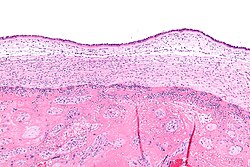
The fetal membranes are the four extraembryonic membranes, associated with the developing embryo, and fetus in humans and other mammals. They are the amnion, chorion, allantois, and yolk sac. The amnion and the chorion are the chorioamniotic membranes that make up the amniotic sac which surrounds and protects the embryo. The fetal membranes are four of six accessory organs developed by the conceptus that are not part of the embryo itself, the other two are the placenta, and the umbilical cord.

<span class="mw-page-title-main">Circumvallate placenta</span> Medical condition

Circumvallate placenta is a rare condition affecting about 1-2% of pregnancies, in which the amnion and chorion fetal membranes essentially "double back" on the fetal side around the edges of the placenta. After delivery, a circumvallate placenta has a thick ring of membranes on its fetal surface. Circumvallate placenta is a placental morphological abnormality associated with increased fetal morbidity and mortality due to the restricted availability of nutrients and oxygen to the developing fetus.

Amnioinfusion is a method in which isotonic fluid is instilled into the uterine cavity.

Neonatal sepsis is a type of neonatal infection and specifically refers to the presence in a newborn baby of a bacterial blood stream infection (BSI) in the setting of fever. Older textbooks may refer to neonatal sepsis as "sepsis neonatorum". Criteria with regards to hemodynamic compromise or respiratory failure are not useful clinically because these symptoms often do not arise in neonates until death is imminent and unpreventable. Neonatal sepsis is divided into two categories: early-onset sepsis (EOS) and late-onset sepsis (LOS). EOS refers to sepsis presenting in the first 7 days of life, with LOS referring to presentation of sepsis after 7 days. Neonatal sepsis is the single most common cause of neonatal death in hospital as well as community in developing country.

Artificial rupture of membranes (AROM), also known as an amniotomy, is performed by a midwife or obstetrician and was once thought to be an effective means to induce or accelerate labor. The membranes can be ruptured using a specialized tool, such as an amnihook or amnicot, or they may be ruptured by the proceduralist's finger. The different techniques for artificial rupture of membranes have not been extensively compared in the literature. In one study comparing amnihook versus amnicot for artificial rupture of membranes, use of an amnicot was associated with fewer neonatal scalp lacerations.

<span class="mw-page-title-main">Neonatal meningitis</span> Medical condition

Neonatal meningitis is a serious medical condition in infants that is rapidly fatal if untreated. Meningitis, an inflammation of the meninges, the protective membranes of the central nervous system, is more common in the neonatal period than any other time in life, and is an important cause of morbidity and mortality globally. Mortality is roughly half in developing countries and ranges from 8%-12.5% in developed countries.

<span class="mw-page-title-main">Neonatal infection</span> Human disease

Neonatal infections are infections of the neonate (newborn) acquired during prenatal development or within the first four weeks of life. Neonatal infections may be contracted by mother to child transmission, in the birth canal during childbirth, or after birth. Neonatal infections may present soon after delivery, or take several weeks to show symptoms. Some neonatal infections such as HIV, hepatitis B, and malaria do not become apparent until much later. Signs and symptoms of infection may include respiratory distress, temperature instability, irritability, poor feeding, failure to thrive, persistent crying and skin rashes.

<span class="mw-page-title-main">Operative vaginal delivery</span>

Operative vaginal delivery, also known as assisted or instrumental vaginal delivery, is a vaginal delivery that is assisted by the use of forceps or a vacuum extractor.

References