Twin-to-twin transfusion syndrome

Last updated

Twin-to-twin transfusion syndrome
Twin to Twin transfusion syndrome.svg
Twin-to-twin transfusion syndrome
Specialty Perinatology, neonatology
Usual onsetUsually weeks 16–25 of gestation
Types Twin anemia–polycythemia sequence
Treatment Fetoscopy and laser ablation; serial amniocentesis
Prognosis 0–20% survival of one or all fetuses without treatment; [1]
66–85% survival of at least one fetus with treatment, with 6–15% risk of cerebral palsy
FrequencyAround 5–15% of identical twin pregnancies [2]

Twin-to-twin transfusion syndrome (TTTS), also known as feto-fetal transfusion syndrome (FFTS), twin oligohydramnios-polyhydramnios sequence (TOPS) and stuck twin syndrome, is a complication of monochorionic multiple pregnancies (the most common form of identical twin pregnancy) in which there is disproportionate blood supply between the fetuses. This leads to unequal levels of amniotic fluid between each fetus and usually leads to death of the undersupplied twin and, without treatment, usually death or a range of birth defects or disabilities for a surviving twin, such as underdeveloped, damaged or missing limbs, digits or organs (including the brain), especially cerebral palsy. [3]

Contents

The condition occurs when the vein–artery connections within the fetuses' shared placenta allow the blood flow between each fetus to become progressively imbalanced. [4] It usually develops between week 16 and 25 of pregnancy, during peak placental growth. The cause of the developmental effects on a surviving fetus may include necrotic embolisms from a dead fetus, low blood volume due to pooling in the dead fetus or velamentous cord insertion (insertion of the umbilical cord into the chorioamniotic membranes). [3]

The primary treatment of TTTS is fetoscopy and laser ablation of the interconnecting blood vessels to cut off the exchange of blood between the fetuses. This treatment is associated with an 85% survival rate of at least one fetus. [5] Other treatments include periodic removal of amniotic fluid (serial amniocentesis), which is associated with a 66% survival rate of at least one fetus. Without treatment, there is an almost 100% mortality rate of one or all fetuses. [1] Even with treatment, the condition is associated with premature birth and a risk of cerebral palsy in a surviving fetus.

Around 5–15% of identical twin fetuses will go on to develop TTTS. [2] The condition was first described by German obstetrician Friedrich Schatz in 1875. [6]

Cause

As a result of sharing a single placenta, the blood supplies of monochorionic twin fetuses can become connected, so that they share blood circulation: although each fetus uses its own portion of the placenta, the connecting blood vessels within the placenta allow blood to pass from one twin to the other. It is thought that most monochorionic placentae have these "shared connections" that cross the placenta, with the net flow volumes being equal between them. This state is sometimes referred to as "flow balance". When the placenta has deep veinartery connection, this can cause blood flow to become unbalanced. [4]

Depending on the number, type and direction of the interconnecting blood vessels (anastomoses), blood can be transferred disproportionately from one twin (the "donor") to the other (the "recipient"), due to a state of "flow imbalance" imparted by new blood vessel growth across the placental "equator", the line that divides each baby's proportion of the shared placenta. This state of transfusion causes the donor twin to have decreased blood volume, retarding the donor's development and growth, and also decreased urinary output, leading to a lower than normal level of amniotic fluid (becoming oligohydramnios). The blood volume of the recipient twin is increased, which can strain the fetus's heart and eventually lead to heart failure, and also higher than normal urinary output, which can lead to excess amniotic fluid (becoming polyhydramnios). The demise of the fetus is typically a result of ischemia related to the lack of blood flow. The lack of blood flow causes bowel atresia, brain damage, and kidney failure. [3]

TTTS usually develops during the period of peak placental growth, starting in week 16 and proceeding through week 25; after this point, the placenta's growth decelerates, essentially stopping just after week 30. While TTTS has occasionally been detected beyond this timepoint, it is thought that its occurrence beyond week 30 may be due to a placental embolism that upsets the flow balance of the shared connections between the babies. TTTS is potentially lethal to either or both twins, no matter when it is detected. However, when detected past week 25, emergency delivery may be considered to rescue the babies if the TTTS is severe.[ citation needed ]

Other than requiring a monochorionic twin (or higher multiple) pregnancy, the underlying causes of TTTS are not known. It is not known to be hereditary or genetic.[ citation needed ]

Effects to the surviving fetus

The fetal demise of one of the twins during the second trimester of a monochorionic pregnancy can result in serious complications to the surviving fetus. Complications include gangrenous limbs, hands and feet, cerebral palsy and IQ deficits, constriction rings of limbs and digits, reduced digits, skin defects, brain cysts, hydranencephaly, multicystic encephalomalacia, microencephaly, renal agenesis and bowel atresia. There are three hypotheses explaining these complications. [3]

  1. Embolic Theory: The demise of the donor twin allows necrotic tissue, presumably thrombosis-plastin like material, to pass through the vasculature of the surviving twin via the placental vessels. The necrotic tissue embolizes the surviving twin's mesenteric artery, resulting in bowel atresia, the renal artery, resulting in renal agenesis, or the peripheral arteries, resulting in limb ischemia and skin abnormalities. [3]
  2. Ischemic Theory: The demise of the donor twin creates a low-pressure state resulting in the shunting of blood into the low-resistance vascular system of the dead fetus. This state of low pressure acutely causes hypovolemia, resulting in ischemia and poor end-organ perfusion. [3]
  3. Placentation Theory: The incidence of velamentous cord insertion, an abnormal insertion of the umbilical cord into the placenta, is higher in TTTS. The exact mechanism of effect this has on fetal damage is unknown. [3]

Diagnosis

Twin-to-twin transfusion syndrome (TTTS) illustration. The donor twin (D) is confined within the amniotic sac, whereas the recipient twin (R) has freedom of movement. Tweeling transfusiesyndroom.jpg
Twin-to-twin transfusion syndrome (TTTS) illustration. The donor twin (D) is confined within the amniotic sac, whereas the recipient twin (R) has freedom of movement.

Quintero stages

A staging system is commonly used to classify the severity of TTTS. [7]

Stage I: A small amount of amniotic fluid (oligohydramnios) is found around the donor twin and a large amount of amniotic fluid (polyhydramnios) is found around the recipient twin.

Stage II: In addition to the description above, the ultrasound is not able to identify the bladder in the donor twin.

Stage III: In addition to the characteristics of Stages I and II, there is abnormal blood flow in the umbilical cords of the twins.

Stage IV: In addition to all of the above findings, the recipient twin has swelling under the skin and appears to be experiencing heart failure (fetal hydrops).

Stage V: In addition to all of the above findings, one of the twins has died. This can happen to either twin. The risk to either the donor or the recipient is roughly equal & is quite high in Stage II or higher TTTS.

The Quintero staging does not provide information about prognosis, and other staging systems have been proposed. [8]

Treatment

Various treatments exist for TTTS.

Non treatment

This is equivalent of zero intervention. It has been associated with almost 100% mortality rate of one or all fetuses. Exceptions to this include patients that are still in Stage 1 TTTS and are past 22 weeks' gestation.[ citation needed ]

Adjustment of amniotic fluid

Serial amniocentesis

This procedure involves removal of amniotic fluid periodically throughout the pregnancy under the assumption that the extra fluid in the recipient twin can cause preterm labor, perinatal mortality, or tissue damage. In the case that the fluid does not reaccumulate, the reduction of amniotic fluid stabilizes the pregnancy. Otherwise, the treatment is repeated as necessary. There is no standard procedure for how much fluid is removed each time. There is a danger that if too much fluid is removed, the recipient twin could die. This procedure is associated with a 66% survival rate of at least one fetus, with a 15% risk of cerebral palsy, and average delivery occurring at 29 weeks' gestation.[ citation needed ]

Septostomy, or iatrogenic disruption of the dividing membrane

This procedure involves the tearing of the dividing membrane between fetuses such that the amniotic fluid of both twins mixes, under the assumption that pressure is different in either amniotic sac and that its equilibration will ameliorate progression of the disease. It has not been proven that pressures are different in either amniotic sac. Use of this procedure can preclude use of other procedures as well as make difficult the monitoring of disease progression. In addition, tearing the dividing membrane has contributed to cord entanglement and demise of fetuses through physical complications.[ citation needed ]

Adjustment of blood supply

Laser therapy (fetoscopic laser photocoagulation)

Illustration of a fetoscopy and laser ablation of connecting vessels in Twin-to-twin transfusion syndrome TTTS laser cartoon.JPG
Illustration of a fetoscopy and laser ablation of connecting vessels in Twin-to-twin transfusion syndrome

This procedure involves endoscopic surgery using laser to interrupt the vessels that allow exchange of blood between fetuses under the assumption that the unequal sharing of blood through these vascular communications leads to unequal levels of amniotic fluid. Each fetus remains connected to its primary source of blood and nutrition, the placenta, through the umbilical cord. This procedure is conducted once, with the exception of all vessels not having been found. The use of endoscopic instruments allows for short recovery time. This procedure has been associated with 85% survival rate of at least one fetus, with a 6–7% risk of cerebral palsy and average delivery occurring at 32–33 weeks' gestation. [5]

Twin anemia–polycythemia sequence (TAPS) may occur after laser surgery for TTTS (post-laser form). The spontaneous form of TAPS complicates approximately 3 to 5% of monochorionic twin pregnancies, whereas the post-laser form occurs in 2 to 13% of TTTS cases. [9] [10] The pathogenesis of TAPS is based on the presence of few, minuscule arterio-venous (AV) placental anastomoses (diameter <1mm) [11] allowing a slow transfusion of blood from the donor to the recipient and leading gradually to highly discordant Hb levels. [12]

A 2014 review found that laser coagulation resulted in fewer fetal and perinatal deaths than amnioreduction and septostomy, and recommended its use for all states of TTTS. [4]

Selective reduction

Selective termination of one of the fetuses is usually not considered until TTFS has reached either stage III or IV, [13] and is indicated when the death of one twin is imminent, but the fetus is too premature to deliver, as the death of one twin will cause the death of the second twin due to the shared blood supply if not immediately delivered. [13]

One procedure for selective reduction is umbilical cord occlusion. This procedure involves the ligation or otherwise occlusion of one twin's umbilical cord to interrupt the exchange of blood between the fetuses. The procedure is typically offered in cases where one of the fetuses is presumed moribund and endangering the life or health of the other twin through resultant hypotension. [14] Use of this treatment has decreased as TTTS is identified and treated in earlier stages and with better outcomes. When used, it is associated with an 85% survival rate of the remaining fetuses with 5% risk of cerebral palsy and a 33–39 weeks of gestation at delivery. [15] [16]

Voluntary termination

Some women choose to have an abortion of both fetuses as the health of both fetuses is impacted by TTFS. [13]

Epidemiology

Based on recent (2005) US NCHS data, the rate of multiple births is now approximately 3.4% (4,138,349 total births, of which 139,816 were twins or higher-order multiple births). The majority of identical twins share a common (monochorionic) placenta, and of these approximately 15% go on to develop TTTS. By extrapolating the number of expected identical twins (about one-third) from annual multiple births, and the number of twins with monochorionic placentae (about two-thirds), and from these the number thought to develop TTTS (about 15%), there are at least 4,500 TTTS cases per year in the U.S. alone: 139,816 × .33 × .66 × .15 = 4,568 cases of TTTS per year in U.S. (involving more than 9,000 babies.) Since spontaneous pregnancy losses and terminations that occur prior to 20 weeks go uncounted by the C.D.C., this estimate of TTTS cases may be very conservative. Although infertility treatments have increased the rate of multiple birth, they have not appreciably diluted the expected incidence of identical twins. Studies show a higher rate of identical twins (up to 20 times with IVF) using these treatments versus spontaneous pregnancy rates.[ citation needed ]

One Australian study, however, noted an occurrence of only 1 in 4,170 pregnancies, or 1 in 58 twin gestations. This distinction could be partly explained by the "hidden mortality" associated with MC multifetal pregnancies—instances lost due to premature rupture of membrane (PROM) or intrauterine fetal demise before a thorough diagnosis of TTTS can be made. [6]

History

TTTS was first described by a German obstetrician, Friedrich Schatz, in 1875. Once defined by neonatal parameters—differences in birth weight and cord hemoglobin at the time of delivery—TTTS is now defined differently. Today, it is known that discordant fetal weights will most likely be a late manifestation, and fetal hemoglobin through cordocentesis is often equivalent in the twin pair even in severe TTTS. [6]

In art

De Wikkelkinderen (The Swaddled Children), 1617, by an unknown artist, is thought to depict TTTS. De Wikkellkinderen.jpeg
De Wikkelkinderen (The Swaddled Children), 1617, by an unknown artist, is thought to depict TTTS.

A painting known as the De Wikkelkinderen (The Swaddled Children), from 1617, is thought to represent a depiction of TTTS. [17] The drawing shows twins that appear to be identical, but one is pale, while the other is red. Analysis of the family histories of the owners of the painting suggests that the twins did not survive to adulthood, although whether that is due to TTTS is uncertain.[ citation needed ]

See also

Related Research Articles

<span class="mw-page-title-main">Amniocentesis</span> Sampling of amniotic fluid done mainly to detect fetal chromosomal abnormalities

Amniocentesis is a medical procedure used primarily in the prenatal diagnosis of genetic conditions. It has other uses such as in the assessment of infection and fetal lung maturity. Prenatal diagnostic testing, which includes amniocentesis, is necessary to conclusively diagnose the majority of genetic disorders, with amniocentesis being the gold-standard procedure after 15 weeks' gestation.

<span class="mw-page-title-main">Amnion</span> Innermost membranous sac that surrounds and protects the developing embryo

The amnion is a membrane that closely covers the human and various other embryos when first formed. It fills with amniotic fluid, which causes the amnion to expand and become the amniotic sac that provides a protective environment for the developing embryo. The amnion, along with the chorion, the yolk sac and the allantois protect the embryo. In birds, reptiles and monotremes, the protective sac is enclosed in a shell. In marsupials and placental mammals, it is enclosed in a uterus.

<span class="mw-page-title-main">Chorion</span> Outermost fetal membrane around the embryo in amniotes

The chorion is the outermost fetal membrane around the embryo in mammals, birds and reptiles (amniotes). It develops from an outer fold on the surface of the yolk sac, which lies outside the zona pellucida, known as the vitelline membrane in other animals. In insects, it is developed by the follicle cells while the egg is in the ovary. Some mollusks also have chorions as part of their eggs. For example, fragile octopus eggs have only a chorion as their envelope.

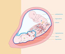
Oligohydramnios is a medical condition in pregnancy characterized by a deficiency of amniotic fluid, the fluid that surrounds the fetus in the abdomen, in the amniotic sac. It is typically diagnosed by ultrasound when the amniotic fluid index (AFI) measures less than 5 cm or when the single deepest pocket (SDP) of amniotic fluid measures less than 2 cm. Amniotic fluid is necessary to allow for normal fetal movement, lung development, and cushioning from uterine compression. Low amniotic fluid can be attributed to a maternal, fetal, placental or idiopathic cause and can result in poor fetal outcomes including death. The prognosis of the fetus is dependent on the etiology, gestational age at diagnosis, and the severity of the oligohydramnios.

<span class="mw-page-title-main">Placental abruption</span> Medical condition

Placental abruption is when the placenta separates early from the uterus, in other words separates before childbirth. It occurs most commonly around 25 weeks of pregnancy. Symptoms may include vaginal bleeding, lower abdominal pain, and dangerously low blood pressure. Complications for the mother can include disseminated intravascular coagulopathy and kidney failure. Complications for the baby can include fetal distress, low birthweight, preterm delivery, and stillbirth.

<span class="mw-page-title-main">Fetoscopy</span> Endoscopic procedure during pregnancy

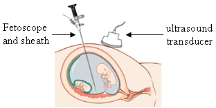
Fetoscopy is an endoscopic procedure during pregnancy to allow surgical access to the fetus, the amniotic cavity, the umbilical cord, and the fetal side of the placenta. A small (3–4 mm) incision is made in the abdomen, and an endoscope is inserted through the abdominal wall and uterus into the amniotic cavity. Fetoscopy allows for medical interventions such as a biopsy or a laser occlusion of abnormal blood vessels or the treatment of spina bifida.

<span class="mw-page-title-main">Hydrops fetalis</span> Human disease of fetuses

Hydrops fetalis or hydrops foetalis is a condition in the fetus characterized by an accumulation of fluid, or edema, in at least two fetal compartments. By comparison, hydrops allantois or hydrops amnion is an accumulation of excessive fluid in the allantoic or amniotic space, respectively.

<span class="mw-page-title-main">EXIT procedure</span>

The EXIT procedure, or ex utero intrapartum treatment procedure, is a specialized surgical delivery procedure used to deliver babies who have airway compression. Causes of airway compression in newborn babies result from a number of rare congenital disorders, including bronchopulmonary sequestration, congenital cystic adenomatoid malformation, mouth or neck tumor such as teratoma, and lung or pleural tumor such as pleuropulmonary blastoma. Airway compression discovered at birth is a medical emergency. In many cases, however, the airway compression is discovered during prenatal ultrasound exams, permitting time to plan a safe delivery using the EXIT procedure or other means.

Placental insufficiency or utero-placental insufficiency is the failure of the placenta to deliver sufficient nutrients to the fetus during pregnancy, and is often a result of insufficient blood flow to the placenta. The term is also sometimes used to designate late decelerations of fetal heart rate as measured by cardiotocography or an NST, even if there is no other evidence of reduced blood flow to the placenta, normal uterine blood flow rate being 600mL/min.

<span class="mw-page-title-main">Percutaneous umbilical cord blood sampling</span>

Percutaneous umbilical cord blood sampling (PUBS), also called cordocentesis, fetal blood sampling, or umbilical vein sampling is a diagnostic genetic test that examines blood from the fetal umbilical cord to detect fetal abnormalities. Fetal and maternal blood supply are typically connected in utero with one vein and two arteries to the fetus. The umbilical vein is responsible for delivering oxygen rich blood to the fetus from the mother; the umbilical arteries are responsible for removing oxygen poor blood from the fetus. This allows for the fetus’ tissues to properly perfuse. PUBS provides a means of rapid chromosome analysis and is useful when information cannot be obtained through amniocentesis, chorionic villus sampling, or ultrasound ; this test carries a significant risk of complication and is typically reserved for pregnancies determined to be at high risk for genetic defect. It has been used with mothers with immune thrombocytopenic purpura.

<span class="mw-page-title-main">Velamentous cord insertion</span> Velamentous placenta

Velamentous cord insertion is a complication of pregnancy where the umbilical cord is inserted in the fetal membranes. It is a major cause of antepartum hemorrhage that leads to loss of fetal blood and associated with high perinatal mortality. In normal pregnancies, the umbilical cord inserts into the middle of the placental mass and is completely encased by the amniotic sac. The vessels are hence normally protected by Wharton's jelly, which prevents rupture during pregnancy and labor. In velamentous cord insertion, the vessels of the umbilical cord are improperly inserted in the chorioamniotic membrane, and hence the vessels traverse between the amnion and the chorion towards the placenta. Without Wharton's jelly protecting the vessels, the exposed vessels are susceptible to compression and rupture.

<span class="mw-page-title-main">Twin reversed arterial perfusion</span> Rare complication of monochorionic twin pregnancies

Twin reversed arterial perfusion sequence, also called TRAP sequence, TRAPS, or acardiac twinning, is a rare complication of monochorionic twin pregnancies. It is a severe variant of twin-to-twin transfusion syndrome (TTTS). In addition to the twins' blood systems being connected instead of independent, one twin, called the acardiac twin, TRAP fetus or acardius, is severely malformed. The heart is missing or deformed, hence the name "acardiac", as are the upper structures of the body. The legs may be partially present or missing, and internal structures of the torso are often poorly formed. The other twin is usually normal in appearance. The normal twin, called the pump twin, drives blood through both fetuses. It is called "reversed arterial perfusion" because in the acardiac twin the blood flows in a reversed direction.

An obstetric labor complication is a difficulty or abnormality that arises during the process of labor or delivery.

<span class="mw-page-title-main">Monoamniotic twins</span> Identical twins sharing the same amniotic sac in the womb

Monoamniotic twins are identical or semi-identical twins that share the same amniotic sac within their mother's uterus. Monoamniotic twins are always monochorionic and are usually termed Monoamniotic-Monochorionic twins. They share the placenta, but have two separate umbilical cords. Monoamniotic twins develop when an embryo does not split until after formation of the amniotic sac, at about 9–13 days after fertilization. Monoamniotic triplets or other monoamniotic multiples are possible, but extremely rare. Other obscure possibilities include multiples sets where monoamniotic twins are part of a larger gestation such as triplets, quadruplets, or more.

<span class="mw-page-title-main">Monochorionic twins</span> Identical twins that share the same placenta

Monochorionic twins are monozygotic (identical) twins that share the same placenta. If the placenta is shared by more than two twins, these are monochorionic multiples. Monochorionic twins occur in 0.3% of all pregnancies. Seventy-five percent of monozygotic twin pregnancies are monochorionic; the remaining 25% are dichorionic diamniotic. If the placenta divides, this takes place before the third day after fertilization.

<span class="mw-page-title-main">Circumvallate placenta</span> Medical condition

Circumvallate placenta is a rare condition affecting about 1-2% of pregnancies, in which the amnion and chorion fetal membranes essentially "double back" on the fetal side around the edges of the placenta. After delivery, a circumvallate placenta has a thick ring of membranes on its fetal surface. Circumvallate placenta is a placental morphological abnormality associated with increased fetal morbidity and mortality due to the restricted availability of nutrients and oxygen to the developing fetus.

Amnioinfusion is a method in which isotonic fluid is instilled into the uterine cavity.

<span class="mw-page-title-main">Twin anemia-polycythemia sequence</span> Medical condition

Twin anemia-polycythemia sequence(TAPS) is a chronic type of unbalanced fetal transfusion in monochorionic twins that results in polycythemia in the TAPS recipient and anemia in the TAPS donor due to tiny placental anastomoses. Post-laser TAPS and spontaneous TAPS are the two forms of TAPS. Unlike twin-twin transfusion syndrome, which arises when twin oligohydramnios polyhydramnios sequence (TOPS) is absent, TAPS develops in its absence.

<span class="mw-page-title-main">Emergency childbirth</span>

Emergency childbirth is the precipitous birth of an infant in an unexpected setting. In planned childbirth, mothers choose the location and obstetric team ahead of time. Options range from delivering at home, at a hospital, a medical facility or a birthing center. Sometimes, birth can occur on the way to these facilities, without a healthcare team. The rates of unplanned childbirth are low. If the birth is imminent, emergency measures may be needed. Emergency services can be contacted for help in some countries.

An Intrauterine transfusion (IUT) is a procedure that provides blood to a fetus, most commonly through the umbilical cord. It is used in cases of severe fetal anemia, such as when fetal red blood cells are being destroyed by maternal antibodies. IUTs are performed by perinatologists at hospitals or specialized centers.

References

  1. 1 2 Zach T, Ford SP. "Twin-to-Twin Transfusion Syndrome". EMedicine. Retrieved July 22, 2006.
  2. 1 2 "Twin-Twin Transfusion Syndrome". NORD (National Organization for Rare Disorders). Retrieved October 4, 2021.
  3. 1 2 3 4 5 6 7 Holmes, Lewis B. (2011). Common Malformations. Oxford University Press. ISBN   978-0-19-513602-9.[ page needed ]
  4. 1 2 3 Roberts D, Neilson JP, Kilby M, Gates S. (2014) Interventions for the treatment of twin–twin transfusion syndrome. Cochrane Database Syst Rev
  5. 1 2 Benoit RM, Baschat AA. (2014) Twin-to-twin transfusion syndrome: prenatal diagnosis and treatment. American J of Perinatology
  6. 1 2 3 Johnson, A.; Moise, K. J. "Improving Survival in Twin-Twin Transfusion Syndrome. Contemporary OB/GYN December 2006" (PDF). Archived from the original (PDF) on July 28, 2011.
  7. "Understanding the Stages of TTTS". UC San Diego. Archived from the original on April 10, 2012. Retrieved April 23, 2012.
  8. Cristina, Rossi (2009). "The Efficacy of Quintero Staging System to Assess Severity of Twin-Twin Transfusion Syndrome Treated with Laser Therapy: A Systematic Review with Meta-Analysis". American Journal of Perinatology. Thieme Medical Publishers. 26 (7): 537–544. doi:10.1055/s-0029-1215430. PMID   19283655. S2CID   5913636.
  9. Robyr R, Lewi L, Salomon LJ, Yamamoto M, Bernard JP, Deprest J et al. Prevalence and management of late fetal complications following successful selective laser coagulation of chorionic plate anastomoses in twin-to-twin transfusion syndrome. Am J Obstet Gynecol 2006; 194(3):796-803.
  10. Habli M, Bombrys A, Lewis D, Lim FY, Polzin W, Maxwell R et al. Incidence of complications in twin-twin transfusion syndrome after selective fetoscopic laser photocoagulation: a single-center experience. Am J Obstet Gynecol 2009; 201(4):417.
  11. Lopriore E, Deprest J, Slaghekke F, Oepkes D, Middeldorp JM, Vandenbussche FP et al. Placental characteristics in monochorionic twins with and without twin anemia-polycythemia sequence. Obstet Gynecol 2008; 112(4):753-758.
  12. Slaghekke F, Kist WJ, Oepkes D, Pasman SA, Middeldorp JM, Klumper FJ et al. Twin anemia-polycythemia sequence: diagnostic criteria, classification, perinatal management and outcome. Fetal Diagn Ther 2010; 27(4):181-190.
  13. 1 2 3 Borse, Vyvian; Shanks, Anthony L. (2024), "Twin-to-Twin Transfusion Syndrome", StatPearls, Treasure Island (FL): StatPearls Publishing, PMID   33085280 , retrieved February 21, 2024
  14. Rossi, A Cristina (September 1, 2012). "Treatment of twin–twin transfusion syndrome". Expert Review of Obstetrics & Gynecology. 7 (5): 477–482. doi: 10.1586/eog.12.45 .
  15. Nakata, M.; Chmait, R. H.; Quintero, R. A. (May 2004). "Umbilical cord occlusion of the donor versus recipient fetus in twin-twin transfusion syndrome: Cord occlusion in TTTS". Ultrasound in Obstetrics and Gynecology. 23 (5): 446–450. doi:10.1002/uog.1034. PMID   15133793. S2CID   25531930.
  16. Taylor, M. J. O.; Shalev, E.; Tanawattanacharoen, S.; Jolly, M.; Kumar, S.; Weiner, E.; Cox, P. M.; Fisk, N. M (January 2001). "Ultrasound-guided umbilical cord occlusion using bipolar diathermy for Stage III/IV twin-twin transfusion syndrome". Prenatal Diagnosis. 22 (1): 70–76. doi:10.1002/pd.256. PMID   11810656. S2CID   19469579.
  17. Berger H, de Waard F, Molenaar Y (2000). "A case of twin-to-twin transfusion in 1617". Lancet. 356 (9232): 847–8. doi:10.1016/S0140-6736(00)02665-9. PMID   11022944. S2CID   5155034.