Hypovolemia

Last updated
Hypovolemia
Other namesOligemia, hypovolaemia, oligaemia, hypovolæmia, volume depletion
Capillary microcirculation.svg
A diagram showing the formation of interstitial fluid from the bloodstream
Specialty Emergency medicine
Symptoms Headache, fatigue, nausea, profuse sweating, dizziness
Complications Hypovolemic shock
Differential diagnosis Dehydration

Hypovolemia, also known as volume depletion or volume contraction, is a state of abnormally low extracellular fluid in the body. [1] This may be due to either a loss of both salt and water or a decrease in blood volume. [2] [3] Hypovolemia refers to the loss of extracellular fluid and should not be confused with dehydration. [4]

Contents

Hypovolemia is caused by a variety of events, but these can be simplified into two categories: those that are associated with kidney function and those that are not. [5] The signs and symptoms of hypovolemia worsen as the amount of fluid lost increases. [6] Immediately or shortly after mild fluid loss (from blood donation, diarrhea, vomiting, bleeding from trauma, etc.), one may experience headache, fatigue, weakness, dizziness, or thirst. Untreated hypovolemia or excessive and rapid losses of volume may lead to hypovolemic shock. [7] Signs and symptoms of hypovolemic shock include increased heart rate, low blood pressure, pale or cold skin, and altered mental status. When these signs are seen, immediate action should be taken to restore the lost volume.

Signs and symptoms

Signs and symptoms of hypovolemia progress with increased loss of fluid volume. [5]

Early symptoms of hypovolemia include headache, fatigue, weakness, thirst, and dizziness. The more severe signs and symptoms are often associated with hypovolemic shock. These include oliguria, cyanosis, abdominal and chest pain, hypotension, tachycardia, cold hands and feet, and progressively altering mental status.[ citation needed ]

Causes

The causes of hypovolemia can be characterized into two categories: [5]

Kidney

Other

Pathophysiology

Pathophysiology of hypovolemia 170225 CVS Hypovolemic Shock Pathophysiology of Disease.png
Pathophysiology of hypovolemia

The signs and symptoms of hypovolemia are primarily due to the consequences of decreased circulating volume and a subsequent reduction in the amount of blood reaching the tissues of the body. [9] In order to properly perform their functions, tissues require the oxygen transported in the blood. [10] A decrease in circulating volume can lead to a decrease in bloodflow to the brain, resulting in headache and dizziness.[ citation needed ]

Baroreceptors in the body (primarily those located in the carotid sinuses and aortic arch) sense the reduction of circulating fluid and send signals to the brain to increase sympathetic response (see also: baroreflex ). [11] This sympathetic response is to release epinephrine and norepinephrine, which results in peripheral vasoconstriction (reducing size of blood vessels) in order to conserve the circulating fluids for organs vital to survival (i.e. brain and heart). Peripheral vasoconstriction accounts for the cold extremities (hands and feet), increased heart rate, increased cardiac output (and associated chest pain). Eventually, there will be less perfusion to the kidneys, resulting in decreased urine output.[ citation needed ]

Diagnosis

Hypovolemia can be recognized by a fast heart rate, low blood pressure, [12] and the absence of perfusion as assessed by skin signs (skin turning pale) and/or capillary refill on forehead, lips and nail beds. The patient may feel dizzy, faint, nauseated, or very thirsty. These signs are also characteristic of most types of shock. [13]

In children, compensation can result in an artificially high blood pressure despite hypovolemia (a decrease in blood volume). Children typically are able to compensate (maintain blood pressure despite hypovolemia) for a longer period than adults, but deteriorate rapidly and severely once they are unable to compensate (decompensate). [14] Consequently, any possibility of internal bleeding in children should be treated aggressively. [15] [16]

Signs of external bleeding should be assessed, noting that individuals can bleed internally without external blood loss or otherwise apparent signs. [16]

There should be considered possible mechanisms of injury that may have caused internal bleeding, such as ruptured or bruised internal organs. If trained to do so and if the situation permits, there should be conducted a secondary survey and checked the chest and abdomen for pain, deformity, guarding, discoloration or swelling. Bleeding into the abdominal cavity can cause the classical bruising patterns of Grey Turner's sign (bruising along the sides) or Cullen's sign (around the navel). [17]

Investigation

In a hospital, physicians respond to a case of hypovolemic shock by conducting these investigations:[ citation needed ]

Stages

Untreated hypovolemia can lead to shock (see also: hypovolemic shock). Most sources state that there are 4 stages of hypovolemia and subsequent shock; [18] however, a number of other systems exist with as many as 6 stages. [19]

The 4 stages are sometimes known as the "Tennis" staging of hypovolemic shock, as the stages of blood loss (under 15% of volume, 15–30% of volume, 30–40% of volume and above 40% of volume) mimic the scores in a game of tennis: 15, 15–30, 30–40 and 40. [20] It is basically the same as used in classifying bleeding by blood loss.[ citation needed ]

The signs and symptoms of the major stages of hypovolemic shock include: [21] [22]

Stage 1Stage 2Stage 3Stage 4
Blood lossUp to 15% (750 mL)15–30% (750–1500 mL)30–40% (1500–2000 mL)Over 40% (over 2000 mL)
Blood pressureNormal (Maintained
by vasoconstriction)
Increased diastolic BP Systolic BP < 100Systolic BP < 70
Heart rateNormalSlight tachycardia (> 100 bpm)Tachycardia (> 120 bpm)Extreme tachycardia (> 140 bpm) with weak pulse
Respiratory rateNormalIncreased (> 20) Tachypneic (> 30)Extreme tachypnea
Mental statusNormalSlight anxiety, restless Altered, confusedDecreased LOC, lethargy, coma
Skin Pale Pale, cool, clammyIncreased diaphoresis Extreme diaphoresis; mottling possible
Capillary refill NormalDelayedDelayedAbsent
Urine outputNormal20–30 mL/h20 mL/hNegligible

Treatment

Field care

The most important step in treatment of hypovolemic shock is to identify and control the source of bleeding. [23]

Medical personnel should immediately supply emergency oxygen to increase efficiency of the patient's remaining blood supply. This intervention can be life-saving. [24]

Also, the respiratory pump is especially important during hypovolemia as spontaneous breathing may help reduce the effect of this loss of blood pressure on stroke volume by increasing venous return. [25]

The use of intravenous fluids (IVs) may help compensate for lost fluid volume, but IV fluids cannot carry oxygen the way blood does—however, researchers are developing blood substitutes that can. Infusing colloid or crystalloid IV fluids also dilutes clotting factors in the blood, increasing the risk of bleeding. Current best practice allow permissive hypotension in patients with hypovolemic shock, [26] both avoid overly diluting clotting factors and avoid artificially raising blood pressure to a point where it "blows off" clots that have formed. [27] [28]

Hospital treatment

Fluid replacement is beneficial in hypovolemia of stage 2, and is necessary in stage 3 and 4. [21] See also the discussion of shock and the importance of treating reversible shock while it can still be countered.

The following interventions are carried out:

Vasopressors (such as dopamine and noradrenaline) should generally be avoided, as they may result in further tissue ischemia and don't correct the primary problem. Fluids are the preferred choice of therapy. [29]

History

In cases where loss of blood volume is clearly attributable to bleeding (as opposed to, e.g., dehydration), most medical practitioners prefer the term exsanguination for its greater specificity and descriptiveness, with the effect that the latter term is now more common in the relevant context. [30]

See also

Related Research Articles

<span class="mw-page-title-main">Shock (circulatory)</span> Medical condition of insufficient blood flow

Shock is the state of insufficient blood flow to the tissues of the body as a result of problems with the circulatory system. Initial symptoms of shock may include weakness, fast heart rate, fast breathing, sweating, anxiety, and increased thirst. This may be followed by confusion, unconsciousness, or cardiac arrest, as complications worsen.

<span class="mw-page-title-main">Bleeding</span> Loss of blood escaping from the circulatory system

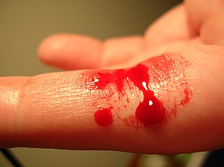
Bleeding, hemorrhage, haemorrhage or blood loss is blood escaping from the circulatory system from damaged blood vessels. Bleeding can occur internally, or externally either through a natural opening such as the mouth, nose, ear, urethra, vagina or anus, or through a puncture in the skin. Hypovolemia is a massive decrease in blood volume, and death by excessive loss of blood is referred to as exsanguination. Typically, a healthy person can endure a loss of 10–15% of the total blood volume without serious medical difficulties. The stopping or controlling of bleeding is called hemostasis and is an important part of both first aid and surgery.

<span class="mw-page-title-main">Dehydration</span> Deficit of total body water

In physiology, dehydration is a lack of total body water, with an accompanying disruption of metabolic processes. It occurs when free water loss exceeds free water intake, usually due to exercise, disease, or high environmental temperature. Mild dehydration can also be caused by immersion diuresis, which may increase risk of decompression sickness in divers.

Iron poisoning typically occurs from ingestion of excess iron that results in acute toxicity. Mild symptoms which occur within hours include vomiting, diarrhea, abdominal pain, and drowsiness. In more severe cases, symptoms can include tachypnea, low blood pressure, seizures, or coma. If left untreated, iron poisoning can lead to multi-organ failure resulting in permanent organ damage or death.

<span class="mw-page-title-main">Fluid replacement</span>

Fluid replacement or fluid resuscitation is the medical practice of replenishing bodily fluid lost through sweating, bleeding, fluid shifts or other pathologic processes. Fluids can be replaced with oral rehydration therapy (drinking), intravenous therapy, rectally such as with a Murphy drip, or by hypodermoclysis, the direct injection of fluid into the subcutaneous tissue. Fluids administered by the oral and hypodermic routes are absorbed more slowly than those given intravenously.

<span class="mw-page-title-main">Hypotension</span> Abnormally low blood pressure

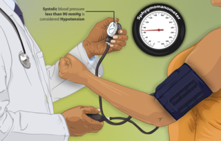
Hypotension, also known as low blood pressure, is a cardiovascular condition characterized by abnormally reduced blood pressure. Blood pressure is the force of blood pushing against the walls of the arteries as the heart pumps out blood and is indicated by two numbers, the systolic blood pressure and the diastolic blood pressure, which are the maximum and minimum blood pressures within the cardiac cycle, respectively. A systolic blood pressure of less than 90 millimeters of mercury (mmHg) or diastolic of less than 60 mmHg is generally considered to be hypotension. Different numbers apply to children. However, in practice, blood pressure is considered too low only if noticeable symptoms are present.

<span class="mw-page-title-main">Thirst</span> Craving for potable fluids experienced by animals

Thirst is the craving for potable fluids, resulting in the basic instinct of animals to drink. It is an essential mechanism involved in fluid balance. It arises from a lack of fluids or an increase in the concentration of certain osmolites, such as sodium. If the water volume of the body falls below a certain threshold or the osmolite concentration becomes too high, structures in the brain detect changes in blood constituents and signal thirst.

Hematemesis is the vomiting of blood. It can be confused with hemoptysis or epistaxis (nosebleed), which are more common. The source is generally the upper gastrointestinal tract, typically above the suspensory muscle of duodenum. It may be caused by ulcers, tumors of the stomach or esophagus, varices, prolonged and vigorous retching, gastroenteritis, ingested blood, or certain drugs.

<span class="mw-page-title-main">Internal bleeding</span> Medical condition

Internal bleeding is a loss of blood from a blood vessel that collects inside the body, and is not usually visible from the outside. It can be a serious medical emergency but the extent of severity depends on bleeding rate and location of the bleeding. Severe internal bleeding into the chest, abdomen, pelvis, or thighs can cause hemorrhagic shock or death if proper medical treatment is not received quickly. Internal bleeding is a medical emergency and should be treated immediately by medical professionals.

<span class="mw-page-title-main">Hypovolemic shock</span> Medical condition

Hypovolemic shock is a form of shock caused by severe hypovolemia. It could be the result of severe dehydration through a variety of mechanisms or blood loss. Hypovolemic shock is a medical emergency; if left untreated, the insufficient blood flow can cause damage to organs, leading to multiple organ failure.

Oliguria or hypouresis is the low output of urine specifically more than 80 ml/day but less than 400ml/day. The decreased output of urine may be a sign of dehydration, kidney failure, hypovolemic shock, hyperosmolar hyperglycemic nonketotic syndrome (HHNS), multiple organ dysfunction syndrome, urinary obstruction/urinary retention, diabetic ketoacidosis (DKA), pre-eclampsia, and urinary tract infections, among other conditions.

<span class="mw-page-title-main">Neurogenic shock</span> Insufficient blood flow due to autonomic nervous system damage

Neurogenic shock is a distributive type of shock resulting in hypotension, often with bradycardia, caused by disruption of autonomic nervous system pathways. It can occur after damage to the central nervous system, such as spinal cord injury and traumatic brain injury. Low blood pressure occurs due to decreased systemic vascular resistance resulting from loss of sympathetic tone, which in turn causes blood pooling within the extremities rather than being available to circulate throughout the body. The slowed heart rate results from a vagal response unopposed by a sympathetic nervous system (SNS) response. Such cardiovascular instability is exacerbated by hypoxia, or treatment with endotracheal or endobronchial suction used to prevent pulmonary aspiration.

Lightheadedness is a common and typically unpleasant sensation of dizziness or a feeling that one may faint. The sensation of lightheadedness can be short-lived, prolonged, or, rarely, recurring. In addition to dizziness, the individual may feel as though their head is weightless. The individual may also feel as though the room is "spinning" or moving (vertigo). Most causes of lightheadedness are not serious and either cure themselves quickly or are easily treated.

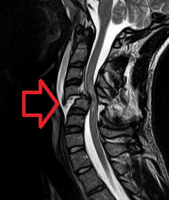
Intracerebral hemorrhage (ICH), also known as hemorrhagic stroke, is a sudden bleeding into the tissues of the brain, into its ventricles, or into both. An ICH is a type of bleeding within the skull and one kind of stroke. Symptoms can vary dramatically depending on the severity, acuity, and location (anatomically) but can include headache, one-sided weakness, numbness, tingling, or paralysis, speech problems, vision or hearing problems, memory loss, attention problems, coordination problems, balance problems, dizziness or lightheadedness or vertigo, nausea/vomiting, seizures, decreased level of consciousness or total loss of consciousness, neck stiffness, and fever.

End organ damage is severe impairment of major body organs due to high blood pressure or states of low blood pressure or low blood volume. This can present as a heart attack or heart failure, pulmonary edema, neurologic deficits including a stroke, or acute kidney failure.

<span class="mw-page-title-main">Amniotic fluid embolism</span> Potentially fatal complication of pregnancy

An amniotic fluid embolism (AFE) is a life-threatening childbirth (obstetric) emergency in which amniotic fluid enters the blood stream of the mother, triggering a serious reaction which results in cardiorespiratory collapse and massive bleeding (coagulopathy). The rate at which it occurs is 1 instance per 20,000 births and it comprises 10% of all maternal deaths.

In medicine, intravascular volume status refers to the volume of blood in a patient's circulatory system, and is essentially the blood plasma component of the overall volume status of the body, which otherwise includes both intracellular fluid and extracellular fluid. Still, the intravascular component is usually of primary interest, and volume status is sometimes used synonymously with intravascular volume status.

Pediatric advanced life support (PALS) is a course offered by the American Heart Association (AHA) for health care providers who take care of children and infants in the emergency room, critical care and intensive care units in the hospital, and out of hospital. The course teaches healthcare providers how to assess injured and sick children and recognize and treat respiratory distress/failure, shock, cardiac arrest, and arrhythmias.

Obstructive shock is one of the four types of shock, caused by a physical obstruction in the flow of blood. Obstruction can occur at the level of the great vessels or the heart itself. Causes include pulmonary embolism, cardiac tamponade, and tension pneumothorax. These are all life-threatening. Symptoms may include shortness of breath, weakness, or altered mental status. Low blood pressure and tachycardia are often seen in shock. Other symptoms depend on the underlying cause.

Permissive hypotension or hypotensive resuscitation is the use of restrictive fluid therapy, specifically in the trauma patient, that increases systemic blood pressure without reaching normotension. The goal blood pressure for these patients is a mean arterial pressure of 40-50 mmHg or systolic blood pressure of less than or equal to 80. This goes along with certain clinical criteria. Following traumatic injury, some patients experience hypotension that is usually due to blood loss (hemorrhage) but can be due to other causes as well. In the past, physicians were very aggressive with fluid resuscitation to try to bring the blood pressure to normal values. Recent studies have found that there is some benefit to allowing specific patients to experience some degree of hypotension in certain settings. This concept does not exclude therapy by means of i.v. fluid, inotropes or vasopressors, the only restriction is to avoid completely normalizing blood pressure in a context where blood loss may be enhanced. When a person starts to bleed the body starts a natural coagulation process that eventually stops the bleed. Issues with fluid resuscitation without control of bleeding are thought to be secondary to dislodgement of the thrombus that is helping to control further bleeding. Thrombus dislodgement was found to occur at a systolic pressure greater than 80mm Hg. In addition, fluid resuscitation will dilute coagulation factors that help form and stabilize a clot, hence making it harder for the body to use its natural mechanisms to stop the bleeding. These factors are aggravated by hypothermia.

References

  1. McGee S (2018). Evidence-based physical diagnosis. Philadelphia, PA: Elsevier. ISBN   978-0-323-39276-1. OCLC   959371826. The term hypovolemia refers collectively to two distinct disorders: (1) volume depletion, which describes the loss of sodium from the extracellular space (i.e., intravascular and interstitial fluid) that occurs during gastrointestinal hemorrhage, vomiting, diarrhea, and diuresis; and (2) dehydration, which refers to the loss of intracellular water (and total body water) that ultimately causes cellular desiccation and elevates the plasma sodium concentration and osmolality.
  2. "Hypovolemia definition – MedicineNet". Medterms.com. 2012-03-19. Archived from the original on 2014-01-23. Retrieved 2015-11-01.
  3. "Hypovolemia | definition of hypovolemia by Medical dictionary". Medical-dictionary.thefreedictionary.com. Retrieved 2015-11-01.
  4. Bhave G, Neilson EG (August 2011). "Volume depletion versus dehydration: how understanding the difference can guide therapy". American Journal of Kidney Diseases. 58 (2): 302–09. doi:10.1053/j.ajkd.2011.02.395. PMC   4096820 . PMID   21705120.
  5. 1 2 3 Jameson, J. Larry; Kasper, Dennis L.; Longo, Dan L.; Fauci, Anthony S.; Hauser, Stephen L.; Loscalzo, Joseph, eds. (2018). Harrison's principles of internal medicine (20th ed.). New York: McGraw-Hill Education. ISBN   9781259644030. OCLC   1029074059.
  6. "Hypovolemic shock: MedlinePlus Medical Encyclopedia". medlineplus.gov. Retrieved 2019-09-02.
  7. Kolecki P (October 13, 2016). "Hypovolemic Shock". Medscape.
  8. Danic B, Gouézec H, Bigant E, Thomas T (June 2005). "[Incidents of blood donation]". Transfusion Clinique et Biologique (in French). 12 (2): 153–59. doi:10.1016/j.tracli.2005.04.003. PMID   15894504.
  9. Taghavi S, Askari R (2019), "Hypovolemic Shock", StatPearls, StatPearls Publishing, PMID   30020669 , retrieved 2019-09-02
  10. Carreau A, El Hafny-Rahbi B, Matejuk A, Grillon C, Kieda C (June 2011). "Why is the partial oxygen pressure of human tissues a crucial parameter? Small molecules and hypoxia". Journal of Cellular and Molecular Medicine. 15 (6): 1239–53. doi:10.1111/j.1582-4934.2011.01258.x. PMC   4373326 . PMID   21251211.
  11. Armstrong M, Moore RA (2019). "Physiology, Baroreceptors". StatPearls. StatPearls Publishing. PMID   30844199 . Retrieved 2019-09-02.
  12. "Stage 3: Compensated Shock". Archived from the original on 2010-06-11.
  13. Alpert JS, Ewy GA (2002). Manual of Cardiovascular Diagnosis and Therapy. Lippincott Williams & Wilkins. p. 101. ISBN   978-0-7817-2803-4.
  14. Henry MC, Stapleton ER, Edgerly D (2011). EMT Prehospital Care. Jones & Bartlett Publishers. pp. 471–. ISBN   978-0-323-08533-5.
  15. Assuma Beevi (2012). Pediatric Nursing Care Plans. JP Medical Ltd. pp. 47–. ISBN   978-93-5025-868-2.
  16. 1 2 Clement I (2013). Textbook on First Aid and Emergency Nursing. Jaypee Brothers Publishers. pp. 113–. ISBN   978-93-5025-987-0.
  17. Blaber A, Harris G (2011). Assessment Skills For Paramedics. McGraw-Hill Education. pp. 83–. ISBN   978-0-335-24199-6.
  18. Hudson, Kristi. "Hypovolemic Shock – 1 Nursing CE". Archived from the original on 2009-06-06.
  19. "Stage 1: Anticipation stage (a new paradigm)". Archived from the original on 2010-01-16.
  20. Greaves I, Porter K, Hodgetts T, et al., eds. (2006). Emergency Care: A Textbook for Paramedics. Elsevier Health Sciences. p. 229. ISBN   9780702025860.
  21. 1 2 Agabegi ED, Steven S A (2008). Step-Up to Medicine (Step-Up Series) . Hagerstwon, MD: Lippincott Williams & Wilkins. ISBN   978-0-7817-7153-5.
  22. Kumar, Vinay; Abbas, Abul K.; Aster, Jon C., eds. (2015). Robbins and Cotran pathologic basis of disease. Illustrated by Perkins, James A. (9th ed.). Philadelphia, PA: Saunders. ISBN   9781455726134. OCLC   879416939.
  23. Bulger, E. M.; et al. (2014). "An evidence-based prehospital guideline for external hemorrhage control: American College of Surgeons Committee on Trauma". Prehospital Emergency Care. 18 (2): 163–173. doi:10.3109/10903127.2014.896962. PMID   24641269. S2CID   15742568.
  24. Takasu, A.; Prueckner, S.; Tisherman, S. A.; Stezoski, S. W.; Stezoski, J.; Safar, P. (2000). "Effects of increased oxygen breathing in a volume controlled hemorrhagic shock outcome model in rats". Resuscitation. 45 (3): 209–220. doi:10.1016/s0300-9572(00)00183-0. PMID   10959021.
  25. Skytioti M, Søvik S, Elstad M (May 2018). "Respiratory pump maintains cardiac stroke volume during hypovolemia in young, healthy volunteers". J Appl Physiol. 124 (5): 1319–1325. doi:10.1152/japplphysiol.01009.2017. hdl: 10852/72205 . PMID   29494288. S2CID   3626450.
  26. "Permissive Hypotension". Trauma.Org. 1997-08-31. Archived from the original on 2013-11-27. Retrieved 2015-11-01.
  27. Kennamer M, American Academy of Orthopaedic Surgeons (AAOS) (2013). Intravenous Therapy for Prehospital Providers. Jones & Bartlett Publishers. pp. 63–. ISBN   978-1-4496-4204-4.
  28. de Franchis R, Dell'Era A (2014). Variceal Hemorrhage. Springer Science & Business Media. pp. 113–. ISBN   978-1-4939-0002-2.
  29. Nordin, A. J.; Mäkisalo, H.; Höckerstedt, K. A. (1996-08-31). "Failure of dobutamine to improve liver oxygenation during resuscitation with a crystalloid solution after experimental haemorrhagic shock". The European Journal of Surgery = Acta Chirurgica. 162 (12). Pubmed-NCBI: 973–979. PMID   9001880 . Retrieved 2017-11-21.
  30. Geeraedts LM, Kaasjager HA, van Vugt AB, Frölke JP (January 2009). "Exsanguination in trauma: A review of diagnostics and treatment options". Injury. 40 (1): 11–20. doi:10.1016/j.injury.2008.10.007. PMID   19135193.