Jugular venous pressure

Last updated
A man with congestive heart failure and marked jugular venous distention. External jugular vein marked by an arrow; however, JVP is not measured by looking at the external jugular vein even but is instead measured by pulsations of the skin from the internal jugular vein, which is not visible in this image. Elevated JVP.JPG
A man with congestive heart failure and marked jugular venous distention. External jugular vein marked by an arrow; however, JVP is not measured by looking at the external jugular vein even but is instead measured by pulsations of the skin from the internal jugular vein, which is not visible in this image.

The jugular venous pressure (JVP, sometimes referred to as jugular venous pulse) is the indirectly observed pressure over the venous system via visualization of the internal jugular vein. It can be useful in the differentiation of different forms of heart and lung disease. Classically three upward deflections and two downward deflections have been described.

Contents

Method

Visualization

The veins of the neck, viewed from in front. Gray558.png
The veins of the neck, viewed from in front.

The patient is positioned at a 45° incline, and the filling level of the external jugular vein determined. [1] The internal jugular vein is visualised when looking for the pulsation. In healthy people, the filling level of the jugular vein should be less than 4 centimetres vertical height above the sternal angle. [2] A pen-light can aid in discerning the jugular filling level by providing tangential light. [3]

The JVP is easiest to observe if one looks along the surface of the sternocleidomastoid muscle, as it is easier to appreciate the movement relative to the neck when looking from the side (as opposed to looking at the surface at a 90 degree angle). Like judging the movement of an automobile from a distance, it is easier for an observer to see the movement of an automobile when it is crossing the observer's path at 90 degrees (i.e., moving left to right or right to left), as opposed to coming towards the observer.[ citation needed ]

Pulses in the JVP are rather hard to observe, but trained cardiologists do try to discern these as signs of the state of the right atrium.

Differentiation from the carotid pulse

The JVP and carotid pulse can be differentiated several ways:[ citation needed ]

JVP waveform

A JVP waveform JVP waveform.png
A JVP waveform

The jugular venous pulsation has a biphasic waveform.

Quantification

A classical method for quantifying the JVP was described by Borst & Molhuysen in 1952. [4] It has since been modified in various ways. A venous arch may be used to measure the JVP more accurately.

Moodley's sign

This sign is used to determine which waveform you are viewing. Feel the radial pulse while simultaneously watching the JVP. The waveform that is seen immediately after the arterial pulsation is felt is the 'v wave' of the JVP[ citation needed ].

Abdominojugular test

The term "hepatojugular reflux" was previously used as it was thought that compression of the liver resulted in "reflux" of blood out of the hepatic sinusoids into the inferior vena cava, thereby elevating right atrial pressure and visualized as jugular venous distention. The exact physiologic mechanism of jugular venous distention with a positive test is much more complex and the commonly accepted term is now "abdominojugular test".[ citation needed ]

In a prospective randomized study involving 86 patients who underwent right and left cardiac catheterization, the abdominojugular test was shown to correlate best with the pulmonary arterial wedge pressure. Furthermore, patients with a positive response had lower left ventricular ejection fractions and stroke volumes, higher left ventricular filling pressure, higher mean pulmonary arterial, and higher right atrial pressures. [5]

The abdominojugular test, when done in a standardized fashion, correlates best with the pulmonary arterial wedge pressure, and therefore, is probably a reflection of an increased central blood volume. In the absence of isolated right ventricular failure, seen in some patients with right ventricular infarction, a positive abdominojugular test suggests a pulmonary artery wedge pressure of 15 mm Hg or greater. [5]

Interpretation

An elevated JVP is the classic sign of venous hypertension (e.g. right-sided heart failure). JVP elevation can be visualized as jugular venous distension, whereby the JVP is visualized at a level of the neck that is higher than normal. The jugular venous pressure is often used to assess the central venous pressure in the absence of invasive measurements (e.g. with a central venous catheter, which is a tube inserted in the neck veins). A 1996 systematic review concluded that a high jugular venous pressure makes a high central venous pressure more likely, but does not significantly help confirm a low central venous pressure. The study also found that agreement between doctors on the jugular venous pressure can be poor, calling into question its reliability as a clinical decision-making tool. [6] Similarly, a 2016 study examined the use of JVP measurements by clinical examination in the evaluation of central venous pressure in patients with heart failure. [7] This study found that JVP examination was not consistent with actual central venous pressures, such that it was unreliable both for ruling in and ruling out heart failure. JVP measurement was especially unreliable in patients with high body fat. Additionally, it was noted that clinicians seemed to "extrapolate" JVP measurements from other, more easily examinable findings (like lung auscultation, body weight, heart rate, brachial blood pressure, and chest radiography findings).

The paradoxical increase of the JVP with inspiration (instead of the expected decrease) is referred to as the Kussmaul sign, and indicates impaired filling of the right ventricle. The differential diagnosis of Kussmaul's sign includes constrictive pericarditis, restrictive cardiomyopathy, pericardial effusion, and severe right-sided heart failure.[ citation needed ]

Certain wave form abnormalities, include cannon a-waves , or increased amplitude 'a' waves, are associated with AV dissociation (third degree heart block), when the atrium is contracting against a closed tricuspid valve, or even in ventricular tachycardia. Another abnormality, "c-v waves", can be a sign of tricuspid regurgitation. The absence of 'a' waves may be seen in atrial fibrillation. [8]

An exaggerated "y" wave or diastolic collapse of the neck veins from constrictive pericarditis is referred to as Friedreich's sign. [9] [10]

Abnormal JVP waveforms
AbnormalityCauses
Raised JVP, normal waveform
Raised JVP, absent pulsation
Large 'a' wave (increased atrial contraction pressure)
Cannon 'a' wave (atria contracting against closed tricuspid valve)
Absent 'a' wave (no unifocal atrial depolarisation)
  • Atrial fibrillation
Large 'v' wave (c–v wave)
  • Tricuspid regurgitation
Absent 'x' descent
  • Tricuspid regurgitation (sometimes 'x' wave is replaced by a positive wave)
Prominent 'x' descent
  • Cardiac tamponade
Slow 'y' descent
Prominent & deep 'y' descent
  • Constrictive pericarditis
Parodoxical JVP (Kussmaul's sign: JVP rises with inspiration, drops with expiration)

See also

Related Research Articles

<span class="mw-page-title-main">Systole</span> Part of the cardiac cycle when a heart chamber contracts

Systole is the part of the cardiac cycle during which some chambers of the heart contract after refilling with blood. The term originates, via New Latin, from Ancient Greek συστολή (sustolē), from συστέλλειν, and is similar to the use of the English term to squeeze.

<span class="mw-page-title-main">Diastole</span> Part of the cardiac cycle

Diastole is the relaxed phase of the cardiac cycle when the chambers of the heart are re-filling with blood. The contrasting phase is systole when the heart chambers are contracting. Atrial diastole is the relaxing of the atria, and ventricular diastole the relaxing of the ventricles.

<span class="mw-page-title-main">Mitral stenosis</span> Heart disease with narrowing of valve

Mitral stenosis is a valvular heart disease characterized by the narrowing of the opening of the mitral valve of the heart. It is almost always caused by rheumatic valvular heart disease. Normally, the mitral valve is about 5 cm2 during diastole. Any decrease in area below 2 cm2 causes mitral stenosis. Early diagnosis of mitral stenosis in pregnancy is very important as the heart cannot tolerate increased cardiac output demand as in the case of exercise and pregnancy. Atrial fibrillation is a common complication of resulting left atrial enlargement, which can lead to systemic thromboembolic complications like stroke.

Kussmaul's sign is a paradoxical rise in jugular venous pressure (JVP) on inspiration, or a failure in the appropriate fall of the JVP with inspiration. It can be seen in some forms of heart disease and is usually indicative of limited right ventricular filling due to right heart dysfunction.

<span class="mw-page-title-main">Mitral regurgitation</span> Form of valvular heart disease

Mitral regurgitation(MR), also known as mitral insufficiency or mitral incompetence, is a form of valvular heart disease in which the mitral valve is insufficient and does not close properly when the heart pumps out blood. It is the abnormal leaking of blood backwards – regurgitation from the left ventricle, through the mitral valve, into the left atrium, when the left ventricle contracts. Mitral regurgitation is the most common form of valvular heart disease.

<span class="mw-page-title-main">Jugular vein</span> Veins that bring deoxygenated blood from the head back to the heart

The jugular veins are veins that take deoxygenated blood from the head back to the heart via the superior vena cava. The internal jugular vein descends next to the internal carotid artery and continues posteriorly to the sternocleidomastoid muscle.

<span class="mw-page-title-main">Atrium (heart)</span> Part of the human heart

The atrium is one of two upper chambers in the heart that receives blood from the circulatory system. The blood in the atria is pumped into the heart ventricles through the atrioventricular valves.

<span class="mw-page-title-main">Internal jugular vein</span> Blood vessel that drains the head

The internal jugular vein is a paired jugular vein that collects blood from the brain and the superficial parts of the face and neck. This vein runs in the carotid sheath with the common carotid artery and vagus nerve.

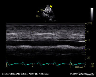
A transthoracic echocardiogram (TTE) is the most common type of echocardiogram, which is a still or moving image of the internal parts of the heart using ultrasound. In this case, the probe is placed on the chest or abdomen of the subject to get various views of the heart. It is used as a non-invasive assessment of the overall health of the heart, including a patient's heart valves and degree of heart muscle contraction. The images are displayed on a monitor for real-time viewing and then recorded.

<span class="mw-page-title-main">Cardiac cycle</span> Performance of the human heart

The cardiac cycle is the performance of the human heart from the beginning of one heartbeat to the beginning of the next. It consists of two periods: one during which the heart muscle relaxes and refills with blood, called diastole, following a period of robust contraction and pumping of blood, called systole. After emptying, the heart immediately relaxes and expands to receive another influx of blood returning from the lungs and other systems of the body, before again contracting to pump blood to the lungs and those systems. A normally performing heart must be fully expanded before it can efficiently pump again. Assuming a healthy heart and a typical rate of 70 to 75 beats per minute, each cardiac cycle, or heartbeat, takes about 0.8 second to complete the cycle. There are two atrial and two ventricle chambers of the heart; they are paired as the left heart and the right heart—that is, the left atrium with the left ventricle, the right atrium with the right ventricle—and they work in concert to repeat the cardiac cycle continuously. At the start of the cycle, during ventricular diastole–early, the heart relaxes and expands while receiving blood into both ventricles through both atria; then, near the end of ventricular diastole–late, the two atria begin to contract, and each atrium pumps blood into the ventricle below it. During ventricular systole the ventricles are contracting and vigorously pulsing two separated blood supplies from the heart—one to the lungs and one to all other body organs and systems—while the two atria are relaxed. This precise coordination ensures that blood is efficiently collected and circulated throughout the body.

The abdominojugular test, also known as abdominojugular reflux (AJR), is a physical examination test useful in diagnosing right ventricle dysfunction, particularly right ventricular failure.

<span class="mw-page-title-main">Right ventricular hypertrophy</span> Medical condition

Right ventricular hypertrophy (RVH) is a condition defined by an abnormal enlargement of the cardiac muscle surrounding the right ventricle. The right ventricle is one of the four chambers of the heart. It is located towards the lower-end of the heart and it receives blood from the right atrium and pumps blood into the lungs.

<span class="mw-page-title-main">Tricuspid regurgitation</span> Type of valvular heart disease

Tricuspid regurgitation (TR), also called tricuspid insufficiency, is a type of valvular heart disease in which the tricuspid valve of the heart, located between the right atrium and right ventricle, does not close completely when the right ventricle contracts (systole). TR allows the blood to flow backwards from the right ventricle to the right atrium, which increases the volume and pressure of the blood both in the right atrium and the right ventricle, which may increase central venous volume and pressure if the backward flow is sufficiently severe.

The following outline is provided as an overview of and topical guide to cardiology, the branch of medicine dealing with disorders of the human heart. The field includes medical diagnosis and treatment of congenital heart defects, coronary artery disease, heart failure, valvular heart disease and electrophysiology. Physicians who specialize in cardiology are called cardiologists.

<span class="mw-page-title-main">Cannon A waves</span>

Cannon A waves, or cannon atrial waves, are waves seen occasionally in the jugular vein of humans with certain cardiac arrhythmias. When the atria and ventricles happen to contract simultaneously, the right atrium contracts against a closed tricuspid valve, resulting in back pressure into the venous system that can be seen in the jugular venous pulse as a high-amplitude "cannon wave". It is associated with heart block, in particular third-degree (complete) heart block. It is also seen in pulmonary hypertension. Cannon A waves may also be seen in ventricular tachycardia due to the inherent AV dissociation of the arrhythmia.

<span class="mw-page-title-main">Pacemaker syndrome</span> Medical condition

Pacemaker syndrome is a condition that represents the clinical consequences of suboptimal atrioventricular (AV) synchrony or AV dyssynchrony, regardless of the pacing mode, after pacemaker implantation. It is an iatrogenic disease—an adverse effect resulting from medical treatment—that is often underdiagnosed. In general, the symptoms of the syndrome are a combination of decreased cardiac output, loss of atrial contribution to ventricular filling, loss of total peripheral resistance response, and nonphysiologic pressure waves.

The E/A ratio is a marker of the function of the left ventricle of the heart. It represents the ratio of peak velocity blood flow from left ventricular relaxation in early diastole to peak velocity flow in late diastole caused by atrial contraction. It is calculated using Doppler echocardiography, an ultrasound-based cardiac imaging modality. Abnormalities in the E/A ratio suggest that the left ventricle, which pumps blood into the systemic circulation, cannot fill with blood properly in the period between contractions. This phenomenon is referred to as diastolic dysfunction and can eventually lead to the symptoms of heart failure.

Cardiac physiology or heart function is the study of healthy, unimpaired function of the heart: involving blood flow; myocardium structure; the electrical conduction system of the heart; the cardiac cycle and cardiac output and how these interact and depend on one another.

Tissue Doppler echocardiography (TDE) is a medical ultrasound technology, specifically a form of echocardiography that measures the velocity of the heart muscle (myocardium) through the phases of one or more heartbeats by the Doppler effect of the reflected ultrasound. The technique is the same as for flow Doppler echocardiography measuring flow velocities. Tissue signals, however, have higher amplitude and lower velocities, and the signals are extracted by using different filter and gain settings. The terms tissue Doppler imaging (TDI) and tissue velocity imaging (TVI) are usually synonymous with TDE because echocardiography is the main use of tissue Doppler.

References

  1. "Cardiovascular Examination - Cardiovascular Disorders".
  2. "Evaluation of the Pulmonary Patient - Pulmonary Disorders".
  3. Gopal, S.; Nagalli, S. (2022). "Jugular Venous Distention". StatPearls. StatPearls. PMID   31971738.
  4. Borst J, Molhuysen J (1952). "Exact determination of the central venous pressure by a simple clinical method". Lancet. 2 (7): 304–9. doi:10.1016/S0140-6736(52)92474-4. PMID   14955978.
  5. 1 2 Ewy GA (September 1988). "The abdominojugular test: technique and hemodynamic correlates". Annals of Internal Medicine. 109 (6): 456–60. doi:10.7326/0003-4819-109-6-456. PMID   3415106.
  6. Cook DJ, Simel DL (February 1996). "The Rational Clinical Examination. Does this patient have abnormal central venous pressure?". JAMA. 275 (8): 630–4. doi:10.1001/jama.1996.03530320054034. PMID   8594245.
  7. Breidthardt, Tobias; Moreno-Weidmann, Zoraida; Uthoff, Heiko; Sabti, Zaid; Aeppli, Sven; Puelacher, Christian; Stallone, Fabio; Twerenbold, Raphael; Wildi, Karin; Kozhuharov, Nikola; Wussler, Desiree (2018). "How accurate is clinical assessment of neck veins in the estimation of central venous pressure in acute heart failure? Insights from a prospective study". European Journal of Heart Failure. 20 (7): 1160–1162. doi: 10.1002/ejhf.1111 . ISSN   1879-0844. PMID   29314487. S2CID   3581825.
  8. Conover, Mary Boudreau (2003). "Bedside Diagnosis". Understanding electrocardiography. St. Louis: Mosby. p. 82. ISBN   0-323-01905-6.
  9. "Friedreich's sign". BMJ Case Reports.
  10. Pittinger, Brook (2018). "Friedreich's sign". BMJ Case Reports. 2018. doi:10.1136/bcr-2018-226820. PMC   6203036 . PMID   30333203.