QT interval

Last updated
QT interval
QT interval.jpg
Electrocardiogram showing QT interval calculated by tangent method
ICD-10-PCS R94.31
ICD-9-CM 89.52
MeSH D004562
MedlinePlus 003868

The QT interval is a measurement made on an electrocardiogram used to assess some of the electrical properties of the heart. It is calculated as the time from the start of the Q wave to the end of the T wave, and approximates to the time taken from when the cardiac ventricles start to contract to when they finish relaxing. An abnormally long or abnormally short QT interval is associated with an increased risk of developing abnormal heart rhythms and sudden cardiac death. Abnormalities in the QT interval can be caused by genetic conditions such as long QT syndrome, by certain medications such as sotalol or pitolisant, by disturbances in the concentrations of certain salts within the blood such as hypokalaemia, or by hormonal imbalances such as hypothyroidism.

Contents

Measurement

Illustrations of the tangent and threshold methods of measuring the QT interval QT interval measurement.jpg
Illustrations of the tangent and threshold methods of measuring the QT interval

The QT interval is most commonly measured in lead II for evaluation of serial ECGs, with leads I and V5 being comparable alternatives to lead II. Leads III, aVL and V1 are generally avoided for measurement of QT interval. [1] The accurate measurement of the QT interval is subjective [2] because the end of the T wave is not always clearly defined and usually merges gradually with the baseline. QT interval in an ECG complex can be measured manually by different methods, such as the threshold method, in which the end of the T wave is determined by the point at which the component of the T wave merges with the isoelectric baseline, or the tangent method, in which the end of the T wave is determined by the intersection of a tangent line extrapolated from the T wave at the point of maximum downslope to the isoelectric baseline. [3]

With the increased availability of digital ECGs with simultaneous 12-channel recording, QT measurement may also be done by the 'superimposed median beat' method. In the superimposed median beat method, a median ECG complex is constructed for each of the 12 leads. The 12 median beats are superimposed on each other and the QT interval is measured either from the earliest onset of the Q wave to the latest offset of the T wave or from the point of maximum convergence for the Q wave onset to the T wave offset. [4]

Correction for heart rate

The QT interval changes in response to the heart rate - as heart rate increase the QT interval shortens. These changes make it harder to compare QT intervals measured at different heart rates. To account for this, and thereby improve the reliability of QT measurement, the QT interval can be corrected for heart rate (QTc) using a variety of mathematical formulae, a process often performed automatically by modern ECG recorders.

Bazett's formula

The most commonly used QT correction formula is the Bazett's formula, [5] named after physiologist Henry Cuthbert Bazett (1885–1950), [6] calculating the heart rate-corrected QT interval (QTcB).

Bazett's formula is based on observations from a study in 1920. Bazett's formula is often given in a form that returns QTc in dimensionally suspect units, square root of seconds. The mathematically correct form of Bazett's formula is:

where QTcB is the QT interval corrected for heart rate, and RR is the interval from the onset of one QRS complex to the onset of the next QRS complex. This mathematically correct formula returns the QTc in the same units as QT, generally milliseconds. [7]

In some popular forms of this formula, it is assumed that QT is measured in milliseconds and that RR is measured in seconds, often derived from the heart rate (HR) as 60/HR. Therefore, the result will be given in seconds per square root of milliseconds. [8] However, reporting QTc using this formula creates a "requirement regarding the units in which the original QT and RR are measured." [7]

In either form, Bazett's non-linear QT correction formula is generally not considered accurate, as it over-corrects at high heart rates and under-corrects at low heart rates. [8] Bazett's correction formula is one of the most suitable QT correction formulae for neonates. [9]

Fridericia's formula

Fridericia [10] had proposed an alternative correction formula (QTcF) using the cube-root of RR.

Sagie's formula

The Framingham correction, also called as Sagie's formula based on the Framingham Heart Study, which used long-term cohort data of over 5,000 subjects, is considered a better [11] method. [12]

Again, here QT and QTlc are in milliseconds and RR is measured in seconds.

Comparison of corrections

A retrospective study suggests that Fridericia's method and the Framingham method may produce results most useful for stratifying the 30-day and 1-year risks of mortality. [11]

Upper limit of normal QT interval, corrected for heart rate according to Bazett's formula, Fridericia's formula, and subtracting 0.02 s from QT for every 10 bpm increase in heart rate. Up to 0.42 s (<= 420 ms) is chosen as normal QTc of QTB and QTF in this diagram. QT interval corrected for heart rate.png
Upper limit of normal QT interval, corrected for heart rate according to Bazett's formula, Fridericia's formula, and subtracting 0.02 s from QT for every 10 bpm increase in heart rate. Up to 0.42 s (≤ 420 ms) is chosen as normal QTc of QTB and QTF in this diagram.

Definitions of normal QTc vary from being equal to or less than 0.40 s (≤ 400 ms), [13] 0.41 s (≤ 410 ms), [15] 0.42 s (≤ 420 ms) [14] or 0.44 s (≤ 440 ms). [16] For risk of sudden cardiac death, "borderline QTc" in males is 431–450 ms; and, in females, 451–470 ms. An "abnormal" QTc in males is a QTc above 450 ms; and, in females, above 470 ms. [17]

If there is not a very high or low heart rate, the upper limits of QT can roughly be estimated by taking QT = QTc at a heart rate of 60 beats per minute (bpm), and subtracting 0.02 s from QT for every 10 bpm increase in heart rate. For example, taking normal QTc  0.42 s, QT would be expected to be 0.42 s or less at a heart rate of 60 bpm. For a heart rate of 70 bpm, QT would roughly be expected to be equal to or below 0.40 s. Likewise, for 80 bpm, QT would roughly be expected to be equal to or below 0.38 s. [13]

Abnormal intervals

Prolonged QTc causes premature action potentials during the late phases of depolarization. This increases the risk of developing ventricular arrhythmias, including fatal ventricular fibrillation. [18] Higher rates of prolonged QTc are seen in females, older patients, high systolic blood pressure or heart rate, and short stature. [19] Prolonged QTc is also associated with ECG findings called Torsades de Pointes, which are known to degenerate into ventricular fibrillation, associated with higher mortality rates. There are many causes of prolonged QT intervals, acquired causes being more common than genetic. [20]

Genetic causes

Distribution of QT intervals amongst healthy males and females, and amongst those with congenital long QT syndrome QT distribution.png
Distribution of QT intervals amongst healthy males and females, and amongst those with congenital long QT syndrome

An abnormally prolonged QT interval could be due to long QT syndrome, whereas an abnormally shortened QT interval could be due to short QT syndrome.

The QTc length is associated with variations in the NOS1AP gene. [21] The autosomal recessive syndrome of Jervell and Lange-Nielsen is characterized by a prolonged QTc interval in conjunction with sensorineural hearing loss.

Due to adverse drug reactions

Prolongation of the QT interval may be due to an adverse drug reaction. [22]

Antipsychotics (especially first generation/"typical")

DMARDs and antimalarial drugs

Antibiotics

Other drugs

Some second-generation antihistamines, such as astemizole, have this effect. The mechanism of action of certain antiarrhythmic drugs, like amiodarone or sotalol, involve intentional pharmacological QT prolongation. In addition, high blood alcohol concentrations prolong the QT interval. [30] A possible interaction between selective serotonin reuptake inhibitors and thiazide diuretics is associated with QT prolongation. [31]

Due to pathological conditions

Hypothyroidism, a condition of low function of the thyroid gland, can cause QT prolongation at the electrocardiogram. Acute hypocalcemia causes prolongation of the QT interval, which may lead to ventricular dysrhythmias.

A shortened QT can be associated with hypercalcemia. [32]

Use in drug approval studies

Since 2005, the FDA and European regulators have required that nearly all new molecular entities be evaluated in a Thorough QT (TQT) or similar study to determine a drug's effect on the QT interval. [33] The TQT study serves to assess the potential arrhythmia liability of a drug. Traditionally, the QT interval had been evaluated by having an individual human reader measure approximately nine cardiac beats per clinical timepoint. However, a substantial portion of drug approvals after 2010 have incorporated a partially automated approach, blending automated software algorithms with expert human readers reviewing a portion of the cardiac beats, to enable the assessment of significantly more beats in order to improve precision and reduce cost. [34] In 2014, an industrywide consortium consisting of the FDA, iCardiac Technologies and other organizations released the results of a seminal study indicating how waivers from TQT studies can be obtained by the assessment of early phase data. [35] As the pharmaceutical industry has gained experience in performing TQT studies, it has also become evident that traditional QT correction formulas such as QTcF, QTcB, and QTcLC may not always be suitable for evaluation of drugs impacting autonomic tone. [36]

As a predictor of mortality

Electrocardiography is a safe and noninvasive tool that can be used to identify those with a higher risk of mortality. In the general population, there has been no consistent evidence that prolonged QTc interval in isolation is associated with an increase in mortality from cardiovascular disease. [37] However, several studies[ which? ] have examined prolonged QT interval as a predictor of mortality for diseased subsets of the population.

Rheumatoid arthritis

Rheumatoid arthritis is the most common inflammatory arthritis. [38] Studies have linked rheumatoid arthritis with increased death from cardiovascular disease. [38] In a 2014 study, [18] Panoulas et al. found a 50 ms increase in QTc interval increased the odds of all-cause mortality by 2.17 in patients with rheumatoid arthritis. Patients with the highest QTc interval (> 424 ms) had higher mortality than those with a lower QTc interval. The association was lost when calculations were adjusted for C-reactive protein levels. The researchers proposed that inflammation prolonged the QTc interval and created arrhythmias that were associated with higher mortality rates. However, the mechanism by which C-reactive protein is associated with the QTc interval is still not understood.

Type 1 diabetes

Compared to the general population, type 1 diabetes may increase the risk of mortality, due largely to an increased risk of cardiovascular disease. [19] [39] Almost half of patients with type 1 diabetes have a prolonged QTc interval (> 440 ms). [19] Diabetes with a prolonged QTc interval was associated with a 29% mortality over 10 years in comparison to 19% with a normal QTc interval. [19] Anti-hypertensive drugs increased the QTc interval, but were not an independent predictor of mortality. [19]

Type 2 diabetes

QT interval dispersion (QTd) is the maximum QT interval minus the minimum QT interval, and is linked with ventricular repolarization. [40] A QTd over 80 ms is considered abnormally prolonged. [41] Increased QTd is associated with mortality in type 2 diabetes. [41] QTd is a better predictor of cardiovascular death than QTc, which was unassociated with mortality in type 2 diabetes. [41] QTd higher than 80 ms had a relative risk of 1.26 of dying from cardiovascular disease compared to a normal QTd.

See also

Related Research Articles

<span class="mw-page-title-main">Heart rate</span> Speed of the heartbeat, measured in beats per minute

Heart rate is the frequency of the heartbeat measured by the number of contractions of the heart per minute. The heart rate can vary according to the body's physical needs, including the need to absorb oxygen and excrete carbon dioxide, but is also modulated by numerous factors, including genetics, physical fitness, stress or psychological status, diet, drugs, hormonal status, environment, and disease/illness as well as the interaction between and among these factors. It is usually equal or close to the pulse measured at any peripheral point.

<span class="mw-page-title-main">Sertindole</span> Antipsychotic medication

Sertindole, sold under the brand name Serdolect among others, is an antipsychotic medication. Sertindole was developed by the Danish pharmaceutical company Lundbeck and marketed under license by Abbott Labs. Like other atypical antipsychotics, it has activity at dopamine and serotonin receptors in the brain. It is used in the treatment of schizophrenia. It is classified chemically as a phenylindole derivative.

<span class="mw-page-title-main">Long QT syndrome</span> Medical condition

Long QT syndrome (LQTS) is a condition affecting repolarization (relaxing) of the heart after a heartbeat, giving rise to an abnormally lengthy QT interval. It results in an increased risk of an irregular heartbeat which can result in fainting, drowning, seizures, or sudden death. These episodes can be triggered by exercise or stress. Some rare forms of LQTS are associated with other symptoms and signs including deafness and periods of muscle weakness.

<span class="mw-page-title-main">Hypocalcemia</span> Low calcium levels in ones blood serum

Hypocalcemia is a medical condition characterized by low calcium levels in the blood serum. The normal range of blood calcium is typically between 2.1–2.6 mmol/L while levels less than 2.1 mmol/L are defined as hypocalcemic. Mildly low levels that develop slowly often have no symptoms. Otherwise symptoms may include numbness, muscle spasms, seizures, confusion, or cardiac arrest.

<span class="mw-page-title-main">Dofetilide</span> Antiarrhythmic medication

Dofetilide is a class III antiarrhythmic agent. It is marketed under the trade name Tikosyn by Pfizer, and is available in the United States in capsules containing 125, 250, and 500 µg of dofetilide. It is not available in Europe or Australia.

<span class="mw-page-title-main">Short QT syndrome</span> Medical condition

Short QT syndrome (SQT) is a very rare genetic disease of the electrical system of the heart, and is associated with an increased risk of abnormal heart rhythms and sudden cardiac death. The syndrome gets its name from a characteristic feature seen on an electrocardiogram (ECG) – a shortening of the QT interval. It is caused by mutations in genes encoding ion channels that shorten the cardiac action potential, and appears to be inherited in an autosomal dominant pattern. The condition is diagnosed using a 12-lead ECG. Short QT syndrome can be treated using an implantable cardioverter-defibrillator or medications including quinidine. Short QT syndrome was first described in 2000, and the first genetic mutation associated with the condition was identified in 2004.

<span class="mw-page-title-main">First-degree atrioventricular block</span> Medical condition

First-degree atrioventricular block is a disease of the electrical conduction system of the heart in which electrical impulses conduct from the cardiac atria to the ventricles through the atrioventricular node more slowly than normal. First degree AV block does not generally cause any symptoms, but may progress to more severe forms of heart block such as second- and third-degree atrioventricular block. It is diagnosed using an electrocardiogram, and is defined as a PR interval greater than 200 milliseconds. First degree AV block affects 0.65-1.1% of the population with 0.13 new cases per 1000 persons each year.

<span class="mw-page-title-main">Torsades de pointes</span> Type of abnormal heart rhythm

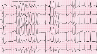
Torsades de pointes, torsade de pointes or torsades des pointes (TdP) is a specific type of abnormal heart rhythm that can lead to sudden cardiac death. It is a polymorphic ventricular tachycardia that exhibits distinct characteristics on the electrocardiogram (ECG). It was described by French physician François Dessertenne in 1966. Prolongation of the QT interval can increase a person's risk of developing this abnormal heart rhythm, occurring in between 1% and 10% of patients who receive QT-prolonging antiarrhythmic drugs.

<span class="mw-page-title-main">Ventricular tachycardia</span> Medical condition of the heart

Ventricular tachycardia is a fast heart rate arising from the lower chambers of the heart. Although a few seconds of VT may not result in permanent problems, longer periods are dangerous; and multiple episodes over a short period of time are referred to as an electrical storm. Short periods may occur without symptoms, or present with lightheadedness, palpitations, or chest pain. Ventricular tachycardia may result in ventricular fibrillation (VF) and turn into cardiac arrest. This conversion of the VT into VF is called the degeneration of the VT. It is found initially in about 7% of people in cardiac arrest.

<span class="mw-page-title-main">Jervell and Lange-Nielsen syndrome</span> Medical condition

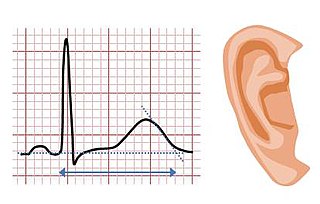
Jervell and Lange-Nielsen syndrome (JLNS) is a rare type of long QT syndrome associated with severe, bilateral sensorineural hearing loss. Those with JLNS are at risk of abnormal heart rhythms called arrhythmias, which can lead to fainting, seizures, or sudden death. JLNS, like other forms of long QT syndrome, causes the cardiac muscle to take longer than usual to recharge between beats. It is caused by genetic variants responsible for producing ion channels that carry transport potassium out of cells. The condition is usually diagnosed using an electrocardiogram, but genetic testing can also be used. Treatment includes lifestyle measures, beta blockers, and implantation of a defibrillator in some cases. It was first described by Anton Jervell and Fred Lange-Nielsen in 1957.

<span class="mw-page-title-main">Romano–Ward syndrome</span> Medical condition

Romano–Ward syndrome is the most common form of congenital Long QT syndrome (LQTS), a genetic heart condition that affects the electrical properties of heart muscle cells. Those affected are at risk of abnormal heart rhythms which can lead to fainting, seizures, or sudden death. Romano–Ward syndrome can be distinguished clinically from other forms of inherited LQTS as it affects only the electrical properties of the heart, while other forms of LQTS can also affect other parts of the body.

<span class="mw-page-title-main">Andersen–Tawil syndrome</span> Rare autosomal dominant genetic disorder

Andersen–Tawil syndrome, also called Andersen syndrome and long QT syndrome 7, is a rare genetic disorder affecting several parts of the body. The three predominant features of Andersen–Tawil syndrome include disturbances of the electrical function of the heart characterised by an abnormality seen on an electrocardiogram and a tendency to abnormal heart rhythms, physical characteristics including low-set ears and a small lower jaw, and intermittent periods of muscle weakness known as hypokalaemic periodic paralysis.

<span class="mw-page-title-main">Dronedarone</span> Drug

Dronedarone, sold under the brand name Multaq, is a medication by Sanofi-Aventis, mainly for the indication of cardiac arrhythmias. It was approved by the FDA on July 2, 2009. It was recommended as an alternative to amiodarone for the treatment of atrial fibrillation and atrial flutter in people whose hearts have either returned to normal rhythm or who undergo drug therapy or electric shock treatment i.e. direct current cardioversion (DCCV) to maintain normal rhythm. It is a class III antiarrhythmic drug. In the United States, the FDA approved label includes a claim for reducing hospitalization, but not for reducing mortality, as a reduction in mortality was not demonstrated in the clinical development program. A trial of the drug in heart failure was stopped as an interim analysis showed a possible increase in heart failure deaths, in patients with moderate to severe CHF.

<span class="mw-page-title-main">Tricyclic antidepressant overdose</span> Medical condition

Tricyclic antidepressant overdose is poisoning caused by excessive medication of the tricyclic antidepressant (TCA) type. Symptoms may include elevated body temperature, blurred vision, dilated pupils, sleepiness, confusion, seizures, rapid heart rate, and cardiac arrest. If symptoms have not occurred within six hours of exposure they are unlikely to occur.

<span class="mw-page-title-main">Vectorcardiography</span>

Vectorcardiography (VCG) is a method of recording the magnitude and direction of the electrical forces that are generated by the heart by means of a continuous series of vectors that form curving lines around a central point.

A Poincaré plot, named after Henri Poincaré, is a type of recurrence plot used to quantify self-similarity in processes, usually periodic functions. It is also known as a return map. Poincaré plots can be used to distinguish chaos from randomness by embedding a data set in a higher-dimensional state space.

Fabio Badilini is an Italian scientist and business man. He has made major contributions to noninvasive electrocardiography not only through his individual contributions, but also through his truly remarkable ability to foster collaborations across scientific disciplines, academic institutions, governmental agencies, device manufacturers and industries around the world.

Heart rhythm disturbances have been seen among astronauts. Most of these have been related to cardiovascular disease, but it is not clear whether this was due to pre-existing conditions or effects of space flight. It is hoped that advanced screening for coronary disease has greatly mitigated this risk. Other heart rhythm problems, such as atrial fibrillation, can develop over time, necessitating periodic screening of crewmembers’ heart rhythms. Beyond these terrestrial heart risks, some concern exists that prolonged exposure to microgravity may lead to heart rhythm disturbances. Although this has not been observed to date, further surveillance is warranted.

QT prolongation is a measure of delayed ventricular repolarisation, which means the heart muscle takes longer than normal to recharge between beats. It is an electrical disturbance which can be seen on an electrocardiogram (ECG). Excessive QT prolongation can trigger tachycardias such as torsades de pointes (TdP). QT prolongation is an established side effect of antiarrhythmics, but can also be caused by a wide range of non-cardiac medicines, including antibiotics, antidepressants, antihistamines, opioids, and complementary medicines. On an ECG, the QT interval represents the summation of action potentials in cardiac muscle cells, which can be caused by an increase in inward current through sodium or calcium channels, or a decrease in outward current through potassium channels. By binding to and inhibiting the “rapid” delayed rectifier potassium current protein, certain drugs are able to decrease the outward flow of potassium ions and extend the length of phase 3 myocardial repolarization, resulting in QT prolongation.

<span class="mw-page-title-main">QT interval variability</span>

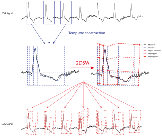
QT interval variability (QTV) refers to the physiological phenomenon of beat-to-beat fluctuations in QT interval of electrocardiograms. Increased QTV appears to be a marker of arrhythmic and cardiovascular death; it may also play a role for noninvasive assessment of sympathetic nervous system activity.

References

  1. Panicker GK, Salvi V, Karnad DR, Chakraborty S, Manohar D, Lokhandwala Y, Kothari S (2014). "Drug-induced QT prolongation when QT interval is measured in each of the 12 ECG leads in men and women in a thorough QT study". Journal of Electrocardiology. 47 (2): 155–157. doi:10.1016/j.jelectrocard.2013.11.004. PMID   24388488.
  2. Panicker GK, Karnad DR, Joshi R, Shetty S, Vyas N, Kothari S, Narula D (January 2009). "Z-score for benchmarking reader competence in a central ECG laboratory". Annals of Noninvasive Electrocardiology. 14 (1): 19–25. doi:10.1111/j.1542-474X.2008.00269.x. PMC   6932360 . PMID   19149789.
  3. Panicker GK, Karnad DR, Natekar M, Kothari S, Narula D, Lokhandwala Y (2009). "Intra- and interreader variability in QT interval measurement by tangent and threshold methods in a central electrocardiogram laboratory". Journal of Electrocardiology. 42 (4): 348–352. doi:10.1016/j.jelectrocard.2009.01.003. PMID   19261293.
  4. Salvi V, Karnad DR, Panicker GK, Natekar M, Hingorani P, Kerkar V, et al. (2011). "Comparison of 5 methods of QT interval measurements on electrocardiograms from a thorough QT/QTc study: effect on assay sensitivity and categorical outliers". Journal of Electrocardiology. 44 (2): 96–104. doi:10.1016/j.jelectrocard.2010.11.010. PMID   21238976.
  5. 1 2 Bazett HC (1920). "An analysis of the time-relations of electrocardiograms". Heart (7): 353–370.
  6. Roguin A (March 2011). "Henry Cuthbert Bazett (1885-1950)--the man behind the QT interval correction formula". Pacing and Clinical Electrophysiology. 34 (3): 384–388. doi:10.1111/j.1540-8159.2010.02973.x. PMID   21091739. S2CID   43739886.
  7. 1 2 Molnar J, Weiss JS, Rosenthal JE (March 1995). "The missing second: what is the correct unit for the Bazett corrected QT interval?". The American Journal of Cardiology. 75 (7): 537–538. doi:10.1016/S0002-9149(99)80603-1. PMID   7864010.
  8. 1 2 Salvi V, Karnad DR, Panicker GK, Kothari S (January 2010). "Update on the evaluation of a new drug for effects on cardiac repolarization in humans: issues in early drug development". British Journal of Pharmacology. 159 (1): 34–48. doi:10.1111/j.1476-5381.2009.00427.x. PMC   2823350 . PMID   19775279.
  9. Stramba-Badiale M, Karnad DR, Goulene KM, Panicker GK, Dagradi F, Spazzolini C, et al. (August 2018). "For neonatal ECG screening there is no reason to relinquish old Bazett's correction". European Heart Journal. 39 (31): 2888–2895. doi: 10.1093/eurheartj/ehy284 . PMID   29860404.
  10. 1 2 Fridericia LS (1920). "The duration of systole in the electrocardiogram of normal subjects and of patients with heart disease". Acta Medica Scandinavica (53): 469–486.
  11. 1 2 Vandenberk B, Vandael E, Robyns T, Vandenberghe J, Garweg C, Foulon V, Ector J, Willems R (2016-06-17), "Which QT Correction Formulae to Use for QT Monitoring?", Journal of the American Heart Association, 5 (6), doi:10.1161/JAHA.116.003264, PMC   4937268 , PMID   27317349
  12. Sagie A, Larson MG, Goldberg RJ, Bengtson JR, Levy D (September 1992). "An improved method for adjusting the QT interval for heart rate (the Framingham Heart Study)". The American Journal of Cardiology. 70 (7): 797–801. doi:10.1016/0002-9149(92)90562-D. PMID   1519533.[ need full text ]
  13. 1 2 3 Yanowitz FG. "Lesson III. Characteristics of the Normal ECG". University of Utah School of Medicine. Retrieved 23 March 2010.
  14. 1 2 Jenkins D, Gerred S. "A normal adult 12-lead ECG". ecglibrary.com. Retrieved 28 January 2018.
  15. Fitz M. "Medicine I". Loyola University Chicago Stritch School of Medicine. Retrieved 23 March 2010.
  16. Klabunde RE. "Image for Cardiovascular Physiology Concepts > Electrocardiogram (EKG, ECG)".
  17. "QTc Prolongation and Risk of Sudden Cardiac Death: Is the Debate Over?". CRM News. 3 February 2006 via medscape.com.
  18. 1 2 Panoulas VF, Toms TE, Douglas KM, Sandoo A, Metsios GS, Stavropoulos-Kalinoglou A, Kitas GD (January 2014). "Prolonged QTc interval predicts all-cause mortality in patients with rheumatoid arthritis: an association driven by high inflammatory burden". Rheumatology. 53 (1): 131–137. doi: 10.1093/rheumatology/ket338 . PMID   24097136.
  19. 1 2 3 4 5 Rossing P, Breum L, Major-Pedersen A, Sato A, Winding H, Pietersen A, et al. (March 2001). "Prolonged QTc interval predicts mortality in patients with Type 1 diabetes mellitus". Diabetic Medicine. 18 (3): 199–205. doi:10.1046/j.1464-5491.2001.00446.x. PMID   11318840. S2CID   38600053.
  20. van Noord C, Eijgelsheim M, Stricker BH (July 2010). "Drug- and non-drug-associated QT interval prolongation". British Journal of Clinical Pharmacology. 70 (1): 16–23. doi:10.1111/j.1365-2125.2010.03660.x. PMC   2909803 . PMID   20642543.
  21. Arking DE, Pfeufer A, Post W, Kao WH, Newton-Cheh C, Ikeda M, et al. (June 2006). "A common genetic variant in the NOS1 regulator NOS1AP modulates cardiac repolarization". Nature Genetics. 38 (6): 644–651. doi:10.1038/ng1790. PMID   16648850. S2CID   12942685.
  22. Leitch A, McGinness P, Wallbridge D (September 2007). "Calculate the QT interval in patients taking drugs for dementia". BMJ. 335 (7619): 557. doi:10.1136/bmj.39020.710602.47. PMC   1976518 . PMID   17855324.
  23. "Information for Healthcare Professionals: Haloperidol (marketed as Haldol, Haldol Decanoate and Haldol Lactate)". Food and Drug Administration . Archived from the original on 2007-10-11. Retrieved 2007-09-18.
  24. 1 2 3 Washington NB, Brahm NC, Kissack J (1 October 2012). "Which psychotropics carry the greatest risk of QTc prolongation?". Current Psychiatry. 11 (10): 36–39. Retrieved 2020-05-19.
  25. Lewis R, Bagnall AM, Leitner M (July 2005). "Sertindole for schizophrenia". The Cochrane Database of Systematic Reviews (3): CD001715. doi:10.1002/14651858.CD001715.pub2. PMC   7025766 . PMID   16034864.
  26. 1 2 3 Malloy T (March 25, 2020). "Guidance on patients at risk of drug-induced sudden cardiac death from off-label COVID-19 treatments". Mayo Clinic News Network. Retrieved 2020-05-19.
  27. Briasoulis A, Agarwal V, Pierce WJ (2011). "QT prolongation and torsade de pointes induced by fluoroquinolones: infrequent side effects from commonly used medications". Cardiology. 120 (2): 103–110. doi:10.1159/000334441. PMID   22156660. S2CID   39533896.
  28. Haigney M. "Cardiotoxicity of methadone" (PDF). Director of Cardiology. Retrieved 21 February 2013.
  29. https://wakix.com "WAKIX prolongs the QT interval; avoid use of WAKIX in patients with known QT prolongation or in combination with other drugs known to prolong QT interval."
  30. Aasebø W, Erikssen J, Jonsbu J, Stavem K (April 2007). "ECG changes in patients with acute ethanol intoxication". Scandinavian Cardiovascular Journal. 41 (2): 79–84. doi: 10.1080/14017430601091698 . PMID   17454831. S2CID   11828588.
  31. Tatonetti NP, Ye PP, Daneshjou R, Altman RB (March 2012). "Data-driven prediction of drug effects and interactions". Science Translational Medicine. 4 (125): 125ra31. doi:10.1126/scitranslmed.3003377. PMC   3382018 . PMID   22422992.
  32. Skugor M (June 2019). "Hypercalcemia". The Cleveland Clinic.
  33. "E14 Clinical Evaluation of QT/QTc Interval Prolongation and Proarrhythmic Potential for Non-Antiarrhythmic Drugs" (PDF). Guidance for Industry. U.S. Food and Drug Administration. October 2005. Archived from the original (PDF) on March 6, 2010. Retrieved December 9, 2009.
  34. "iCardiac Applies Automated Approach to Thorough QT Study for a Leading Pharmaceutical Company – Applied Clinical Trials". 5 October 2011. Archived from the original on 5 October 2011. Retrieved 19 March 2018.{{cite web}}: CS1 maint: bot: original URL status unknown (link)
  35. Or A (2014-12-16). "Norwest-Backed iCardiac to Expand Its Market Thanks to Breakthrough". Wall Street Journal. ISSN   0099-9660 . Retrieved 2021-01-26.
  36. Garnett C (29 April 2009). "CSRC White Paper: QT/QTc Evaluation for Drugs with Autonomic Effects" (PDF). THEW Annual Meeting. Retrieved 6 June 2014.
  37. Montanez A, Ruskin JN, Hebert PR, Lamas GA, Hennekens CH (May 2004). "Prolonged QTc interval and risks of total and cardiovascular mortality and sudden death in the general population: a review and qualitative overview of the prospective cohort studies". Archives of Internal Medicine. 164 (9): 943–948. doi:10.1001/archinte.164.9.943. PMID   15136301.
  38. 1 2 Solomon DH, Karlson EW, Rimm EB, Cannuscio CC, Mandl LA, Manson JE, et al. (March 2003). "Cardiovascular morbidity and mortality in women diagnosed with rheumatoid arthritis". Circulation. 107 (9): 1303–1307. doi: 10.1161/01.cir.0000054612.26458.b2 . PMID   12628952.
  39. Borch-Johnsen K, Andersen PK, Deckert T (August 1985). "The effect of proteinuria on relative mortality in type 1 (insulin-dependent) diabetes mellitus". Diabetologia. 28 (8): 590–596. doi: 10.1007/bf00281993 . PMID   4054448.
  40. Okin PM, Devereux RB, Howard BV, Fabsitz RR, Lee ET, Welty TK (2000). "Assessment of QT interval and QT dispersion for prediction of all-cause and cardiovascular mortality in American Indians: The Strong Heart Study". Circulation. 101 (1): 61–66. doi: 10.1161/01.cir.101.1.61 . PMID   10618305.
  41. 1 2 3 Giunti S, Gruden G, Fornengo P, Barutta F, Amione C, Ghezzo G, et al. (March 2012). "Increased QT interval dispersion predicts 15-year cardiovascular mortality in type 2 diabetic subjects: the population-based Casale Monferrato Study". Diabetes Care. 35 (3): 581–583. doi:10.2337/dc11-1397. PMC   3322722 . PMID   22301117.