Carotid body

Last updated
Carotid body
Gray1186.png
Section of part of human carotid body. Highly magnified. Numerous blood vessels are seen in section among the cells.
Gray474.png
Diagram showing the origins of the main branches of the carotid arteries.
Details
Nerve Branch of glossopharyngeal nerve to carotid sinus
Identifiers
Latin glomus caroticum
MeSH D002344
TA98 A12.2.04.007
TA2 3886
FMA 50095
Anatomical terminology

The carotid body is a small cluster of chemoreceptor cells and supporting sustentacular cells situated at bifurcation of each common carotid artery in its adventitia. [1] [2]

Contents

The carotid body detects changes in the composition of arterial blood flowing through it, mainly the partial pressure of arterial oxygen, but also of carbon dioxide. It is also sensitive to changes in blood pH, and temperature.

Structure

The carotid body is situated on the posterior aspect of the bifurcation of the common carotid artery. [3]

The carotid body is made up of two types of cells, called glomus cells: glomus type I cells are peripheral chemoreceptors, and glomus type II cells are sustentacular supportive cells.

Function

The carotid body functions as a sensor: it responds to a stimulus, primarily O2 partial pressure, which is detected by the type I (glomus) cells, and triggers an action potential through the afferent fibers of the glossopharyngeal nerve, which relays the information to the central nervous system.

Stimulus

The carotid body peripheral chemoreceptors are primarily sensitive to decreases in the partial pressure of oxygen (PO2). This is in contrast to the central chemoreceptors in the medulla oblongata that are primarily sensitive to changes in pH and PCO2 (a decrease in pH and an increase in PCO2). The carotid body chemoreceptors are also sensitive to pH and PCO2, but only secondarily. More specifically, the sensitivity of carotid body chemoreceptors to decreased PO2 is greater when pH is decreased and PCO2 is increased.

Impulse rate for carotid bodies is particularly sensitive to changes in arterial PO2 in the range of 60 down to 30 mm Hg, a range in which hemoglobin saturation with oxygen decreases rapidly. [5]

The output of the carotid bodies is low at an oxygen partial pressure above about 100mmHg (13,3 kPa) (at normal physiological pH), but below 60mmHg the activity of the type I (glomus) cells increases rapidly due to a decrease in hemoglobin-oxygen saturation below 90%.

Detection

The mechanism for detecting reductions in PO2 has yet to be identified, there may be multiple mechanisms and could vary between species. [6] Hypoxia detection has been shown to depend upon increased hydrogen sulfide generation produced by cystathionine gamma-lyase as hypoxia detection is reduced in mice in which this enzyme is knocked out or pharmacologically inhibited. The process of detection involves the interaction of cystathionine gamma-lyase with hemeoxygenase-2 and the production of carbon monoxide. [7] Yet, some studies show that physiologic concentration of hydrogen sulfide may not be strong enough to trigger such responses.

Other theories suggest it may involve mitochondrial oxygen sensors and the haem-containing cytochromes that undergo reversible one-electron reduction during oxidative-phosphorylation. Haem reversibly binds O2 with an affinity similar to that of the carotid body, suggesting that haem containing proteins may have a role in O2, potentially this could be one of the complexes involved in oxidative-phosphorylation. This leads to increases in reactive oxygen species and rises in intracellular Ca2+. However, whether hypoxia leads to an increase or decrease in reactive oxygen species is unknown. The role of reactive oxygen species in hypoxia sensing is also under question. [8]

The oxygen dependent enzyme haem-oxidase has also been put forward as a hypoxia sensor. In normoxia, haem-oxygenase generates carbon monoxide (CO), CO activates the large conductance calcium-activated potassium channel, BK. Falls in CO that occur as a consequence of hypoxia would lead to closure of this potassium channel and this would lead to membrane depolarisation and consequence activation of the carotid body. [9] A role for the "energy sensor" AMP-activated protein kinase (AMPK) has also been proposed in hypoxia sensing. This enzyme is activated during times of net energy usage and metabolic stress, including hypoxia. AMPK has a number of targets and it appears that, in the carotid body, when AMPK is activated by hypoxia, it leads to downstream potassium channel closure of both O2-sentive TASK-like and BK channels [10]

An increased PCO2 is detected because the CO2 diffuses into the cell, where it increases the concentration of carbonic acid and thus protons. The precise mechanism of CO2 sensing is unknown, however it has been demonstrated that CO2 and low pH inhibit a TASK-like potassium conductance, reducing potassium current. This leads to depolarisation of the cell membrane which leads to Ca2+ entry, excitation of glomus cells and consequent neurotransmitter release. [11]

Arterial acidosis (either metabolic or from altered PCO2) inhibits acid-base transporters (e.g. Na+-H+) which raise intracellular pH, and activates transporters (e.g. Cl-HCO3) which decrease it. Changes in proton concentration caused by acidosis (or the opposite from alkalosis) inside the cell stimulates the same pathways involved in PCO2 sensing.

Another mechanism is through oxygen sensitive potassium channels. A drop in dissolved oxygen lead to closing of these channels which results in depolarization. This leads to release of the neurotransmitter dopamine in the glossopharyngeal and vagus afferente to the vasomotor area.

Action potential

The type I (glomus) cells in the carotid (and aortic bodies) are derived from neuroectoderm and are thus electrically excitable. A decrease in oxygen partial pressure, an increase in carbon dioxide partial pressure, and a decrease in arterial pH can all cause depolarization of the cell membrane, and they affect this by blocking potassium currents. This reduction in the membrane potential opens voltage-gated calcium channels, which causes a rise in intracellular calcium concentration. This causes exocytosis of vesicles containing a variety of neurotransmitters, including acetylcholine, noradrenaline, dopamine, adenosine, ATP, substance P, and met-enkephalin. These act on receptors on the afferent nerve fibres which lie in apposition to the glomus cell to cause an action potential.

Relay

The feedback from the carotid body is sent to the cardiorespiratory centers in the medulla oblongata via the afferent branches of the glossopharyngeal nerve. (The afferent fibers of the aortic body chemoreceptors are relayed by the vagus nerve.) These centers, in turn, regulate breathing and blood pressure, with hypoxia causing an increase in ventilation.

Clinical significance

Micrograph of a carotid body tumor. Carotid body tumour 2 intermed mag.jpg
Micrograph of a carotid body tumor.

Paraganglioma

A paraganglioma is a tumor that may involve the carotid body and is usually benign. Rarely, a malignant neuroblastoma may originate from the carotid body.

See also

List of distinct cell types in the adult human body

Related Research Articles

<span class="mw-page-title-main">Hypoxia (medicine)</span> Medical condition of lack of oxygen in the tissues

Hypoxia is a condition in which the body or a region of the body is deprived of adequate oxygen supply at the tissue level. Hypoxia may be classified as either generalized, affecting the whole body, or local, affecting a region of the body. Although hypoxia is often a pathological condition, variations in arterial oxygen concentrations can be part of the normal physiology, for example, during strenuous physical exercise.

In biology, homeostasis is the state of steady internal physical and chemical conditions maintained by living systems. This is the condition of optimal functioning for the organism and includes many variables, such as body temperature and fluid balance, being kept within certain pre-set limits. Other variables include the pH of extracellular fluid, the concentrations of sodium, potassium, and calcium ions, as well as the blood sugar level, and these need to be regulated despite changes in the environment, diet, or level of activity. Each of these variables is controlled by one or more regulators or homeostatic mechanisms, which together maintain life.

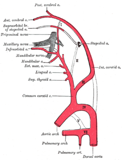
Baroreceptors are sensors located in the carotid sinus and in the aortic arch. They sense the blood pressure and relay the information to the brain, so that a proper blood pressure can be maintained.

<span class="mw-page-title-main">Glossopharyngeal nerve</span> Cranial nerve IX, for the tongue and pharynx

The glossopharyngeal nerve, also known as the ninth cranial nerve, cranial nerve IX, or simply CN IX, is a cranial nerve that exits the brainstem from the sides of the upper medulla, just anterior to the vagus nerve. Being a mixed nerve (sensorimotor), it carries afferent sensory and efferent motor information. The motor division of the glossopharyngeal nerve is derived from the basal plate of the embryonic medulla oblongata, whereas the sensory division originates from the cranial neural crest.

<span class="mw-page-title-main">Diving reflex</span> The physiological responses to immersion of air-breathing vertebrates

The diving reflex, also known as the diving response and mammalian diving reflex, is a set of physiological responses to immersion that overrides the basic homeostatic reflexes, and is found in all air-breathing vertebrates studied to date. It optimizes respiration by preferentially distributing oxygen stores to the heart and brain, enabling submersion for an extended time.

A chemoreceptor, also known as chemosensor, is a specialized sensory receptor which transduces a chemical substance to generate a biological signal. This signal may be in the form of an action potential, if the chemoreceptor is a neuron, or in the form of a neurotransmitter that can activate a nerve fiber if the chemoreceptor is a specialized cell, such as taste receptors, or an internal peripheral chemoreceptor, such as the carotid bodies. In physiology, a chemoreceptor detects changes in the normal environment, such as an increase in blood levels of carbon dioxide (hypercapnia) or a decrease in blood levels of oxygen (hypoxia), and transmits that information to the central nervous system which engages body responses to restore homeostasis.

The control of ventilation is the physiological mechanisms involved in the control of breathing, which is the movement of air into and out of the lungs. Ventilation facilitates respiration. Respiration refers to the utilization of oxygen and balancing of carbon dioxide by the body as a whole, or by individual cells in cellular respiration.

<span class="mw-page-title-main">Solitary nucleus</span> Sensory nuclei in medulla oblongata

The solitary nucleus is a series of sensory nuclei forming a vertical column of grey matter in the medulla oblongata of the brainstem. It receives general visceral and/or special visceral inputs from the facial nerve, glossopharyngeal nerve and vagus nerve ; it receives and relays stimuli related to taste and visceral sensation. It sends outputs to various parts of the brain, such as the hypothalamus, thalamus, and reticular formation. Neuron cell bodies of the SN are roughly somatotopically arranged along its length according to function.

<span class="mw-page-title-main">Sensory neuron</span> Nerve cell that converts environmental stimuli into corresponding internal stimuli

Sensory neurons, also known as afferent neurons, are neurons in the nervous system, that convert a specific type of stimulus, via their receptors, into action potentials or graded receptor potentials. This process is called sensory transduction. The cell bodies of the sensory neurons are located in the dorsal ganglia of the spinal cord.

<span class="mw-page-title-main">Glomus cell</span>

Glomus cells are the cell type mainly located in the carotid bodies and aortic bodies. Glomus type I cells are peripheral chemoreceptors which sense the oxygen, carbon dioxide and pH levels of the blood. When there is a decrease in the blood's pH, a decrease in oxygen (pO2), or an increase in carbon dioxide (pCO2), the carotid bodies and the aortic bodies signal the dorsal respiratory group in the medulla oblongata to increase the volume and rate of breathing. The glomus cells have a high metabolic rate and good blood perfusion and thus are sensitive to changes in arterial blood gas tension. Glomus type II cells are sustentacular cells having a similar supportive function to glial cells.

<span class="mw-page-title-main">Aortic body</span>

The aortic bodies are one of several small clusters of peripheral chemoreceptors located along the aortic arch. They are important in measuring partial pressures of oxygen and carbon dioxide in the blood, and blood pH.

<span class="mw-page-title-main">Inferior ganglion of glossopharyngeal nerve</span>

The inferior ganglion of the glossopharyngeal nerve is a sensory ganglion. It is larger than and inferior to the superior ganglion of the glossopharyngeal nerve. It is located within the jugular foramen.

<span class="mw-page-title-main">Carotid sinus nerve</span>

The carotid branch of the glossopharyngeal nerve is a small branch of the glossopharyngeal nerve that innervates the carotid sinus, and carotid body.

Peripheral chemoreceptors are so named because they are sensory extensions of the peripheral nervous system into blood vessels where they detect changes in chemical concentrations. As transducers of patterns of variability in the surrounding environment, carotid and aortic bodies count as chemosensors in a similar way as taste buds and photoreceptors. However, because carotid and aortic bodies detect variation within the body's internal organs, they are considered interoceptors. Taste buds, olfactory bulbs, photoreceptors, and other receptors associated with the five traditional sensory modalities, by contrast, are exteroceptors in that they respond to stimuli outside the body. The body also contains proprioceptors, which respond to the amount of stretch within the organ, usually muscle, that they occupy.

Central chemoreceptors of the central nervous system, located on the ventrolateral medullary surface in the vicinity of the exit of the 9th and 10th cranial nerves, are sensitive to the pH of their environment.

Hypoxic ventilatory response (HVR) is the increase in ventilation induced by hypoxia that allows the body to take in and transport lower concentrations of oxygen at higher rates. It is initially elevated in lowlanders who travel to high altitude, but reduces significantly over time as people acclimatize. In biological anthropology, HVR also refers to human adaptation to environmental stresses resulting from high altitude.

<span class="mw-page-title-main">Breathing</span> Process of moving air in and out of the lungs

Breathing is the rhythmical process of moving air into and out of the lungs to facilitate gas exchange with the internal environment, mostly to flush out carbon dioxide and bring in oxygen.

<span class="mw-page-title-main">Oxygen saturation (medicine)</span> Medical measurement

Oxygen saturation is the fraction of oxygen-saturated haemoglobin relative to total haemoglobin in the blood. The human body requires and regulates a very precise and specific balance of oxygen in the blood. Normal arterial blood oxygen saturation levels in humans are 96–100 percent. If the level is below 90 percent, it is considered low and called hypoxemia. Arterial blood oxygen levels below 80 percent may compromise organ function, such as the brain and heart, and should be promptly addressed. Continued low oxygen levels may lead to respiratory or cardiac arrest. Oxygen therapy may be used to assist in raising blood oxygen levels. Oxygenation occurs when oxygen molecules enter the tissues of the body. For example, blood is oxygenated in the lungs, where oxygen molecules travel from the air and into the blood. Oxygenation is commonly used to refer to medical oxygen saturation.

Blood gas tension refers to the partial pressure of gases in blood. There are several significant purposes for measuring gas tension. The most common gas tensions measured are oxygen tension (PxO2), carbon dioxide tension (PxCO2) and carbon monoxide tension (PxCO). The subscript x in each symbol represents the source of the gas being measured: "a" meaning arterial, "A" being alveolar, "v" being venous, and "c" being capillary. Blood gas tests (such as arterial blood gas tests) measure these partial pressures.

Fish are exposed to large oxygen fluctuations in their aquatic environment since the inherent properties of water can result in marked spatial and temporal differences in the concentration of oxygen. Fish respond to hypoxia with varied behavioral, physiological, and cellular responses to maintain homeostasis and organism function in an oxygen-depleted environment. The biggest challenge fish face when exposed to low oxygen conditions is maintaining metabolic energy balance, as 95% of the oxygen consumed by fish is used for ATP production releasing the chemical energy of nutrients through the mitochondrial electron transport chain. Therefore, hypoxia survival requires a coordinated response to secure more oxygen from the depleted environment and counteract the metabolic consequences of decreased ATP production at the mitochondria.

References

  1. "Carotid Body and Carotid Sinus -- General Information". Iowa Head and Neck Protocols. medicine.uiowa.edu. Retrieved 23 October 2019.
  2. Hall, John Edward (20 May 2015). Guyton and Hall textbook of medical physiology (13th ed.). Philadelphia, PA. ISBN   978-1-4557-7005-2. OCLC   900869748.{{cite book}}: CS1 maint: location missing publisher (link)
  3. Kadasne, D. K. (2009). Kadasne's Textbook of Anatomy (1st ed.). New Delhi: Jaypee Brothers Medical Publishers. p. 916. ISBN   978-81-8448-455-7. OCLC   682534511.
  4. Gonzalez C, Almaraz L, Obeso A, Rigual R (1994). "Carotid body chemoreceptors: from natural stimuli to sensory discharges". Physiol. Rev. 74 (4): 829–98. doi:10.1152/physrev.1994.74.4.829. PMID   7938227.
  5. Hall, John Edward (20 May 2015). Guyton and Hall textbook of medical physiology (13th ed.). Philadelphia, PA. ISBN   978-1-4557-7005-2. OCLC   900869748.{{cite book}}: CS1 maint: location missing publisher (link)
  6. Ward JP (2008). "Oxygen sensors in context". Biochim Biophys Acta. 1777 (1): 1–14. doi: 10.1016/j.bbabio.2007.10.010 . PMID   18036551.
  7. Peng Y-J, Nanduri J, Raghuraman G, Souvannakitti D, Gadalla M.M, Kumar GK, Snyder SH, Prabhakar NR. (2010). H2S mediates O2 sensing in the carotid body PNAS 107 (23) 10719-10724. doi : 10.1073/pnas.1005866107
  8. Gonzalez C, Sanz-Alfayate G, Agapito MT, Gomez-Niño A, Rocher A, Obeso A (2002). "Oxygen sensors in context". Respir Physiol Neurobiol. 132 (1): 17–41. doi:10.1016/S1569-9048(02)00047-2. PMID   12126693. S2CID   25674998.
  9. Williams SE, Wootton P, Mason HS, Bould J, Iles DE, Riccardi D, Peers C, Kemp PJ (2004). "Hemoxygenase-2 is an oxygen sensor for a calcium-sensitive potassium channel". Science. 306 (5704): 2093–7. Bibcode:2004Sci...306.2093W. doi: 10.1126/science.1105010 . PMID   15528406. S2CID   41811182.
  10. Wyatt CN, Mustard KJ, Pearson SA, Dallas ML, Atkinson L, Kumar P, Peers C, Hardie DG, Evans AM (2007). "AMP-activated Protein Kinase Mediates Carotid Body Excitation by Hypoxia". J Biol Chem. 282 (11): 8092–8. doi: 10.1074/jbc.M608742200 . PMC   1832262 . PMID   17179156.
  11. Buckler KJ, Williams BA, Honore E (2000). "An oxygen-, acid- and anaesthetic-sensitive TASK-like background potassium channel in rat arterial chemoreceptor cells". J. Physiol. 525 (1): 135–142. doi:10.1111/j.1469-7793.2000.00135.x. PMC   2269923 . PMID   10811732.