Respiratory center

Last updated

Respiratory center
2327 Respiratory Centers of the Brain.jpg
Respiratory groups in the respiratory center and their influence
Identifiers
MeSH D012125
Anatomical terminology

The respiratory center is located in the medulla oblongata and pons, in the brainstem. The respiratory center is made up of three major respiratory groups of neurons, two in the medulla and one in the pons. In the medulla they are the dorsal respiratory group, and the ventral respiratory group. In the pons, the pontine respiratory group includes two areas known as the pneumotaxic center and the apneustic center.

Contents

The respiratory center is responsible for generating and maintaining the rhythm of respiration, and also of adjusting this in homeostatic response to physiological changes. The respiratory center receives input from chemoreceptors, mechanoreceptors, the cerebral cortex, and the hypothalamus in order to regulate the rate and depth of breathing. Input is stimulated by altered levels of oxygen, carbon dioxide, and blood pH, by hormonal changes relating to stress and anxiety from the hypothalamus, and also by signals from the cerebral cortex to give a conscious control of respiration.

Injury to respiratory groups can cause various breathing disorders that may require mechanical ventilation, and is usually associated with a poor prognosis.

Respiratory groups

The respiratory center is divided into three major groups, two in the medulla and one in the pons. The two groups in the medulla are the dorsal respiratory group and the ventral respiratory group. In the pons, the pontine respiratory group is made up of two areas – the pneumotaxic center and the apneustic center. The dorsal and ventral medullary groups control the basic rhythm of respiration. [1] [2] The groups are paired with one on each side of the brainstem. [3]

Dorsal respiratory group

Solitary tract nucleus in the dorsal respiratory group and nucleus ambiguous of the ventral respiratory group shown in their positions on the medulla oblongata. Gray696.svg
Solitary tract nucleus in the dorsal respiratory group and nucleus ambiguous of the ventral respiratory group shown in their positions on the medulla oblongata.

The dorsal respiratory group (DRG) has the most fundamental role in the control of respiration, initiating inspiration (inhalation). The DRG is a collection of neurons forming an elongated mass that extends most of the length of the dorsal medulla. They are near to the central canal of the spinal cord, and just behind the ventral group. They set and maintain the rate of respiration. [4] [5]

Most of the neurons are located in the nucleus of the solitary tract. Other important neurons are found in the adjacent areas including the reticular substance of the medulla. The solitary nucleus is the end-point for sensory information arriving from the pontine respiratory group, and from two cranial nerves – the vagus nerve, and the glossopharyngeal nerve. The solitary nucleus sends signals to the respiratory center from peripheral chemoreceptors, baroreceptors, and other types of receptors in the lungs in particular the stretch receptors. Thus, the dorsal respiratory group is seen as an integrating center that gives the ventral respiratory group output to modify the breathing rhythm. [4] [5]

Ventral respiratory group

The VRG maintains a constant breathing rhythm by stimulating the diaphragm and external intercostal muscles to contract, resulting in inspiration. [6]

In the medulla, the ventral respiratory group (VRG) consists of four groups of neurons that make up the exhalation (expiratory) area of respiratory control. This area is in the ventrolateral part of the medulla, about 5 mm anterior and lateral to the dorsal respiratory group. The neurons involved include those in the nucleus ambiguus, the nucleus retroambiguus, and the interneurons in the pre-Bötzinger complex.

The VRG contains both inspiratory and expiratory neurons. [7] [4] The ventral respiratory group of neurons are active in forceful breathing and inactive during quiet, restful respirations. [1] The VRG sends inhibitory impulses to the apneustic center.

Pontine respiratory group

In the pontine tegmentum in the pons, the pontine respiratory group (PRG) includes the pneumotaxic and apneustic centers. These have connections between them, and from both to the solitary nucleus. [8]

Pneumotaxic center

The pneumotaxic center is located in the upper part of the pons. Its nuclei are the subparabrachial nucleus and the medial parabrachial nucleus. [9] The pneumotaxic center controls both the rate and the pattern of breathing. The pneumotaxic center is considered an antagonist to the apneustic center (which produces abnormal breathing during inhalation), cyclically inhibiting inhalation. The pneumotaxic center is responsible for limiting inspiration, providing an inspiratory off-switch (IOS). [10] It limits the burst of action potentials in the phrenic nerve, effectively decreasing the tidal volume and regulating the respiratory rate. Absence of the center results in an increase in depth of respiration and a decrease in respiratory rate.

The pneumotaxic center regulates the amount of air that can be taken into the body in each breath. The dorsal respiratory group has rhythmic bursts of activity that are constant in duration and interval. [11] When a faster rate of breathing is needed the pneumotaxic center signals the dorsal respiratory group to speed up. When longer breaths are needed the bursts of activity are elongated. All the information that the body uses to help respiration happens in the pneumotaxic center. If this was damaged or in any way harmed it would make breathing almost impossible.

One study on this subject was on anaesthetized paralyzed cats before and after bilateral vagotomy. Ventilation was monitored in awake and anaesthetized cats breathing air or CO2. Ventilation was monitored both before and after lesions to the pneumatic center region and after subsequent bilateral vagotomy. Cats with pontine lesions had a prolonged inhalation duration. [12] In cats, after anaesthesia and vagotomy, pontine transaction has been described as evoking a long sustained inspiratory discharges interrupted by short expiratory pauses.[ jargon ] In rats on the other hand, after anaesthesia, vagotomy and pontine transaction, this breathing pattern was not observed, either in vivo or in vitro. These results suggest interspecies differences between rat and cat in the pontine influences on the medullary respiratory center. [13]

Apneustic center

The apneustic center of the lower pons appears to promote inhalation by constant stimulation of the neurons in the medulla oblongata. The apneustic center sends signals to the dorsal group in the medulla to delay the 'switch off, the inspiratory off switch (IOS) signal of the inspiratory ramp provided by the pneumotaxic center. It controls the intensity of breathing, giving positive impulses to the neurons involved with inhalation. The apneustic center is inhibited by pulmonary stretch receptors and also by the pneumotaxic center. It also discharges an inhibitory impulse to the pneumotaxic center.

Respiratory rhythm

Breathing is the repetitive process of bringing air into the lungs and taking waste products out. The oxygen brought in from the air is a constant, on-going need of an organism to maintain life. This need is still there during sleep so that the functioning of this process has to be automatic and be part of the autonomic nervous system. The in-breath is followed by the out-breath, giving the respiratory cycle of inhalation and exhalation. There are three phases of the respiratory cycle: inspiration, post-inspiration or passive expiration, and late or active expiration. [14] [15]

The number of cycles per minute is the respiratory rate. The respiratory rate is set in the respiratory center by the dorsal respiratory group, in the medulla, and these neurons are mostly concentrated in the solitary nucleus that extends the length of the medulla. [4]

The basic rhythm of respiration is that of quiet, restful breathing known as eupnea. Quiet breathing only requires the activity of the dorsal group which activates the diaphragm, and the external intercostal muscles. Exhalation is passive and relies on the elastic recoil of the lungs. When the metabolic need for oxygen increases, inspiration becomes more forceful and the neurons in the ventral group are activated to bring about forceful exhalation. [1] Shortness of breath is termed dyspnea – the opposite of eupnea.

Clinical significance

Depression of the respiratory center can be caused by: brain trauma, brain damage, a brain tumour, or ischemia. A depression can also be caused by drugs including opioids, and sedatives.

The respiratory center can be stimulated by amphetamine, to produce faster and deeper breaths. [16] Normally at therapeutic doses, this effect is not noticeable, but may be evident when respiration is already compromised. [16]

See also

Related Research Articles

<span class="mw-page-title-main">Respiratory system</span> Biological system in animals and plants for gas exchange

The respiratory system is a biological system consisting of specific organs and structures used for gas exchange in animals and plants. The anatomy and physiology that make this happen varies greatly, depending on the size of the organism, the environment in which it lives and its evolutionary history. In land animals, the respiratory surface is internalized as linings of the lungs. Gas exchange in the lungs occurs in millions of small air sacs; in mammals and reptiles, these are called alveoli, and in birds, they are known as atria. These microscopic air sacs have a very rich blood supply, thus bringing the air into close contact with the blood. These air sacs communicate with the external environment via a system of airways, or hollow tubes, of which the largest is the trachea, which branches in the middle of the chest into the two main bronchi. These enter the lungs where they branch into progressively narrower secondary and tertiary bronchi that branch into numerous smaller tubes, the bronchioles. In birds, the bronchioles are termed parabronchi. It is the bronchioles, or parabronchi that generally open into the microscopic alveoli in mammals and atria in birds. Air has to be pumped from the environment into the alveoli or atria by the process of breathing which involves the muscles of respiration.

<span class="mw-page-title-main">Medulla oblongata</span> Structure of the brain stem

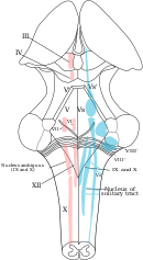
The medulla oblongata or simply medulla is a long stem-like structure which makes up the lower part of the brainstem. It is anterior and partially inferior to the cerebellum. It is a cone-shaped neuronal mass responsible for autonomic (involuntary) functions, ranging from vomiting to sneezing. The medulla contains the cardiac, respiratory, vomiting and vasomotor centers, and therefore deals with the autonomic functions of breathing, heart rate and blood pressure as well as the sleep–wake cycle.

<span class="mw-page-title-main">Pons</span> Part of the brainstem in humans and other bipeds

The pons is part of the brainstem that in humans and other mammals, lies inferior to the midbrain, superior to the medulla oblongata and anterior to the cerebellum.

<span class="mw-page-title-main">Brainstem</span> Posterior part of the brain, adjoining and structurally continuous

The brainstem is the stalk-like part of the brain that interconnects the cerebrum and diencephalon with the spinal cord. In the human brain, the brainstem is composed of the midbrain, the pons, and the medulla oblongata. The midbrain is continuous with the thalamus of the diencephalon through the tentorial notch.

<span class="mw-page-title-main">Exhalation</span> Flow of the respiratory current out of an organism

Exhalation is the flow of the breath out of an organism. In animals, it is the movement of air from the lungs out of the airways, to the external environment during breathing. This happens due to elastic properties of the lungs, as well as the internal intercostal muscles which lower the rib cage and decrease thoracic volume. As the thoracic diaphragm relaxes during exhalation it causes the tissue it has depressed to rise superiorly and put pressure on the lungs to expel the air. During forced exhalation, as when blowing out a candle, expiratory muscles including the abdominal muscles and internal intercostal muscles generate abdominal and thoracic pressure, which forces air out of the lungs.

The control of ventilation is the physiological mechanisms involved in the control of breathing, which is the movement of air into and out of the lungs. Ventilation facilitates respiration. Respiration refers to the utilization of oxygen and balancing of carbon dioxide by the body as a whole, or by individual cells in cellular respiration.

<span class="mw-page-title-main">Spirometry</span> Pulmonary function test

Spirometry is the most common of the pulmonary function tests (PFTs). It measures lung function, specifically the amount (volume) and/or speed (flow) of air that can be inhaled and exhaled. Spirometry is helpful in assessing breathing patterns that identify conditions such as asthma, pulmonary fibrosis, cystic fibrosis, and COPD. It is also helpful as part of a system of health surveillance, in which breathing patterns are measured over time.

<span class="mw-page-title-main">Reticular formation</span> Spinal trigeminal nucleus

The reticular formation is a set of interconnected nuclei that are located in the brainstem, hypothalamus, and other regions. It is not anatomically well defined, because it includes neurons located in different parts of the brain. The neurons of the reticular formation make up a complex set of networks in the core of the brainstem that extend from the upper part of the midbrain to the lower part of the medulla oblongata. The reticular formation includes ascending pathways to the cortex in the ascending reticular activating system (ARAS) and descending pathways to the spinal cord via the reticulospinal tracts.

<span class="mw-page-title-main">Pontine tegmentum</span>

The pontine tegmentum, or dorsal pons, is located within the brainstem, and is one of two parts of the pons, the other being the ventral pons or basilar part of the pons. The pontine tegmentum can be defined in contrast to the basilar pons: basilar pons contains the corticospinal tract running craniocaudally and can be considered the rostral extension of the ventral medulla oblongata; however, basilar pons is distinguished from ventral medulla oblongata in that it contains additional transverse pontine fibres that continue laterally to become the middle cerebellar peduncle. The pontine tegmentum is all the material dorsal from the basilar pons to the fourth ventricle. Along with the dorsal surface of the medulla, it forms part of the rhomboid fossa – the floor of the fourth ventricle.

The respiratory rate is the rate at which breathing occurs; it is set and controlled by the respiratory center of the brain. A person's respiratory rate is usually measured in breaths per minute.

<span class="mw-page-title-main">Tegmentum</span>

The tegmentum is a general area within the brainstem. The tegmentum is the ventral part of the midbrain and the tectum is the dorsal part of the midbrain. It is located between the ventricular system and distinctive basal or ventral structures at each level. It forms the floor of the midbrain (mesencephalon) whereas the tectum forms the ceiling. It is a multisynaptic network of neurons that is involved in many subconscious homeostatic and reflexive pathways. It is a motor center that relays inhibitory signals to the thalamus and basal nuclei preventing unwanted body movement.

<span class="mw-page-title-main">Lateral hypothalamus</span>

The lateral hypothalamus (LH), also called the lateral hypothalamic area (LHA), contains the primary orexinergic nucleus within the hypothalamus that widely projects throughout the nervous system; this system of neurons mediates an array of cognitive and physical processes, such as promoting feeding behavior and arousal, reducing pain perception, and regulating body temperature, digestive functions, and blood pressure, among many others. Clinically significant disorders that involve dysfunctions of the orexinergic projection system include narcolepsy, motility disorders or functional gastrointestinal disorders involving visceral hypersensitivity, and eating disorders.

The Hering–Breuer inflation reflex, named for Josef Breuer and Ewald Hering, is a reflex triggered to prevent the over-inflation of the lung. Pulmonary stretch receptors present on the wall of bronchi and bronchioles of the airways respond to excessive stretching of the lung during large inspirations.

The preBötzinger complex, often abbreviated as preBötC, is a functionally and anatomically specialized site in the ventral-lateral region of the lower medulla oblongata. The preBötC is part of the ventral respiratory group of respiratory related interneurons. Its foremost function is to generate the inspiratory breathing rhythm in mammals. In addition, the preBötC is widely and paucisynaptically connected to higher brain centers that regulate arousal and excitability more generally such that respiratory brain function is intimately connected with many other rhythmic and cognitive functions of the brain and central nervous system. Further, the preBötC receives mechanical sensory information from the airways that encode lung volume as well as pH, oxygen, and carbon dioxide content of circulating blood and the cerebrospinal fluid.

In mammals, the Bötzinger complex (BötC) is a group of neurons located in the rostral ventrolateral medulla, and ventral respiratory column. In the medulla, this group is located caudally to the facial nucleus and ventral to nucleus ambiguus.

<span class="mw-page-title-main">Breathing</span> Process of moving air in and out of the lungs

Breathing is the rhythmical process of moving air into and out of the lungs to facilitate gas exchange with the internal environment, mostly to flush out carbon dioxide and bring in oxygen.

Serotonergic cell groups refer to collections of neurons in the central nervous system that have been demonstrated by histochemical fluorescence to contain the neurotransmitter serotonin (5-hydroxytryptamine). Since they are for the most part localized to classical brainstem nuclei, particularly the raphe nuclei, they are more often referred to by the names of those nuclei than by the B1-9 nomenclature. These cells appear to be common across most mammals and have two main regions in which they develop; one forms in the mesencephlon and the rostral pons and the other in the medulla oblongata and the caudal pons.

Many terms are used in mechanical ventilation, some are specific to brand, model, trademark and mode of mechanical ventilation. There is a standardized nomenclature of mechanical ventilation that is specific about nomenclature related to modes, but not settings and variables.

The parabrachial nuclei, also known as the parabrachial complex, are a group of nuclei in the dorsolateral pons that surrounds the superior cerebellar peduncle as it enters the brainstem from the cerebellum. They are named from the Latin term for the superior cerebellar peduncle, the brachium conjunctivum. In the human brain, the expansion of the superior cerebellar peduncle expands the parabrachial nuclei, which form a thin strip of grey matter over most of the peduncle. The parabrachial nuclei are typically divided along the lines suggested by Baxter and Olszewski in humans, into a medial parabrachial nucleus and lateral parabrachial nucleus. These have in turn been subdivided into a dozen subnuclei: the superior, dorsal, ventral, internal, external and extreme lateral subnuclei; the lateral crescent and subparabrachial nucleus along the ventrolateral margin of the lateral parabrachial complex; and the medial and external medial subnuclei

References

  1. 1 2 3 Tortora, G; Derrickson, B (2011). Principles of anatomy & physiology (13th. ed.). Wiley. pp. 906–909. ISBN   9780470646083.
  2. Pocock, Gillian; Richards, Christopher D. (2006). Human physiology : the basis of medicine (3rd ed.). Oxford: Oxford University Press. p. 332. ISBN   978-0-19-856878-0.
  3. Saladin, Kenneth (2012). Anatomy Physiology The Unity of Form and Function. McGraw-Hill Education. pp. 868–871. ISBN   9780073378251.
  4. 1 2 3 4 Hall, John (2011). Guyton and Hall textbook of medical physiology (12th ed.). Philadelphia, Pa.: Saunders/Elsevier. pp. 505–510. ISBN   978-1-4160-4574-8.
  5. 1 2 Saladin, K (2011). Human anatomy (3rd ed.). McGraw-Hill. pp. 646–647. ISBN   9780071222075.
  6. Betts, J. Gordon; Young, Kelly A.; Wise, James A.; Johnson, Eddie; Poe, Brandon; Kruse, Dean H.; Korol, Oksana; Johnson, Jody E.; Womble, Mark (2022-04-20). "22.3 The Process of Breathing - Anatomy and Physiology 2e | OpenStax". openstax.org. Retrieved 2024-03-20.
  7. Koeppen, Bruce M.; Stanton, Bruce A. (18 January 2017). Berne and Levy Physiology E-Book. Elsevier Health Sciences. ISBN   9780323523400.
  8. Song, G; Poon, CS (15 November 2004). "Functional and structural models of pontine modulation of mechanoreceptor and chemoreceptor reflexes". Respiratory Physiology & Neurobiology. 143 (2–3): 281–92. doi:10.1016/j.resp.2004.05.009. PMID   15519561. S2CID   38265906.
  9. Song, Gang; Yu, Yunguo; Poon, Chi-Sang (2006). "Cytoarchitecture of Pneumotaxic Integration of Respiratory and Nonrespiratory Information in the Rat". Journal of Neuroscience. 26 (1): 300–10. doi: 10.1523/JNEUROSCI.3029-05.2006 . PMC   6674322 . PMID   16399700.
  10. Dutschmann, M; Dick, TE (October 2012). "Pontine mechanisms of respiratory control". Comprehensive Physiology. 2 (4): 2443–69. doi:10.1002/cphy.c100015. PMC   4422496 . PMID   23720253.
  11. Dutschmann, Mathias (2011). Comprehensive Physiology. [Bethesda, Md.]: John Wiley and Sons. ISBN   978-0-470-65071-4.
  12. Gautier, H; Bertrand, F (1975). "Respiratory effects of pneumatic center lesions and subsequent vagotomy in chronic cats". Respiration Physiology. 23 (1): 71–85. doi:10.1016/0034-5687(75)90073-0. PMID   1129551.
  13. Monteau, R.; Errchidi, S.; Gauthier, P.; Hilaire, G.; Rega, P. (1989). "Pneumotaxic centre and apneustic breathing: Interspecies differences between rat and cat". Neuroscience Letters. 99 (3): 311–6. doi:10.1016/0304-3940(89)90465-5. PMID   2725956. S2CID   42790256.
  14. Mörschel, M; Dutschmann, M (12 September 2009). "Pontine respiratory activity involved in inspiratory/expiratory phase transition". Philosophical Transactions of the Royal Society of London. Series B, Biological Sciences. 364 (1529): 2517–26. doi:10.1098/rstb.2009.0074. PMC   2865127 . PMID   19651653.
  15. Ramirez, JM; Dashevskiy, T; Marlin, IA; Baertsch, N (December 2016). "Microcircuits in respiratory rhythm generation: commonalities with other rhythm generating networks and evolutionary perspectives". Current Opinion in Neurobiology. 41: 53–61. doi:10.1016/j.conb.2016.08.003. PMC   5495096 . PMID   27589601.
  16. 1 2 Westfall DP, Westfall TC (2010). "Miscellaneous Sympathomimetic Agonists". In Brunton LL, Chabner BA, Knollmann BC (eds.). Goodman & Gilman's Pharmacological Basis of Therapeutics (12th ed.). New York, USA: McGraw-Hill. ISBN   9780071624428.

Further reading