Bradycardia

Last updated

Bradycardia
Other namesBradyarrhythmia, brachycardia
Sinus bradycardia lead2.svg
Sinus bradycardia seen in lead II with a heart rate of about 50BPM
Pronunciation
Specialty Cardiology
Diagnostic method electrocardiogram
Frequency15% (males), 7% (females)

Bradycardia, also called bradyarrhythmia, is a resting heart rate under 60 beats per minute (BPM). [1] While bradycardia can result from various pathologic processes, it is commonly a physiologic response to cardiovascular conditioning or due to asymptomatic type 1 atrioventricular block. Resting heart rates of less than 50 BPM are often normal during sleep in young and healthy adults and athletes. [2] In large population studies of adults without underlying heart disease, resting heart rates of 45-50 BPM appear to be the lower limits of normal, dependent on age and sex. [3] [4] Bradycardia is most likely to be discovered in the elderly, as age and underlying cardiac disease progression contribute to its development. [5]

Contents

Bradycardia may be associated with symptoms of fatigue, dyspnea, dizziness, confusion, and frank syncope due to reduced forward blood flow to the brain, lungs, and skeletal muscle. [6] The types of symptoms often depend on the etiology of the slow heart rate, classified by the anatomic location of a dysfunction within the cardiac conduction system. [2] Generally, these classifications involve the broad categories of sinus node dysfunction (SND), atrioventricular block, and other conduction tissue diseases. [7] However, bradycardia can also result without dysfunction of the native conduction system, arising secondary to medications including beta blockers, calcium channel blockers, antiarrythmics, and other cholinergic drugs. Excess vagus nerve activity or carotid sinus hypersensitivity are neurological causes of transient symptomatic bradycardia. Hypothyroidism and metabolic derangements are other common extrinsic causes of bradycardia. [6]

The management of bradycardia is generally reserved for patients with symptoms, regardless of minimum heart rate during sleep or the presence of concomitant heart rhythm abnormalities (See: Sinus pause), which are common with this condition. [6] Untreated SND has been shown to increase the future risk of heart failure and syncope, sometimes warranting definitive treatment with an implanted pacemaker. [8] [7] In atrioventricular causes of bradycardia, permanent pacemaker implantation is often required when no reversible causes of disease are found. [6] [2] In both SND and atrioventricular blocks, there is little role for medical therapy unless a patient is hemodynamically unstable, which may require the use of medications such as atropine and isoproterenol and interventions such as transcutenous pacing until such time that an appropriate workup can be undertaken and long term treatment selected. [2] While asymptomatic bradycardias rarely require treatment, consultation with a physician is recommended, especially in the elderly.[ citation needed ]

The term "relative bradycardia" can refer to a heart rate lower than expected in a particular disease state, often a febrile illness. [9] "Chronotropic incompetence" (CI) refers to an inadequate rise in heart rate during periods of increased demand, often due to exercise, and is an important sign of SND and an indication for pacemaker implantation. [7] [2]

The word "bradycardia" is from the Greek βραδύς bradys "slow", and καρδία kardia "heart". [10]

Normal cardiac conduction

The heart is a specialized muscle containing repeating units of cardiomyocytes, or heart muscle cells. Like most cells, cardiomyocytes maintain a highly regulated negative voltage at rest and are capable of propagating action potentials, much like neurons. [11] While at rest, the negative cellular voltage of a cardiomyocyte can be raised above a certain threshold (so-called depolarization) by an incoming action potential, causing the myocyte to contract. When these contractions occur in a coordinated fashion, the atria and ventricles of the heart will pump, delivering blood to the rest of the body. [11]

Normally, the origination of the action potential causing cardiomyocyte contraction originates from the sinoatrial node (SA node). This collection of specialized conduction tissue is located in the right atrium, near the entrance of the superior vena cava. [12] The SA node contains pacemaker cells that demonstrate "automaticity" and can generate impulses that travel through the heart and create a steady heartbeat. [12]

At the beginning of the cardiac cycle, the SA node generates an electrical action potential that spreads across the right and left atria, causing the atrial contraction of the cardiac cycle. [12] This electrical impulse carries on to the atrioventricular node (AV node), another specialized grouping of cells located in the base of the right atrium, which is the only anatomically normal electrical connection between the atria and ventricles. Impulses coursing through the AV node are slowed before carrying on to the ventricles, [13] allowing for appropriate filling of the ventricles before contraction. The SA and AV nodes are both closely regulated by the autonomic nervous system's fibres, allowing for adjustment of cardiac output by the central nervous system in times of increased metabolic demand.

Following slowed conduction through the atrioventricular node, the action potential produced initially at the SA node now flows through the His-Purkinje system. The bundle of His originates in the AV node and rapidly splits into a left and right branch, each destined for a different ventricle. Finally, these bundle branches terminate in the small Purkinje fibers that innervate myocardial tissue. The His-Purkinje system conducts action potentials much faster than can be propagated between myocardial cells, allowing the entire ventricular myocardium to contract in less time, improving pump function. [12]

Classification

Illustration comparing the ECGs of a healthy person (top) and a person with bradycardia (bottom): The points on the heart where the ECG signals are measured are also shown. Blausen 0099 bradycardia.png
Illustration comparing the ECGs of a healthy person (top) and a person with bradycardia (bottom): The points on the heart where the ECG signals are measured are also shown.

Most pathological causes of bradycardia result from damage to this normal cardiac conduction system at various levels: the sinoatrial node, the atrioventricular node, or damage to conduction tissue between or after these nodes.

Sinus node

Bradycardia caused by the alterations of sinus node activity is divided into three types.

Sinus bradycardia

Sinus bradycardia is a sinus rhythm of less than 50 BPM. [14] Cardiac action potentials are generated from the SA node and propagated through an otherwise normal conduction system, but they occur at a slow rate. It is a common condition found in both healthy individuals and those considered well-conditioned athletes. [1] Studies have found that 50–85% of conditioned athletes have benign sinus bradycardia, as compared to 23% of the general population studied. [15] The heart muscle of athletes has a higher stroke volume, requiring fewer contractions to circulate the same volume of blood. [16] Asymptomatic sinus bradycardia decreases in prevalence with age.

Sinus arrhythmia

Sinus arrhythmias are heart rhythm abnormalities characterized by variations in the cardiac cycle length over 120 milliseconds (longest cycle - shortest cycle). [2] These are the most common type of arrhythmia in the general population and usually have no significant consequences. They typically occur in the young, athletes or after administration of medications such as morphine. The types of sinus arrhythmia are separated into the respiratory and non-respiratory categories. [2]

Respiratory sinus arrhythmia

Respiratory sinus arrhythmia refers to the physiologically normal variation in heart rate due to breathing. During inspiration, vagus nerve activity decreases, reducing parasympathetic innervation of the sinoatrial node and causing an increase in heart rate. During expiration, heart rates fall due to the converse occurring. [2]

Non-respiratory sinus arrhythmia

Non-respiratory causes of sinus arrhythmia include sinus pause, sinus arrest, and sinoatrial exit block. Sinus pause and arrest involve slowing or arresting of automatic impulse generation from the sinus node. This can lead to asystole or cardiac arrest if ventricular escape rhythms do not create backup sources of cardiac action potentials. [2]

Sinoatrial exit block is a similar non-respiratory phenomenon of temporarily lost sinoatrial impulses. However, in contrast to a sinus pause, the action potential is still generated at the SA node but is either unable to leave or delayed from leaving the node, preventing or delaying atrial depolarization and subsequent ventricular systole. Therefore, the length of the pause in heartbeats is usually a multiple of the P-P interval, as seen on electrocardiography. Like a sinus pause, a sinoatrial exit block can be symptomatic, especially with prolonged pause length. [2]

Sinus node dysfunction

A syndrome of intrinsic disease of the sinus node, referred to as sick sinus syndrome or sinus node dysfunction, covers conditions that include symptomatic sinus bradycardia or persistent chronotropic incompetence, sinoatrial block, sinus arrest, and tachycardia-bradycardia syndrome. [2] These conditions can be caused by damage to the native sinus node itself and are frequently accompanied by damaged AV node conduction and reduced backup pacemaker activity. [17] The condition can also be caused by dysfunction of the autonomic nervous system that regulates the node and is commonly exacerbated by medications. [2]

Atrioventricular node

Bradycardia can also result from the inhibition of the flow of action potentials through the atrioventricular (AV) node. While this can be normal in young patients due to excessive vagus nerve tone, symptomatic bradycardia due to AV node dysfunction in older people is commonly due to structural heart disease, myocardial ischemia, or age-related fibrosis. [18]

Characteristic ECGs of atrioventricular blocks, organized by degree. Heart block.png
Characteristic ECGs of atrioventricular blocks, organized by degree.

Atrioventricular block

Atrioventricular blocks are divided into three categories, ranked by severity. AV block is diagnosed via surface ECG, which is usually sufficient to locate the causal lesion of the block without the need for an invasive electrophysiology study. [2]

In 1st degree AV block, electrical impulses originating in the SA node (or other ectopic focus above the ventricles) are conducted with significant delay through the AV node. This condition is diagnosed via ECG, with PR intervals in excess of 200 milliseconds. [2] The PR interval represents the length of time between the start of atrial depolarization and the start of ventricular depolarization, representing the flow of electrical impulses between the SA and AV nodes. Despite the term "block," no impulses are fully lost in this conduction but are merely delayed. The location of the causal lesion can be anywhere between the AV node and the His-Purkinje system but is most commonly found in the AV node itself. [19] Generally, isolated PR prolongation in 1st degree AV block is not associated with increased mortality or hospitalization. [20]

2nd degree AV block is characterized by intermittently lost conduction of impulses between the SA node and the ventricles. 2nd degree block is classified into two types. Mobitz type 1 block, otherwise known by the eponym Wenckebach, classically demonstrates grouped patterns of heartbeats on ECG. Throughout the group, the PR interval gradually lengthens until a dropped conduction occurs, resulting in no QRS complex seen on surface ECG following the last P wave. After a delay, the grouping repeats, with the PR interval shortening again to baseline. [21] Type 1 2nd degree AV block due to disease in the AV node (as opposed to in the His-purkinje system) rarely needs intervention with pacemaker implantation. [21]

2nd degree, Mobitz type 2 AV block is another phenomenon of intermittently dropped QRS complexes after characteristic groupings of beats seen on surface ECG. The PR and RR intervals are consistent in this condition, followed by a sudden AV block and dropped QRS complex. [21] Because type 2 blocks are typically due to lesions below the AV node, the ability for ventricular escape rhythms to maintain cardiac output is compromised. Permanent pacemaker implantation is often required. [18]

Junctional rhythms

An AV-junctional rhythm, or atrioventricular nodal bradycardia, is usually caused by the absence of the electrical impulse from the sinus node. This usually appears on an electrocardiogram with a normal QRS complex accompanied by an inverted P wave either before, during, or after the QRS complex. [16]

An AV-junctional escape beat is a delayed heartbeat originating from an ectopic focus somewhere in the AV junction. It occurs when the rate of depolarization of the SA node falls below the rate of the AV node. [16] This dysrhythmia may also occur when the electrical impulses from the SA node fail to reach the AV node because of SA or AV block. [22] This is a protective mechanism for the heart to compensate for an SA node that is no longer handling the pacemaking activity and is one of a series of backup sites that can take over pacemaker function when the SA node fails to do so. This would present with a longer PR interval. An AV-junctional escape complex is a normal response that may result from excessive vagal tone on the SA node. Pathological causes include sinus bradycardia, sinus arrest, sinus exit block, or AV block. [16]

Ventricular

Idioventricular rhythm, also known as atrioventricular bradycardia or ventricular escape rhythm, is a heart rate of less than 50 BPM. This is a safety mechanism when a lack of electrical impulse or stimuli from the atrium occurs. [16] Impulses originating within or below the bundle of His in the AV node will produce a wide QRS complex with heart rates between 20 and 40 BPM. Those above the bundle of His, also known as junctional, will typically range between 40 and 60 BPM with a narrow QRS complex. [23] [24] In a third-degree heart block, about 61% take place at the bundle branch-Purkinje system, 21% at the AV node, and 15% at the bundle of His. [24] AV block may be ruled out with an ECG indicating "a 1:1 relationship between P waves and QRS complexes." [23] Ventricular bradycardias occurs with sinus bradycardia, sinus arrest, and AV block. Treatment often consists of the administration of atropine and cardiac pacing. [16]

Infantile

For infants, bradycardia is defined as a heart rate less than 100 BPM (normal is around 120–160 BPM). Premature babies are more likely than full-term babies to have apnea and bradycardia spells; their cause is not clearly understood. The spells may be related to centers inside the brain that regulate breathing which may not be fully developed. Touching the baby gently or rocking the incubator slightly will almost always get the baby to start breathing again, which increases the heart rate. The neonatal intensive-care unit standard practice is to electronically monitor the heart and lungs.[ citation needed ]

Causes

Bradycardia arrhythmia may have many causes, both cardiac and non-cardiac.

Non-cardiac causes are usually secondary and can involve recreational drug use or abuse, metabolic or endocrine issues, especially hypothyroidism, an electrolyte imbalance, neurological factors, autonomic reflexes, situational factors, such as prolonged bed rest, and autoimmunity. [25] At rest, although tachycardia is more commonly seen in fatty acid oxidation disorders, acute bradycardia can occur more rarely. [26]

Cardiac causes include acute or chronic ischemic heart disease, vascular heart disease, valvular heart disease, or degenerative primary electrical disease. Ultimately, the causes act by three mechanisms: depressed automaticity of the heart, conduction block, or escape pacemakers and rhythms. [27]

In general, two types of problems result in bradycardias: disorders of the SA node and disorders of the AV node. [28]

With SA node dysfunction (sometimes called sick sinus syndrome), there may be disordered automaticity or impaired conduction of the impulse from the SA node into the surrounding atrial tissue (an "exit block"). Second-degree sinoatrial blocks can be detected only by use of a 12-lead ECG. [29] It is difficult and sometimes impossible to assign a mechanism to any particular bradycardia, but the underlying mechanism is not clinically relevant to treatment, which is the same in both cases of sick sinus syndrome: a permanent pacemaker. [27]

AV conduction disturbances (AV block; primary AV block, secondary type I AV block, secondary type II AV block, tertiary AV block) may result from impaired conduction in the AV node or anywhere below it, such as in the bundle of His. The clinical relevance pertaining to AV blocks is greater than that of SA blocks. [29]

Beta blocker medicines also can slow the heart rate and decrease how forcefully the heart contracts. Beta-blockers may slow the heart rate to a dangerous level if prescribed with calcium channel blocker-type medications. [30] Bradycardia is also part of the mammalian diving reflex. [31]

Diagnosis

A diagnosis of bradycardia in adults is based on a heart rate of less than 60 BPM, [1] although some studies use a heart rate of less than 50 BPM. [32] This is usually determined either by palpation or ECG. [1] If symptoms occur, a determining electrolytes may help determine the underlying cause. [30]

Management

The treatment of bradycardia depends on whether the person is stable or unstable. [1] [32]

Stable

Emergency treatment is not needed if the person is asymptomatic or minimally symptomatic. [32]

Unstable

If a person is unstable, the initial recommended treatment is intravenous atropine. [32] Doses less than 0.5 mg should not be used, which may further decrease the rate. [32] If this is ineffective, intravenous inotrope infusion (dopamine, epinephrine) or transcutaneous pacing should be used. [32] Transvenous pacing may be required if the cause of the bradycardia is not rapidly reversible. [32]

In children, giving oxygen, supporting their breathing, and chest compressions are recommended. [33] [34]

Epidemiology

In clinical practice, elderly people over age 65 and young athletes of both sexes may have sinus bradycardia. [1] The US Centers for Disease Control and Prevention reported in 2011 that 15.2% of adult males and 6.9% of adult females had clinically defined bradycardia (a resting pulse rate below 60 BPM). [35]

Society and culture

Records

See also

Related Research Articles

<span class="mw-page-title-main">Electrocardiography</span> Examination of the hearts electrical activity

Electrocardiography is the process of producing an electrocardiogram, a recording of the heart's electrical activity through repeated cardiac cycles. It is an electrogram of the heart which is a graph of voltage versus time of the electrical activity of the heart using electrodes placed on the skin. These electrodes detect the small electrical changes that are a consequence of cardiac muscle depolarization followed by repolarization during each cardiac cycle (heartbeat). Changes in the normal ECG pattern occur in numerous cardiac abnormalities, including:

<span class="mw-page-title-main">Tachycardia</span> Heart rate exceeding normal resting rate

Tachycardia, also called tachyarrhythmia, is a heart rate that exceeds the normal resting rate. In general, a resting heart rate over 100 beats per minute is accepted as tachycardia in adults. Heart rates above the resting rate may be normal or abnormal.

<span class="mw-page-title-main">Sinus node dysfunction</span> Medical condition

Sinus node dysfunction (SND), also known as sick sinus syndrome (SSS), is a group of abnormal heart rhythms (arrhythmias) usually caused by a malfunction of the sinus node, the heart's primary pacemaker. Tachycardia-bradycardia syndrome is a variant of sick sinus syndrome in which the arrhythmia alternates between fast and slow heart rates.

<span class="mw-page-title-main">Third-degree atrioventricular block</span> Medical condition

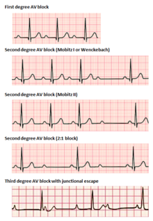
Third-degree atrioventricular block is a medical condition in which the electrical impulse generated in the sinoatrial node in the atrium of the heart can not propagate to the ventricles.

<span class="mw-page-title-main">Cardiac conduction system</span> Aspect of heart function

The cardiac conduction system transmits the signals generated by the sinoatrial node – the heart's pacemaker, to cause the heart muscle to contract, and pump blood through the body's circulatory system. The pacemaking signal travels through the right atrium to the atrioventricular node, along the bundle of His, and through the bundle branches to Purkinje fibers in the walls of the ventricles. The Purkinje fibers transmit the signals more rapidly to stimulate contraction of the ventricles.

<span class="mw-page-title-main">Second-degree atrioventricular block</span> Medical condition

Second-degree atrioventricular block is a disease of the electrical conduction system of the heart. It is a conduction block between the atria and ventricles. The presence of second-degree AV block is diagnosed when one or more of the atrial impulses fail to conduct to the ventricles due to impaired conduction. It is classified as a block of the AV node, falling between first-degree and third degree blocks.

<span class="mw-page-title-main">Supraventricular tachycardia</span> Abnormally fast heart rhythm

Supraventricular tachycardia (SVT) is an umbrella term for fast heart rhythms arising from the upper part of the heart. This is in contrast to the other group of fast heart rhythms – ventricular tachycardia, which start within the lower chambers of the heart. There are four main types of SVT: atrial fibrillation, atrial flutter, paroxysmal supraventricular tachycardia (PSVT), and Wolff–Parkinson–White syndrome. The symptoms of SVT include palpitations, feeling of faintness, sweating, shortness of breath, and/or chest pain.

A junctional escape beat is a delayed heartbeat originating not from the atrium but from an ectopic focus somewhere in the atrioventricular junction. It occurs when the rate of depolarization of the sinoatrial node falls below the rate of the atrioventricular node. This dysrhythmia also may occur when the electrical impulses from the SA node fail to reach the AV node because of SA or AV block. It is a protective mechanism for the heart, to compensate for the SA node no longer handling the pacemaking activity, and is one of a series of backup sites that can take over pacemaker function when the SA node fails to do so. It can also occur following a premature ventricular contraction or blocked premature atrial contraction.

<span class="mw-page-title-main">Atrioventricular block</span> Medical condition

Atrioventricular block is a type of heart block that occurs when the electrical signal traveling from the atria, or the upper chambers of the heart, to ventricles, or the lower chambers of the heart, is impaired. Normally, the sinoatrial node produces an electrical signal to control the heart rate. The signal travels from the SA node to the ventricles through the atrioventricular node. In an AV block, this electrical signal is either delayed or completely blocked. When the signal is completely blocked, the ventricles produce their own electrical signal to control the heart rate. The heart rate produced by the ventricles is much slower than that produced by the SA node.

Premature atrial contraction (PAC), also known as atrial premature complexes (APC) or atrial premature beats (APB), are a common cardiac dysrhythmia characterized by premature heartbeats originating in the atria. While the sinoatrial node typically regulates the heartbeat during normal sinus rhythm, PACs occur when another region of the atria depolarizes before the sinoatrial node and thus triggers a premature heartbeat, in contrast to escape beats, in which the normal sinoatrial node fails, leaving a non-nodal pacemaker to initiate a late beat.

<span class="mw-page-title-main">Ventricular escape beat</span>

In cardiology, a ventricular escape beat is a self-generated electrical discharge initiated by, and causing contraction of the ventricles of the heart; normally the heart rhythm is begun in the atria of the heart and is subsequently transmitted to the ventricles. The ventricular escape beat follows a long pause in ventricular rhythm and acts to prevent cardiac arrest. It indicates a failure of the electrical conduction system of the heart to stimulate the ventricles.

<span class="mw-page-title-main">Multifocal atrial tachycardia</span> Fast heart rhythm associated with exacerbations of COPD

Multifocal atrial tachycardia (MAT) is an abnormal heart rhythm, specifically a type of supraventricular tachycardia, that is particularly common in older people and is associated with exacerbations of chronic obstructive pulmonary disease (COPD). Normally, the heart rate is controlled by a cluster of cells called the sinoatrial node. When a number of different clusters of cells outside the SA node take over control of the heart rate, and the rate exceeds 100 beats per minute, this is called multifocal atrial tachycardia.

Sinoatrial arrest is a medical condition wherein the sinoatrial node of the heart transiently ceases to generate the electrical impulses that normally stimulate the myocardial tissues to contract and thus the heart to beat. It is defined as lasting from 2.0 seconds to several minutes. Since the heart contains multiple pacemakers, this interruption of the cardiac cycle generally lasts only a few seconds before another part of the heart, such as the atrio-ventricular junction or the ventricles, begins pacing and restores the heart action. This condition can be detected on an electrocardiogram (ECG) as a brief period of irregular length with no electrical activity before either the sinoatrial node resumes normal pacing, or another pacemaker begins pacing. If a pacemaker other than the sinoatrial node is pacing the heart, this condition is known as an escape rhythm. If no other pacemaker begins pacing during an episode of sinus arrest it becomes a cardiac arrest. This condition is sometimes confused with sinoatrial block, a condition in which the pacing impulse is generated, but fails to conduct through the myocardium. Differential diagnosis of the two conditions is possible by examining the exact length of the interruption of cardiac activity. If the next available pacemaker takes over, it is in the following order:

<span class="mw-page-title-main">Accelerated idioventricular rhythm</span> Medical condition

Accelerated idioventricular rhythm is a ventricular rhythm with a rate of between 40 and 120 beats per minute. Idioventricular means “relating to or affecting the cardiac ventricle alone” and refers to any ectopic ventricular arrhythmia. Accelerated idioventricular arrhythmias are distinguished from ventricular rhythms with rates less than 40 and those faster than 120. Though some other references limit to between 60 and 100 beats per minute. It is also referred to as AIVR and "slow ventricular tachycardia."

<span class="mw-page-title-main">Junctional rhythm</span> Medical condition

Junctional rhythm also called nodal rhythm describes an abnormal heart rhythm resulting from impulses coming from a locus of tissue in the area of the atrioventricular node, the "junction" between atria and ventricles.

<span class="mw-page-title-main">Sinoatrial block</span> Medical condition

A sinoatrial block is a disorder in the normal rhythm of the heart, known as a heart block, that is initiated in the sinoatrial node. The initial action impulse in a heart is usually formed in the sinoatrial node and carried through the atria, down the internodal atrial pathways to the atrioventricular node (AV) node. In normal conduction, the impulse would travel across the bundle of His, down the bundle branches, and into the Purkinje fibers. This would depolarize the ventricles and cause them to contract.

<span class="mw-page-title-main">Ectopic pacemaker</span> Cardiac condition

An ectopic pacemaker, also known as ectopic focus or ectopic foci, is an excitable group of cells that causes a premature heart beat outside the normally functioning SA node of the heart. It is thus a cardiac pacemaker that is ectopic, producing an ectopic beat. Acute occurrence is usually non-life-threatening, but chronic occurrence can progress into tachycardia, bradycardia or ventricular fibrillation. In a normal heart beat rhythm, the SA node usually suppresses the ectopic pacemaker activity due to the higher impulse rate of the SA node. However, in the instance of either a malfunctioning SA node or an ectopic focus bearing an intrinsic rate superior to SA node rate, ectopic pacemaker activity may take over the natural heart rhythm. This phenomenon is called an escape rhythm, the lower rhythm having escaped from the dominance of the upper rhythm. As a rule, premature ectopic beats indicate increased myocyte or conducting tissue excitability, whereas late ectopic beats indicate proximal pacemaker or conduction failure with an escape 'ectopic' beat.

<span class="mw-page-title-main">Junctional ectopic tachycardia</span> Medical condition

Junctional ectopic tachycardia (JET) is a rare syndrome of the heart that manifests in patients recovering from heart surgery. It is characterized by cardiac arrhythmia, or irregular beating of the heart, caused by abnormal conduction from or through the atrioventricular node. In newborns and infants up to 6 weeks old, the disease may also be referred to as His bundle tachycardia or congenital JET.

<span class="mw-page-title-main">Arrhythmia</span> Group of medical conditions characterized by irregular heartbeat

Arrhythmias, also known as cardiac arrhythmias, heart arrhythmias, or dysrhythmias, are irregularities in the heartbeat, including when it is too fast or too slow. A resting heart rate that is too fast – above 100 beats per minute in adults – is called tachycardia, and a resting heart rate that is too slow – below 60 beats per minute – is called bradycardia. Some types of arrhythmias have no symptoms. Symptoms, when present, may include palpitations or feeling a pause between heartbeats. In more serious cases, there may be lightheadedness, passing out, shortness of breath, chest pain, or decreased level of consciousness. While most cases of arrhythmia are not serious, some predispose a person to complications such as stroke or heart failure. Others may result in sudden death.

An idioventricular rhythm is a cardiac rhythm characterized by a rate of <50 beats per minute (bpm), absence of P waves and widening of the QRS complex. In cases where the heart rate is between 50 and 110 bpm, it is known as accelerated idioventricular rhythm and ventricular tachycardia if the rate exceeds 120 bpm. Causes of idioventricular rhythms are varied and can include drugs or a heart defect at birth. It is typically benign and not life-threatening.

References

  1. 1 2 3 4 5 6 Hafeez Y, Grossman SA (9 August 2021). "Sinus bradycardia". StatPearls [Internet]. Treasure Island (FL): StatPearls Publishing. PMID   29630253 . Retrieved 16 January 2022.
  2. 1 2 3 4 5 6 7 8 9 10 11 12 13 14 Patterson KK, Olgin JE (2022). "Bradyarrhythmias and Atrioventricular Block". Braunwald's Heart Disease: A Textbook of Cardiovascular Medicine (12th ed.). Philadelphia, PA: Elsevier. pp. 1312–1320. ISBN   978-0-323-82467-5.
  3. Rijnbeek PR, van Herpen G, Bots ML, Man S, Verweij N, Hofman A, et al. (2014). "Normal values of the electrocardiogram for ages 16-90 years". Journal of Electrocardiology. 47 (6): 914–921. doi:10.1016/j.jelectrocard.2014.07.022. hdl: 1887/117357 . PMID   25194872.
  4. Rijnbeek PR (2012). "Normal ECG values". Rotterdam, The Netherlands.
  5. Kusumoto FM, Schoenfeld MH, Barrett C, Edgerton JR, Ellenbogen KA, Gold MR, et al. (August 2019). "2018 ACC/AHA/HRS Guideline on the Evaluation and Management of Patients With Bradycardia and Cardiac Conduction Delay: A Report of the American College of Cardiology/American Heart Association Task Force on Clinical Practice Guidelines and the Heart Rhythm Society". Circulation. 140 (8): e382–e482. doi: 10.1161/CIR.0000000000000628 . PMID   30586772.
  6. 1 2 3 4 Sidhu S, Marine JE (July 2020). "Evaluating and managing bradycardia". Trends in Cardiovascular Medicine. 30 (5): 265–272. doi: 10.1016/j.tcm.2019.07.001 . PMID   31311698.
  7. 1 2 3 Kusumoto FM, Schoenfeld MH, Barrett C, Edgerton JR, Ellenbogen KA, Gold MR, et al. (August 2019). "2018 ACC/AHA/HRS Guideline on the Evaluation and Management of Patients With Bradycardia and Cardiac Conduction Delay: A Report of the American College of Cardiology/American Heart Association Task Force on Clinical Practice Guidelines and the Heart Rhythm Society". Circulation. 140 (8): e382–e482. doi: 10.1161/CIR.0000000000000628 . PMID   30586772.
  8. Menozzi C, Brignole M, Alboni P, Boni L, Paparella N, Gaggioli G, et al. (November 1998). "The natural course of untreated sick sinus syndrome and identification of the variables predictive of unfavorable outcome". The American Journal of Cardiology. 82 (10): 1205–1209. doi:10.1016/s0002-9149(98)00605-5. PMID   9832095.
  9. Ye F, Hatahet M, Youniss MA, Toklu HZ, Mazza JJ, Yale S (June 2018). "The Clinical Significance of Relative Bradycardia". WMJ. 117 (2): 73–78. PMID   30048576.
  10. Prutchi, David (2005). Design and Development of Medical Electronic Instrumentation. John Wiley & Sons. p. 371. ISBN   9780471681830.
  11. 1 2 Loscalzo J, Keaney JF, MacRae CA (2022). "Basic Biology of the Cardiovascular System". In Loscalzo J, Fauci AS, Kasper DL, Hauser S, Longo D, Jameson JL (eds.). Harrison's Principles of Internal Medicine (21st ed.). McGraw Hill. ISBN   978-1-264-26850-4.
  12. 1 2 3 4 Sauer WH, Koplan BA, Zei PC (2022). "Principles of Clinical Cardiac Electrophysiology". In Loscalzo J, Fauci AS, Kasper DL, Hauser S, Longo D, Jameson JL (eds.). Harrison's Principles of Internal Medicine (21st ed.). McGraw Hill. ISBN   978-1-264-26850-4.
  13. Kurian T, Ambrosi C, Hucker W, Fedorov VV, Efimov IR (June 2010). "Anatomy and electrophysiology of the human AV node". Pacing and Clinical Electrophysiology. 33 (6): 754–762. doi:10.1111/j.1540-8159.2010.02699.x. PMC   2889145 . PMID   20180918.
  14. Kusumoto FM, Schoenfeld MH, Barrett C, Edgerton JR, Ellenbogen KA, Gold MR, et al. (August 2019). "2018 ACC/AHA/HRS Guideline on the Evaluation and Management of Patients With Bradycardia and Cardiac Conduction Delay: A Report of the American College of Cardiology/American Heart Association Task Force on Clinical Practice Guidelines and the Heart Rhythm Society". Circulation. 140 (8): e382–e482. doi: 10.1161/CIR.0000000000000628 . PMID   30586772.
  15. Bryan G, Ward A, Rippe JM (April 1992). "Athletic heart syndrome". Clinics in Sports Medicine. 11 (2). Elsevier: 259–272. doi:10.1016/S0278-5919(20)30529-9. PMID   1591784.
  16. 1 2 3 4 5 6 Allan B. Wolfson, ed. (2005). Harwood-Nuss' Clinical Practice of Emergency Medicine (4th ed.). Lippincott Williams & Wilkins. p. 260. ISBN   978-0-7817-5125-4.
  17. John RM, Kumar S (May 2016). "Sinus Node and Atrial Arrhythmias". Circulation. 133 (19): 1892–1900. doi: 10.1161/CIRCULATIONAHA.116.018011 . PMID   27166347.
  18. 1 2 Sauer WH, Koplan BA (2022). "The Bradyarrhythmias: Disorders of the Atrioventricular Node". In Loscalzo J, Fauci AS, Kasper DL, Hauser S, Longo D, Jameson JL (eds.). Harrison's Principles of Internal Medicine (21st ed.). McGraw Hill. ISBN   978-1-264-26850-4.
  19. Clark BA, Prystowsky EN (December 2021). "Electrocardiography of Atrioventricular Block". Cardiac Electrophysiology Clinics. 13 (4): 599–605. doi:10.1016/j.ccep.2021.07.001. PMID   34689889. S2CID   239091592.
  20. Aro AL, Anttonen O, Kerola T, Junttila MJ, Tikkanen JT, Rissanen HA, et al. (January 2014). "Prognostic significance of prolonged PR interval in the general population". European Heart Journal. 35 (2): 123–129. doi: 10.1093/eurheartj/eht176 . PMID   23677846.
  21. 1 2 3 Clark BA, Prystowsky EN (December 2021). "Electrocardiography of Atrioventricular Block". Cardiac Electrophysiology Clinics. 13 (4): 599–605. doi:10.1016/j.ccep.2021.07.001. PMID   34689889. S2CID   239091592.
  22. "AV Junctional Rhythm Disturbances (for Professionals)". American Heart Association. 4 December 2008. Retrieved 15 December 2009.
  23. 1 2 "Arrhythmias and Conduction Disorders". The Merck Manuals: Online Medical Library. Merck Sharp and Dohme Corp. January 2008. Retrieved 16 December 2009.
  24. 1 2 Adams MG, Pelter MM (September 2003). "Ventricular escape rhythms". American Journal of Critical Care. 12 (5): 477–478. doi:10.4037/ajcc2003.12.5.477. PMID   14503433.
  25. Ye F, Hatahet M, Youniss MA, Toklu HZ, Mazza JJ, Yale S (June 2018). "The Clinical Significance of Relative Bradycardia". WMJ. 117 (2): 73–78. PMID   30048576.
  26. Bonnet D, Martin D, Villain E, Jouvet P, Rabier D, Brivet M, et al. (November 1999). "Arrhythmias and conduction defects as presenting symptoms of fatty acid oxidation disorders in children". Circulation. 100 (22): 2248–2253. doi: 10.1161/01.cir.100.22.2248 . PMID   10577999.
  27. 1 2 "What is Bradycardia?". WebMD. Retrieved 5 July 2021.
  28. "Bradyarrhythmias". The Lecturio Medical Concept Library. Retrieved 5 July 2021.
  29. 1 2 Ufberg JW, Clark JS (February 2006). "Bradydysrhythmias and atrioventricular conduction blocks". Emergency Medicine Clinics of North America. 24 (1): 1–9, v. doi:10.1016/j.emc.2005.08.006. PMID   16308110.
  30. 1 2 "What is Bradycardia?". WebMD. Retrieved 5 July 2021.
  31. Panneton WM (September 2013). "The mammalian diving response: an enigmatic reflex to preserve life?". Physiology. 28 (5): 284–297. doi:10.1152/physiol.00020.2013. PMC   3768097 . PMID   23997188.
  32. 1 2 3 4 5 6 7 Neumar RW, Otto CW, Link MS, Kronick SL, Shuster M, Callaway CW, et al. (November 2010). "Part 8: adult advanced cardiovascular life support: 2010 American Heart Association Guidelines for Cardiopulmonary Resuscitation and Emergency Cardiovascular Care". Circulation. 122 (18 Suppl 3): S729–S767. doi: 10.1161/CIRCULATIONAHA.110.970988 . PMID   20956224.
  33. de Caen AR, Berg MD, Chameides L, Gooden CK, Hickey RW, Scott HF, et al. (November 2015). "Part 12: Pediatric Advanced Life Support: 2015 American Heart Association Guidelines Update for Cardiopulmonary Resuscitation and Emergency Cardiovascular Care". Circulation. 132 (18 Suppl 2): S526–S542. doi:10.1161/cir.0000000000000266. PMC   6191296 . PMID   26473000.
  34. Atkins DL, Berger S, Duff JP, Gonzales JC, Hunt EA, Joyner BL, et al. (November 2015). "Part 11: Pediatric Basic Life Support and Cardiopulmonary Resuscitation Quality: 2015 American Heart Association Guidelines Update for Cardiopulmonary Resuscitation and Emergency Cardiovascular Care". Circulation. 132 (18 Suppl 2): S519–S525. doi: 10.1161/cir.0000000000000265 . PMID   26472999.
  35. Yechiam Ostchega, et al. (24 August 2011). "Resting Pulse Rate Reference Data for Children, Adolescents, and Adults: United States, 1999–2008" (PDF). National Health Statistics Reports. Centers for Disease Control. Archived (PDF) from the original on 10 October 2022. Retrieved 15 December 2018.
  36. "Slowest heart rate: Daniel Green breaks Guinness World Records record". World Record Academy. 29 November 2014. Retrieved 5 August 2015.
  37. "Lowest heart rate". Guinness World Records. 11 August 2005. Retrieved 5 August 2015.
  38. Lovgren S (20 August 2004). "Olympic Gold Begins With Good Genes, Experts Say". National Geographic News. Archived from the original on 20 August 2004. Retrieved 8 September 2014.