Acute pericarditis

Last updated
Acute pericarditis
PericarditisECG.JPG
An ECG showing pericarditis. Note the ST elevation in multiple leads with slight reciprocal ST depression in aVR.
Specialty Cardiology   OOjs UI icon edit-ltr-progressive.svg

Acute pericarditis is a type of pericarditis (inflammation of the sac surrounding the heart, the pericardium) usually lasting less than 6 weeks.[ citation needed ] It is the most common condition affecting the pericardium.

Contents

Signs and symptoms

Chest pain is one of the common symptoms of acute pericarditis. It is usually of sudden onset, occurring in the anterior chest and often has a sharp quality that worsens with breathing in or coughing, due to inflammation of the pleural surface at the same time. The pain may be reduced with sitting up and leaning forward while worsened with lying down, and also may radiate to the back, to one or both trapezius ridges. However, the pain can also be dull and steady, resembling the chest pain in an acute myocardial infarction. As with any chest pain, other causes must also be ruled out, such as GERD, pulmonary embolism, muscular pain, etc.

A pericardial friction rub is a very specific sign of acute pericarditis, meaning the presence of this sign invariably indicates presence of disease. However, absence of this sign does not rule out disease. This rub can be best heard by the diaphragm of the stethoscope at the left sternal border arising as a squeaky or scratching sound, resembling the sound of leather rubbing against each other. This sound should be distinguished from the sound of a murmur, which is similar but sounds more like a "swish" sound than a scratching sound. The pericardial rub is said to be generated from the friction generated by the two inflamed layers of the pericardium; however, even a large pericardial effusion does not necessarily present a rub. The rub is best heard during the maximal movement of the heart within the pericardial sac, namely, during atrial systole, ventricular systole, and the filling phase of early ventricular diastole.

Fever may be present since this is an inflammatory process.

Causes

There are several causes of acute pericarditis. [1] In developed nations, the cause of most (80–90%) cases of acute pericarditis is unknown but a viral cause is suspected in the majority of such cases. [1] The other 10–20% of acute pericarditis cases have various causes including connective tissue diseases (e.g., systemic lupus erythematosus), cancer, or involve an inflammatory reaction of the pericardium following trauma to the heart such as after a heart attack such as Dressler's syndrome. [1] Familial mediterranean fever and TNF receptor associated periodic syndrome are rare inherited autoimmune diseases capable of causing recurring episodes of acute pericarditis. [1]

Pathophysiology

Clinical presentation of diseases of pericardium may vary between: [2] [3]

Diagnosis

ECG showing slight ST elevation in many leads congruent with pericarditis Acute pericarditis.jpg
ECG showing slight ST elevation in many leads congruent with pericarditis

For acute pericarditis to formally be diagnosed, two or more of the following criteria must be present: chest pain consistent with a diagnosis of acute pericarditis (sharp chest pain worsened by breathing in or a cough), a pericardial friction rub, a pericardial effusion, and changes on electrocardiogram (ECG) consistent with acute pericarditis. [1]

A complete blood count may show an elevated white count and a serum C-reactive protein may be elevated. Acute pericarditis is associated with a modest increase in serum creatine kinase MB (CK-MB). [4] and cardiac troponin I (cTnI), [5] [6] both of which are also markers for injury to the muscular layer of the heart. Therefore, it is imperative to also rule out acute myocardial infarction in the face of these biomarkers. The elevation of these substances may occur when inflammation of the heart's muscular layer in addition to acute pericarditis. [1] Also, ST elevation on EKG (see below) is more common in those patients with a cTnI > 1.5 µg/L. [6] Coronary angiography in those patients should indicate normal vascular perfusion. Troponin levels increase in 35-50% of people with pericarditis. [7]

Electrocardiogram (ECG) changes in acute pericarditis mainly indicates inflammation of the epicardium (the layer directly surrounding the heart), since the fibrous pericardium is electrically inert. For example, in uremia, there is no inflammation in the epicardium, only fibrin deposition, and therefore the EKG in uremic pericarditis will be normal. Typical EKG changes in acute pericarditis includes [4] [8]

The two most common clinical conditions where ECG findings may mimic pericarditis are acute myocardial infarction (AMI) and generalized early repolarization. [9] As opposed to pericarditis, AMI usually causes localized convex ST-elevation usually associated with reciprocal ST-depression which may also be frequently accompanied by Q-waves, T-wave inversions (while ST is still elevated unlike pericarditis), arrhythmias and conduction abnormalities. [10] In AMI, PR-depressions are rarely present. Early repolarization usually occurs in young males (age <40 years) and ECG changes are characterized by terminal R-S slurring, temporal stability of ST-deviations and J-height/ T-amplitude ratio in V5 and V6 of <25% as opposed to pericarditis where terminal R-S slurring is very uncommon and J-height/ T-amplitude ratio is ≥ 25%. Very rarely, ECG changes in hypothermia may mimic pericarditis, however differentiation can be helpful by a detailed history and presence of an Osborne wave in hypothermia. [11]

Another important diagnostic electrocardiographic sign in acute pericarditis is the Spodick sign. [12] It signifies to the PR-depressions in a usual (but not always) association with downsloping TP segment in patients with acute pericarditis and is present in up to 80% of the patients affected with acute pericarditis. The sign is often best visualized in lead II and lateral precordial leads. In addition, Spodick's sign may also serve as an important distinguishing electrocardiographic tool between the acute pericarditis and acute coronary syndrome. The presence of a classical Spodick's sign is often a giveaway to the diagnosis.[ citation needed ]

Rarely, electrical alternans may be seen, depending on the size of the effusion.[ citation needed ]

A chest x-ray is usually normal in acute pericarditis but can reveal the presence of an enlarged heart if a pericardial effusion is present and is greater than 200 mL in volume. Conversely, patients with unexplained new onset cardiomegaly should always be worked up for acute pericarditis.[ citation needed ]

An echocardiogram is typically normal in acute pericarditis but can reveal pericardial effusion, the presence of which supports the diagnosis, although its absence does not exclude the diagnosis.[ citation needed ]

Treatment

Patients with uncomplicated acute pericarditis can generally be treated and followed up in an outpatient clinic. However, those with high risk factors for developing complications (see above) will need to be admitted to an inpatient service, most likely an ICU setting. High risk patients include the following: [13]

Pericardiocentesis is a procedure whereby the fluid in a pericardial effusion is removed through a needle. It is performed under the following conditions: [14]

NSAIDs in viral or idiopathic pericarditis. In patients with underlying causes other than viral, the specific etiology should be treated. With idiopathic or viral pericarditis, NSAID is the mainstay treatment. Goal of therapy is to reduce pain and inflammation. The course of the disease may not be affected. The preferred NSAID is ibuprofen because of rare side effects, better effect on coronary flow, and larger dose range. [14] Depending on severity, dosing is between 300 and 800 mg every 6–8 hours for days or weeks as needed. An alternative protocol is aspirin 800 mg every 6–8 hours. [13] Dose tapering of NSAIDs may be needed. In pericarditis following acute myocardial infarction, NSAIDs other than aspirin should be avoided since they can impair scar formation. As with all NSAID use, GI protection should be engaged. Failure to respond to NSAIDs within one week (indicated by persistence of fever, worsening of condition, new pericardial effusion, or continuing chest pain) likely indicates that a cause other than viral or idiopathic is in process.[ citation needed ]

Colchicine, which has been essential to treat recurrent pericarditis, has been supported for routine use in acute pericarditis by recent prospective studies. [15] Colchicine can be given 0.6 mg twice a day (0.6 mg daily for patients <70 kg) for 3 months following an acute attack. It should be considered in all patients with acute pericarditis, preferably in combination with a short-course of NSAIDs. [9] For patients with a first episode of acute idiopathic or viral pericarditis, they should be treated with an NSAID plus colchicine 1–2 mg on first day followed by 0.5 daily or twice daily for three months. [16] [17] [18] [19] [20] It should be avoided or used with caution in patients with severe chronic kidney disease, hepatobiliary dysfunction, blood dyscrasias, and gastrointestinal motility disorders. [9]

Corticosteroids are usually used in those cases that are clearly refractory to NSAIDs and colchicine and a specific cause has not been found. Systemic corticosteroids are usually reserved for those with autoimmune disease. [16]

Prognosis

One of the most feared complications of acute pericarditis is cardiac tamponade. Cardiac tamponade is accumulation of enough fluid in the pericardial space --- pericardial effusion --- to cause serious obstruction to the inflow of blood to the heart. Signs of cardiac tamponade include distended neck veins, muffled heart sounds when listening with a stethoscope, and low blood pressure (together known as Beck's triad). [1] This condition can be fatal if not immediately treated.

Another longer term complication of pericarditis, if it recurs over a longer period of time (normally more than 3 months), is progression to constrictive pericarditis. Recent studies have shown this to be an uncommon complication. [21] The definitive treatment for constrictive pericarditis is pericardial stripping, which is a surgical procedure where the entire pericardium is peeled away from the heart.[ citation needed ]

Related Research Articles

<span class="mw-page-title-main">Pericardium</span> Double-walled sac containing the heart and roots of the great vessels

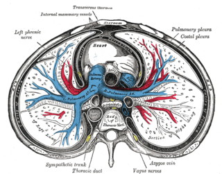
The pericardium, also called pericardial sac, is a double-walled sac containing the heart and the roots of the great vessels. It has two layers, an outer layer made of strong inelastic connective tissue, and an inner layer made of serous membrane. It encloses the pericardial cavity, which contains pericardial fluid, and defines the middle mediastinum. It separates the heart from interference of other structures, protects it against infection and blunt trauma, and lubricates the heart's movements.

<span class="mw-page-title-main">Cardiac tamponade</span> Buildup of fluid around the heart

Cardiac tamponade, also known as pericardial tamponade, is a compression of the heart due to pericardial effusion. Onset may be rapid or gradual. Symptoms typically include those of obstructive shock including shortness of breath, weakness, lightheadedness, and cough. Other symptoms may relate to the underlying cause.

<span class="mw-page-title-main">Pericarditis</span> Medical condition

Pericarditis is inflammation of the pericardium, the fibrous sac surrounding the heart. Symptoms typically include sudden onset of sharp chest pain, which may also be felt in the shoulders, neck, or back. The pain is typically less severe when sitting up and more severe when lying down or breathing deeply. Other symptoms of pericarditis can include fever, weakness, palpitations, and shortness of breath. The onset of symptoms can occasionally be gradual rather than sudden.

<span class="mw-page-title-main">Pericardiocentesis</span> Procedure where fluid is aspirated from the pericardium

Pericardiocentesis (PCC), also called pericardial tap, is a medical procedure where fluid is aspirated from the pericardium.

Dressler syndrome is a secondary form of pericarditis that occurs in the setting of injury to the heart or the pericardium. It consists of fever, pleuritic pain, pericarditis and/or pericardial effusion.

<span class="mw-page-title-main">Pericardial effusion</span> Medical condition

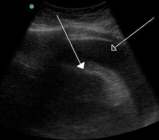
A pericardial effusion is an abnormal accumulation of fluid in the pericardial cavity. The pericardium is a two-part membrane surrounding the heart: the outer fibrous connective membrane and an inner two-layered serous membrane. The two layers of the serous membrane enclose the pericardial cavity between them. This pericardial space contains a small amount of pericardial fluid, normally 15-50 mL in volume. The pericardium, specifically the pericardial fluid provides lubrication, maintains the anatomic position of the heart in the chest, and also serves as a barrier to protect the heart from infection and inflammation in adjacent tissues and organs.

<span class="mw-page-title-main">Pericardial fluid</span>

Pericardial fluid is the serous fluid secreted by the serous layer of the pericardium into the pericardial cavity. The pericardium consists of two layers, an outer fibrous layer and the inner serous layer. This serous layer has two membranes which enclose the pericardial cavity into which is secreted the pericardial fluid. The fluid is similar to the cerebrospinal fluid of the brain which also serves to cushion and allow some movement of the organ.

<span class="mw-page-title-main">Electrical alternans</span> Medical condition

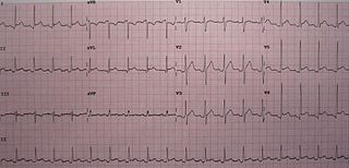
Electrical alternans is an electrocardiographic phenomenon of alternation of QRS complex amplitude or axis between beats and a possible wandering base-line. It is seen in cardiac tamponade and severe pericardial effusion and is thought to be related to changes in the ventricular electrical axis due to fluid in the pericardium, as the heart essentially wobbles in the fluid filled pericardial sac.

Pericardiectomy is the surgical removal of part or most of the pericardium. This operation is most commonly used to relieve constrictive pericarditis, or to remove a pericardium that is calcified and fibrous. It may also be used for severe or recurrent cases of pericardial effusion. Post-operative outcomes and mortality are significantly impacted by the disease it is used to treat.

<span class="mw-page-title-main">Myocardial rupture</span> Medical condition

Myocardial rupture is a laceration of the ventricles or atria of the heart, of the interatrial or interventricular septum, or of the papillary muscles. It is most commonly seen as a serious sequela of an acute myocardial infarction.

Autoimmune heart diseases are the effects of the body's own immune defense system mistaking cardiac antigens as foreign and attacking them leading to inflammation of the heart as a whole, or in parts. The commonest form of autoimmune heart disease is rheumatic heart disease or rheumatic fever.

Obstructive shock is one of the four types of shock, caused by a physical obstruction in the flow of blood. Obstruction can occur at the level of the great vessels or the heart itself. Causes include pulmonary embolism, cardiac tamponade, and tension pneumothorax. These are all life-threatening. Symptoms may include shortness of breath, weakness, or altered mental status. Low blood pressure and tachycardia are often seen in shock. Other symptoms depend on the underlying cause.

<span class="mw-page-title-main">Hemopericardium</span> Medical condition

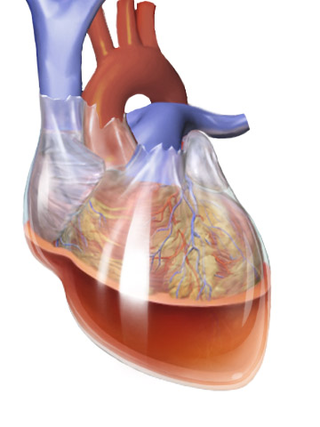
Hemopericardium refers to blood in the pericardial sac of the heart. It is clinically similar to a pericardial effusion, and, depending on the volume and rapidity with which it develops, may cause cardiac tamponade.

Tuberculous pericarditis is a form of pericarditis. It is a condition in which the pericardium surrounding the heart is infected by the bacterial species Mycobacterium tuberculosis. Tuberculous pericarditis accounts for a significant percentage of presentations of tuberculosis worldwide. The condition has four stages of disease which manifests with clinical presentations ranging from acute pericarditis to overt heart failure. Tuberculous pericarditis is an under-diagnosed condition. Diagnosis often requires a range of diagnostic tools, including pericardiocentesis, biochemical tests, and imaging. Treatment of this disease is similar to treatment of pulmonary tuberculosis. Alternative treatment options to reduce cardiac complications are also available.

<span class="mw-page-title-main">Uremic pericarditis</span> Medical condition

Uremic pericarditis is a form of pericarditis. It causes fibrinous pericarditis. The main cause of the disease is poorly understood.

Myocardial infarction complications may occur immediately following a heart attack, or may need time to develop. After an infarction, an obvious complication is a second infarction, which may occur in the domain of another atherosclerotic coronary artery, or in the same zone if there are any live cells left in the infarct.

<span class="mw-page-title-main">Postpericardiotomy syndrome</span> Medical condition

Postpericardiotomy syndrome (PPS) is a medical syndrome referring to an immune phenomenon that occurs days to months after surgical incision of the pericardium. PPS can also be caused after a trauma, a puncture of the cardiac or pleural structures, after percutaneous coronary intervention, or due to pacemaker or pacemaker wire placement.

<span class="mw-page-title-main">Chest pain in children</span> Medical condition

Chest pain in children is the pain felt in the chest by infants, children and adolescents. In most cases the pain is not associated with the heart. It is primarily identified by the observance or report of pain by the infant, child or adolescent by reports of distress by parents or caregivers. Chest pain is not uncommon in children. Many children are seen in ambulatory clinics, emergency departments and hospitals and cardiology clinics. Most often there is a benign cause for the pain for most children. Some have conditions that are serious and possibly life-threatening. Chest pain in pediatric patients requires careful physical examination and a detailed history that would indicate the possibility of a serious cause. Studies of pediatric chest pain are sparse. It has been difficult to create evidence-based guidelines for evaluation.

<span class="mw-page-title-main">Purulent pericarditis</span> Inflammation of the sac surrounding the heart due to bacterial infection.

Purulent pericarditis refers to localized inflammation in the setting of infection of the pericardial sac surrounding the heart. In contrast to other causes of pericarditis which may have a viral etiology, purulent pericarditis refers specifically to bacterial or fungal infection of the pericardial sac. Clinical etiologies of purulent pericarditis may include recent surgery, adjacent infection, trauma, or even primary infection. The onset of purulent pericarditis is usually acute, with most individuals presenting to a medical facility approximately 3 days following the onset of symptoms.

References

  1. 1 2 3 4 5 6 7 LeWinter MM (December 2014). "Clinical practice. Acute pericarditis". New England Journal of Medicine. 371 (25): 2410–6. doi:10.1056/NEJMcp1404070. PMID   25517707.
  2. Imazio, M (May 2012). "Contemporary management of pericardial diseases". Current Opinion in Cardiology. 27 (3): 308–17. doi:10.1097/HCO.0b013e3283524fbe. PMID   22450720. S2CID   25862792.
  3. Khandaker, MH; Espinosa, RE; Nishimura, RA; Sinak, LJ; Hayes, SN; Melduni, RM; Oh, JK (June 2010). "Pericardial disease: diagnosis and management". Mayo Clinic Proceedings. 85 (6): 572–93. doi:10.4065/mcp.2010.0046. PMC   2878263 . PMID   20511488.
  4. 1 2 Spodick DH (2003). "Acute pericarditis: current concepts and practice". JAMA. 289 (9): 1150–3. doi:10.1001/jama.289.9.1150. PMID   12622586.
  5. Bthere is a fondel onnefoy E, Godon P, Kirkorian G, Fatemi M, Chevalier P, Touboul P (2000). "Serum cardiac troponin I and ST-segment elevation in patients with acute pericarditis". Eur Heart J. 21 (10): 832–6. doi: 10.1053/euhj.1999.1907 . PMID   10781355.
  6. 1 2 Imazio M, Demichelis B, Cecchi E, Belli R, Ghisio A, Bobbio M, Trinchero R (2003). "Cardiac troponin I in acute pericarditis". J Am Coll Cardiol. 42 (12): 2144–8. doi: 10.1016/j.jacc.2003.02.001 . PMID   14680742.
  7. Amal Mattu; Deepi Goyal; Barrett, Jeffrey W.; Joshua Broder; DeAngelis, Michael; Peter Deblieux; Gus M. Garmel; Richard Harrigan; David Karras; Anita L'Italien; David Manthey (2007). Emergency medicine: avoiding the pitfalls and improving the outcomes. Malden, Mass: Blackwell Pub./BMJ Books. p. 10. ISBN   978-1-4051-4166-6.
  8. Troughton RW, Asher CR, Klein AL (2004). "Pericarditis". Lancet. 363 (9410): 717–27. doi:10.1016/S0140-6736(04)15648-1. PMID   15001332. S2CID   208789653.
  9. 1 2 3 Chhabra, Lovely (2014). Chhabra L, Spodick DH. Pericardial disease in the elderly. In: Aronow WS, Fleg JL, Rich MW, (ed.). Tresch and Aronow's Cardiovascular Disease in the Elderly. 5th ed. Boca Raton, FL: CRC Press. pp. 644–668. ISBN   978-1842145432.
  10. Chhabra, Lovely; Spodick, David H. (2012). "Ideal Isoelectric Reference Segment in Pericarditis: A Suggested Approach to a Commonly Prevailing Clinical Misconception". Cardiology. 122 (4): 210–212. doi:10.1159/000339758. PMID   22890314. S2CID   5389517.
  11. Chhabra, Lovely; Spodick, David H. (2012). "Hypothermia masquerading as pericarditis: an unusual electrocardiographic analogy". Journal of Electrocardiology . 45 (4): 350–352. doi:10.1016/j.jelectrocard.2012.03.006. PMID   22516141.
  12. Chaubey, Vinod (2014). "Spodick's Sign: A Helpful Electrocardiographic Clue to the Diagnosis of Acute Pericarditis". The Permanente Journal. 18 (1): e122. doi:10.7812/tpp/14-001. PMC   3951045 . PMID   24626086.
  13. 1 2 Imazio M, Demichelis B, Parrini I, Giuggia M, Cecchi E, Gaschino G, Demarie D, Ghisio A, Trinchero R (2004). "Day-hospital treatment of acute pericarditis: a management program for outpatient therapy". J Am Coll Cardiol. 43 (6): 1042–6. doi:10.1016/j.jacc.2003.09.055. PMID   15028364.
  14. 1 2 Maisch B, Seferovic PM, Ristic AD, Erbel R, Rienmuller R, Adler Y, Tomkowski WZ, Thiene G, Yacoub MH (2004). "Guidelines on the diagnosis and management of pericardial diseases executive summary; The Task force on the diagnosis and management of pericardial diseases of the European Society of Cardiology". Eur Heart J. 25 (7): 587–10. doi: 10.1016/j.ehj.2004.02.002 . PMID   15120056.
  15. Chhabra, L.; Spodick, D. H. (2014). "Sign In". American Journal of Health-System Pharmacy. 71 (23): 2012–3. doi:10.2146/ajhp140505. PMID   25404591 . Retrieved 2015-12-22.
  16. 1 2 Chhabra, Lovely; Spodick, David H. (2013-11-05). "Letter by Chhabra and Spodick Regarding Article, "Treatment of Acute and Recurrent Idiopathic Pericarditis"". Circulation. 128 (19): e391. doi: 10.1161/CIRCULATIONAHA.113.003737 . ISSN   0009-7322. PMID   24190942.
  17. Chhabra, Lovely (2015). "What we do not know about the role of colchicine in pericarditis in 2014". Journal of Cardiovascular Medicine. 16 (2): 143–144. doi:10.2459/jcm.0000000000000225. PMID   25539160. S2CID   207381123.
  18. Adler Y, Zandman-Goddard G, Ravid M, Avidan B, Zemer D, Ehrenfeld M, Shemesh J, Tomer Y, Shoenfeld Y (1994). "Usefulness of colchicine in preventing recurrences of pericarditis". Am J Cardiol. 73 (12): 916–7. doi:10.1016/0002-9149(94)90828-1. PMID   8184826.
  19. Imazio M, Bobbio M, Cecchi E, Demarie D, Demichelis B, Pomari F, Moratti M, Gaschino G, Giammaria M, Ghisio A, Belli R, Trinchero R (2005). "Colchicine in addition to conventional therapy for acute pericarditis: results of the COlchicine for acute PEricarditis (COPE) trial". Circulation. 112 (13): 2012–6. doi: 10.1161/CIRCULATIONAHA.105.542738 . PMID   16186437.
  20. Imazio M, Bobbio M, Cecchi E, Demarie D, Pomari F, Moratti M, Ghisio A, Belli R, Trinchero R (2005). "Colchicine as first-choice therapy for recurrent pericarditis: results of the CORE (COlchicine for REcurrent pericarditis) trial". Arch Intern Med. 165 (17): 1987–91. doi: 10.1001/archinte.165.17.1987 . PMID   16186468.
  21. Shabetai R (September 2005). "Recurrent pericarditis: recent advances and remaining questions". Circulation. 112 (13): 1921–3. doi:10.1161/circulationaha.105.569244. PMID   16186432. S2CID   32460894.

Further reading