Junctional ectopic tachycardia

Last updated

Junctional ectopic tachycardia
Other namesHis bundle tachycardia
JEt (CardioNetworks ECGpedia).jpg
Specialty Cardiology
CausesPost-cardiac surgery
Differential diagnosis AV nodal re-entrant tachycardia
TreatmentCorrection of acidosis and electrolyte disturbances, antiarrhythmic medication, catheter ablation
Medication Amiodarone, propranolol, verapamil, flecainide, propafenone

Junctional ectopic tachycardia (JET) is a rare syndrome of the heart that manifests in patients recovering from heart surgery. [1] It is characterized by cardiac arrhythmia, or irregular beating of the heart, caused by abnormal conduction from or through the atrioventricular node (AV node). [2] [3] In newborns and infants up to 6 weeks old, the disease may also be referred to as His bundle tachycardia or congenital JET.

Contents

Pathophysiology

In normal individuals, electrical activity in the heart is initiated in the sinoatrial (SA) node (located in the right atrium), propagates to the atrioventricular (AV) node, and then through the bundle of His to the ventricles of the heart[ citation needed ].

The AV node acts as a gatekeeper, limiting the electrical activity that reaches the ventricles of the heart. This function of the AV node is important, because if the signals generated in the atria of the heart were to increase in rate (as they do during atrial fibrillation or atrial flutter), the AV node will limit the electrical activity that conducts to the ventricles. For instance, if the atria are electrically activated at 300 beats per minute, half those electrical impulses are blocked by the AV node, so that the ventricles are activated at 150 beats per minute (giving a pulse of 150 beats per minute). Another important property of the AV node is that it slows down individual electrical impulses. This is manifest on the ECG as the PR interval, which is about less than 200 milliseconds, the time from activation of the atria (manifest as the P wave) and activation of the ventricles (manifest as the QRS complex).[ citation needed ]

Individuals with JET have a "short-circuit" in their heart, where the electricity bypasses the AV node, causing the heart to beat faster than normal. The cause of the arrhythmia, the ectopic focus, is usually near the AV node in the triangle of Koch (a rough triangle with points at the coronary sinus, the tendon of Todaro, and the tricuspid valve). [4]

Patients of heart surgery may experience an accelerated narrow complex tachycardia, usually within the first 24–48 hours (but occasionally longer) after surgery. There may be atrio-ventricular disassociation with more ventricular signals than atrial signals. The cause of JET is felt to be due to manipulation of the tissue surrounding the AV node during surgery, however debate exists regarding the exact cause, as it is seen after procedures even without significant manipulation of this area.[ citation needed ]

JET-like symptoms can also manifest congenitally and in the first six months of life. This syndrome, which may also referred to as His bundle tachycardia, is resistant to therapy and can be difficult to treat. [3] [5]

Diagnosis

JET in a 2-month-old girl following cardiac surgery. In this case the right bundle branch block was present during tachycardia and during normal sinus rhythm. PMC6092634 Valdes 2018 JET ECG.jpg
JET in a 2-month-old girl following cardiac surgery. In this case the right bundle branch block was present during tachycardia and during normal sinus rhythm.

JET is most commonly diagnosed using a 12-lead ECG. The appearance is usually of a tachycardia with rapid, regular ventricular rates of 170-260 beats per minute. [6] The QRS complexes are usually narrow, but may be broad if a bundle branch block is present. There may a 1:1 relationship between atria and ventricular activity with a short RP interval, or atrioventricular dissociation with slower atrial than ventricular rates if the AV node is unable to conduct from the ventricles to the atria. [7]

The differential diagnosis of JET includes other forms of supraventricular tachycardia, most commonly atrioventricular nodal reentrant tachycardia (AVNRT). These can be distinguished using adenosine. The response to adenosine in JET is a temporary loss of conduction to the atria leading to continuation of the tachycardia but with atrioventricular dissociation. [6] In contrast, administration of adenosine in AVNRT will usually terminate the arrhythmia. [8] The diagnosis can be conclusively proven by performing an invasive electrophysiological study. [8]

Treatment

Treatment is aimed at slowing the rate by correcting acidosis, correcting electrolytes (especially magnesium and calcium), cooling the patient, and antiarrhythmic medications. [6] Occasionally pacing of the atrium at a rate higher than the JET may allow improved cardiac function by allowing atrial and ventricular synchrony. [6]

Medications used to treat JET include beta-adrenoceptor blockers such as propranolol, calcium channel antagonists such as verapamil, and antiarrhythmics such as flecainide, amiodarone, and propafenone. [7] Amiodarone is frequently used in the short term in children experiencing JET following heart surgery, although propanolol, flecainide and propafenone are more commonly recommended for long term use due to the frequency of side effects associated with amiodarone. [7]

In those who experience recurrent episodes of JET, an alternative to long term medical therapy is catheter ablation. [7] In this procedure, the small area in which the cells initiating JET are found can be destroyed by heating or freezing the tissue. [9] This is accomplished using wires passed to the heart via the femoral vein through a small puncture in the groin. [9] The main risk of this procedure is accidental damage to the AV node. This occurs more frequently when treating JET than other forms of supraventricular tachycardia, requiring treatment with a pacemaker in 5-10% of ablations for JET. [7] The risk is lower if the tissue is frozen rather than heated. [7]

For those at risk of developing JET such as children undergoing heart surgery, treatment can also be given prophylactically. A meta-analysis of 9 studies found that sedation with dexmedetomidine reduced the risk of JET occurring post-operatively. [10]

Epidemiology

JET is most commonly seen in children following cardiac surgery. The arrhythmia affects 2-22% of children depending on the type of surgery performed, with higher rates seen following repair of Tetralogy of Fallot, and lower rates following the repair of ventricular septal defects and arterial switch operations. [6]

Etymology

Junctional ectopic tachycardia derives its name from the problem it causes. "Junctional" is used as the abnormal tissue driving the ventricular rate is located close junction between the atria and ventricles, known as the AV node. Ectopic (from the Greek ektopos, meaning "out of place") refers to the fact that the ventricles are being triggered by tissue that is not the normal pacemaker tissue within the heart. Tachycardia (from the Greek takhys, meaning "swift", and kardia, meaning heart) means a swift heart rate. [11]

By this definition, junctional ectopic tachycardia is an abnormally swift heart rhythm due to cells firing within the heart near the AV node.[ citation needed ]

Related Research Articles

<span class="mw-page-title-main">Bradycardia</span> Heart rate below the normal range

Bradycardia is a medical term used to describe a resting heart rate under 60 beats per minute (BPM). While bradycardia can result from a variety of pathologic processes, it is commonly a physiologic response to cardiovascular conditioning, or due to asymptomatic type 1 atrioventricular block. Resting heart rates less than 50 BPM are often normal during sleep in young and healthy adults, and in athletes. In large population studies of adults without underlying heart disease, resting heart rates of 45-50 BPM appear to be the lower limits of normal, dependent on age and sex. Bradycardia is most likely to be discovered in the elderly, as both age and underlying cardiac disease progression contribute to its development.

<span class="mw-page-title-main">Tachycardia</span> Heart rate that exceeds the normal resting rate

Tachycardia, also called tachyarrhythmia, is a heart rate that exceeds the normal resting rate. In general, a resting heart rate over 100 beats per minute is accepted as tachycardia in adults. Heart rates above the resting rate may be normal or abnormal.

<span class="mw-page-title-main">Wolff–Parkinson–White syndrome</span> Medical condition

Wolff–Parkinson–White syndrome (WPWS) is a disorder due to a specific type of problem with the electrical system of the heart involving an accessory pathway able to conduct electrical current between the atria and the ventricles, thus bypassing the atrioventricular node. About 60% of people with the electrical problem developed symptoms, which may include an abnormally fast heartbeat, palpitations, shortness of breath, lightheadedness, or syncope. Rarely, cardiac arrest may occur. The most common type of irregular heartbeat that occurs is known as paroxysmal supraventricular tachycardia.

<span class="mw-page-title-main">Atrial flutter</span> Medical condition

Atrial flutter (AFL) is a common abnormal heart rhythm that starts in the atrial chambers of the heart. When it first occurs, it is usually associated with a fast heart rate and is classified as a type of supraventricular tachycardia. Atrial flutter is characterized by a sudden-onset (usually) regular abnormal heart rhythm on an electrocardiogram (ECG) in which the heart rate is fast. Symptoms may include a feeling of the heart beating too fast, too hard, or skipping beats, chest discomfort, difficulty breathing, a feeling as if one's stomach has dropped, a feeling of being light-headed, or loss of consciousness.

<span class="mw-page-title-main">Cardiac conduction system</span> Aspect of heart function

The cardiac conduction system transmits the signals generated by the sinoatrial node – the heart's pacemaker, to cause the heart muscle to contract, and pump blood through the body's circulatory system. The pacemaking signal travels through the right atrium to the atrioventricular node, along the bundle of His, and through the bundle branches to Purkinje fibers in the walls of the ventricles. The Purkinje fibers transmit the signals more rapidly to stimulate contraction of the ventricles.

<span class="mw-page-title-main">Amiodarone</span> Antiarrhythmic medication used for various types of irregular heartbeats

Amiodarone is an antiarrhythmic medication used to treat and prevent a number of types of cardiac dysrhythmias. This includes ventricular tachycardia (VT), ventricular fibrillation (VF), and wide complex tachycardia, as well as atrial fibrillation and paroxysmal supraventricular tachycardia. Evidence in cardiac arrest, however, is poor. It can be given by mouth, intravenously, or intraosseously. When used by mouth, it can take a few weeks for effects to begin.

<span class="mw-page-title-main">Ventricular tachycardia</span> Medical condition of the heart

Ventricular tachycardia is a cardiovascular disorder in which fast heart rate occurs in the ventricles of the heart. Although a few seconds of VT may not result in permanent problems, longer periods are dangerous; and multiple episodes over a short period of time are referred to as an electrical storm. Short periods may occur without symptoms, or present with lightheadedness, palpitations, or chest pain. Ventricular tachycardia may result in ventricular fibrillation (VF) and turn into cardiac arrest. This conversion of the VT into VF is called the degeneration of the VT. It is found initially in about 7% of people in cardiac arrest.

<span class="mw-page-title-main">Supraventricular tachycardia</span> Abnormally fast heart rhythm

Supraventricular tachycardia (SVT) is an umbrella term for fast heart rhythms arising from the upper part of the heart. This is in contrast to the other group of fast heart rhythms – ventricular tachycardia, which start within the lower chambers of the heart. There are four main types of SVT: atrial fibrillation, atrial flutter, paroxysmal supraventricular tachycardia (PSVT), and Wolff–Parkinson–White syndrome. The symptoms of SVT include palpitations, feeling of faintness, sweating, shortness of breath, and/or chest pain.

<span class="mw-page-title-main">AV nodal reentrant tachycardia</span> Medical condition

AV-nodal reentrant tachycardia (AVNRT) is a type of abnormal fast heart rhythm. It is a type of supraventricular tachycardia (SVT), meaning that it originates from a location within the heart above the bundle of His. AV nodal reentrant tachycardia is the most common regular supraventricular tachycardia. It is more common in women than men. The main symptom is palpitations. Treatment may be with specific physical maneuvers, medications, or, rarely, synchronized cardioversion. Frequent attacks may require radiofrequency ablation, in which the abnormally conducting tissue in the heart is destroyed.

Premature atrial contraction (PAC), also known as atrial premature complexes (APC) or atrial premature beats (APB), are a common cardiac dysrhythmia characterized by premature heartbeats originating in the atria. While the sinoatrial node typically regulates the heartbeat during normal sinus rhythm, PACs occur when another region of the atria depolarizes before the sinoatrial node and thus triggers a premature heartbeat, in contrast to escape beats, in which the normal sinoatrial node fails, leaving a non-nodal pacemaker to initiate a late beat.

<span class="mw-page-title-main">Paroxysmal supraventricular tachycardia</span> Medical condition

Paroxysmal supraventricular tachycardia (PSVT) is a type of supraventricular tachycardia, named for its intermittent episodes of abrupt onset and termination. Often people have no symptoms. Otherwise symptoms may include palpitations, feeling lightheaded, sweating, shortness of breath, and chest pain.

<span class="mw-page-title-main">Ventricular escape beat</span>

In cardiology, a ventricular escape beat is a self-generated electrical discharge initiated by, and causing contraction of the ventricles of the heart; normally the heart rhythm is begun in the atria of the heart and is subsequently transmitted to the ventricles. The ventricular escape beat follows a long pause in ventricular rhythm and acts to prevent cardiac arrest. It indicates a failure of the electrical conduction system of the heart to stimulate the ventricles.

<span class="mw-page-title-main">Wandering atrial pacemaker</span> Medical condition

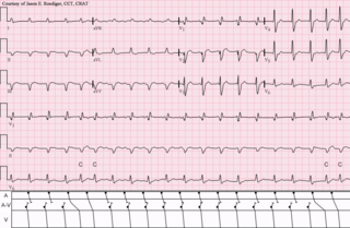
Wandering atrial pacemaker (WAP) is an atrial rhythm where the pacemaking activity of the heart originates from different locations within the atria. This is different from normal pacemaking activity, where the sinoatrial node is responsible for each heartbeat and keeps a steady rate and rhythm. Causes of wandering atrial pacemaker are unclear, but there may be factors leading to its development. It is often seen in the young, the old, and in athletes, and rarely causes symptoms or requires treatment. Diagnosis of wandering atrial pacemaker is made by an ECG.

An accessory pathway is an additional electrical connection between two parts of the heart. These pathways can lead to abnormal heart rhythms or arrhythmias associated with symptoms of palpitations. Some pathways may activate a region of ventricular muscle earlier than would normally occur, referred to as pre-excitation, and this may be seen on an electrocardiogram. The combination of an accessory pathway that causes pre-excitation with arrhythmias is known as Wolff-Parkinson-White syndrome.

<span class="mw-page-title-main">Junctional tachycardia</span> Medical condition

Junctional tachycardia is a form of supraventricular tachycardia characterized by involvement of the AV node. It can be contrasted to atrial tachycardia. It is a tachycardia associated with the generation of impulses in a focus in the region of the atrioventricular node due to an A-V disassociation. In general, the AV junction's intrinsic rate is 40-60 bpm so an accelerated junctional rhythm is from 60-100bpm and then becomes junctional tachycardia at a rate of >100 bpm.

<span class="mw-page-title-main">Ectopic pacemaker</span> Cardiac condition

An ectopic pacemaker, also known as ectopic focus or ectopic foci, is an excitable group of cells that causes a premature heart beat outside the normally functioning SA node of the heart. It is thus a cardiac pacemaker that is ectopic, producing an ectopic beat. Acute occurrence is usually non-life-threatening, but chronic occurrence can progress into tachycardia, bradycardia or ventricular fibrillation. In a normal heart beat rhythm, the SA node usually suppresses the ectopic pacemaker activity due to the higher impulse rate of the SA node. However, in the instance of either a malfunctioning SA node or an ectopic focus bearing an intrinsic rate superior to SA node rate, ectopic pacemaker activity may take over the natural heart rhythm. This phenomenon is called an escape rhythm, the lower rhythm having escaped from the dominance of the upper rhythm. As a rule, premature ectopic beats indicate increased myocyte or conducting tissue excitability, whereas late ectopic beats indicate proximal pacemaker or conduction failure with an escape 'ectopic' beat.

Atrial tachycardia is a type of heart rhythm problem in which the heart's electrical impulse comes from an ectopic pacemaker in the upper chambers (atria) of the heart, rather than from the sinoatrial node, the normal origin of the heart's electrical activity. As with any other form of tachycardia, the underlying mechanism can be either the rapid discharge of an abnormal focus, the presence of a ring of cardiac tissue that gives rise to a circle movement (reentry), or a triggered rapid rhythm due to other pathological circumstances.

<span class="mw-page-title-main">Arrhythmia</span> Group of medical conditions characterized by irregular heartbeat

Arrhythmias, also known as cardiac arrhythmias, heart arrhythmias, or dysrhythmias, are irregularities in the heartbeat, including when it is too fast or too slow. A resting heart rate that is too fast – above 100 beats per minute in adults – is called tachycardia, and a resting heart rate that is too slow – below 60 beats per minute – is called bradycardia. Some types of arrhythmias have no symptoms. Symptoms, when present, may include palpitations or feeling a pause between heartbeats. In more serious cases, there may be lightheadedness, passing out, shortness of breath, chest pain, or decreased level of consciousness. While most cases of arrhythmia are not serious, some predispose a person to complications such as stroke or heart failure. Others may result in sudden death.

<span class="mw-page-title-main">Automatic tachycardia</span> Medical condition

An automatic tachycardia is a cardiac arrhythmia which involves an area of the heart generating an abnormally fast rhythm, sometimes also called enhanced automaticity. These tachycardias, or fast heart rhythms, differ from reentrant tachycardias in which there is an abnormal electrical pathway which gives rise to the pathology. Most automatic tachycardias are supraventricular tachycardias (SVT). It is important to recognize an automatic tachycardia because the treatment will be different to that for a reentrant tachycardia. The most useful clue will be the presence of 'warm up' and 'cool down'. This means that whereas a reentrant tachycardia will both begin and end abruptly as cardiac conduction uses then ceases to use the accessory pathway, an automatic tachycardia will rise and fall gradually in rate as the automatic focus increases and decreases its automatic rate of electrical discharge.

References

  1. Sarubbi B, Vergara P, D'Alto M, Calabro R (2003). "Congenital junctional ectopic tachycardia: presentation and outcome". Indian Pacing Electrophysiol J. 3 (3): 143–7. PMC   1502046 . PMID   16943912. Archived from the original on 28 September 2021. Retrieved 21 December 2008.
  2. "Supraventricular Tachycardia, Junctional Ectopic Tachycardia: Overview - eMedicine" . Retrieved 21 December 2008.
  3. 1 2 Campbell, R. W. F.; Wren, C. (1987). "His bundle tachycardia- arrhythmogenic and antiarrhythmic effects of therapy". European Heart Journal. 8 (6): 647–650. doi:10.1093/oxfordjournals.eurheartj.a062336. PMID   3113958.
  4. Zhivadinovik J, Lazarova D, Gjorgov N (2006). "Dimensions of the Triangle of Koch" (PDF). Bratisl Lek Listy. 107 (4): 107–9. PMID   16796135 . Retrieved 31 January 2013.
  5. Sarubbi B, Musto B, Ducceschi V, D'Onofrio A, Cavallaro C, Vecchione F, Musto C, Calabro R (2002). "Congenital junctional ectopic tachycardia in children and adolescents: a 20 year experience based study". Heart. 88 (2): 188–190. doi:10.1136/heart.88.2.188. PMC   1767240 . PMID   12117855.
  6. 1 2 3 4 5 Haas, N. A.; Plumpton, K.; Justo, R.; Jalali, H.; Pohlner, P. (May 2004). "Postoperative junctional ectopic tachycardia (JET)". Zeitschrift für Kardiologie. 93 (5): 371–380. doi:10.1007/s00392-004-0067-3. ISSN   0300-5860. PMID   15160272. S2CID   5609507.
  7. 1 2 3 4 5 6 Brugada, Josep; Katritsis, Demosthenes G.; Arbelo, Elena; Arribas, Fernando; Bax, Jeroen J.; Blomström-Lundqvist, Carina; Calkins, Hugh; Corrado, Domenico; Deftereos, Spyridon G.; Diller, Gerhard-Paul; Gomez-Doblas, Juan J. (31 August 2019). "2019 ESC Guidelines for the management of patients with supraventricular tachycardiaThe Task Force for the management of patients with supraventricular tachycardia of the European Society of Cardiology (ESC)". European Heart Journal. 41 (5): 655–720. doi: 10.1093/eurheartj/ehz467 . hdl: 1887/3232621 . ISSN   1522-9645. PMID   31504425.
  8. 1 2 Josephson, Mark E. (10 August 2015). Josephson's clinical cardiac electrophysiology : techniques and interpretations. Preceded by: Josephson, Mark E. (Fifth ed.). Baltimore, MD. ISBN   9781496326614. OCLC   938434294.{{cite book}}: CS1 maint: location missing publisher (link)
  9. 1 2 Handbook of cardiac electrophysiology : a practical guide to invasive EP studies and catheter ablation. Murgatroyd, Francis D. London: ReMEDICA Pub. 2002. ISBN   9781901346374. OCLC   48363139.{{cite book}}: CS1 maint: others (link)
  10. Li, Xin; Zhang, Chengxin; Dai, Di; Liu, Haiyuan; Ge, Shenglin (September 2018). "Efficacy of dexmedetomidine in prevention of junctional ectopic tachycardia and acute kidney injury after pediatric cardiac surgery: A meta-analysis". Congenital Heart Disease. 13 (5): 799–807. doi:10.1111/chd.12674. ISSN   1747-0803. PMID   30260073. S2CID   52843168.
  11. "Tachycardia". Online Etymology Dictionary. Retrieved 29 August 2011.