Pericarditis

Last updated
Pericarditis
Pericarditis10.JPG
An ECG showing pericarditis, with ST elevation in multiple leads and slight reciprocal ST depression in aVR.
Specialty Cardiology
Symptoms Sharp chest pain, better sitting up and worse with lying down, fever [1]
Complications Cardiac tamponade, myocarditis, constrictive pericarditis [1] [2]
Usual onsetTypically sudden [1]
DurationFew days to weeks [3]
Causes Viral infection, tuberculosis, uremic pericarditis, following a heart attack, cancer, autoimmune disorders, chest trauma [4] [5]
Diagnostic method Based on symptoms, electrocardiogram, fluid around the heart [6]
Differential diagnosis Heart attack [1]
Treatment NSAIDs, colchicine, corticosteroids [6]
Prognosis Usually good [6] [7]
Frequency3 per 10,000 per year [2]

Pericarditis is inflammation of the pericardium, the fibrous sac surrounding the heart. [8] Symptoms typically include sudden onset of sharp chest pain, which may also be felt in the shoulders, neck, or back. [1] The pain is typically less severe when sitting up and more severe when lying down or breathing deeply. [1] Other symptoms of pericarditis can include fever, weakness, palpitations, and shortness of breath. [1] The onset of symptoms can occasionally be gradual rather than sudden. [8]

Contents

The cause of pericarditis often remains unknown but is believed to be most often due to a viral infection. [4] [8] Other causes include bacterial infections such as tuberculosis, uremic pericarditis, heart attack, cancer, autoimmune disorders, and chest trauma. [4] [5] Diagnosis is based on the presence of chest pain, a pericardial rub, specific electrocardiogram (ECG) changes, and fluid around the heart. [6] A heart attack may produce similar symptoms to pericarditis. [1]

Treatment in most cases is with NSAIDs and possibly the anti-inflammatory medication colchicine. [6] Steroids may be used if these are not appropriate. [6] Symptoms usually improve in a few days to weeks but can occasionally last months. [3] Complications can include cardiac tamponade, myocarditis, and constrictive pericarditis. [1] [2] Pericarditis is an uncommon cause of chest pain. [9] About 3 per 10,000 people are affected per year. [2] Those most commonly affected are males between the ages of 20 and 50. [10] Up to 30% of those affected have more than one episode. [10]

Signs and symptoms

Substernal or left precordial pleuritic chest pain with radiation to the trapezius ridge (the bottom portion of scapula on the back) is the characteristic pain of pericarditis. The pain is usually relieved by sitting up or bending forward, and worsened by lying down (both recumbent and supine positions) or by inspiration (taking a breath in). [11] The pain may resemble that of angina but differs in that pericarditis pain changes with body position, where heart attack pain is generally constant and pressure-like. Other symptoms of pericarditis may include dry cough, fever, fatigue, and anxiety.[ citation needed ]

Due to its similarity to the pain of myocardial infarction (heart attack), pericarditis can be misdiagnosed as a heart attack. Acute myocardial infarction can also cause pericarditis, but the presenting symptoms often differ enough to warrant diagnosis. The following table organizes the clinical presentation of pericarditis differential to myocardial infarction: [11]

CharacteristicPericarditisMyocardial infarction
Pain descriptionSharp, pleuritic, retro-sternal (under the sternum) or left precordial (left chest) painCrushing, pressure-like, heavy pain. Described as "elephant on the chest."
RadiationPain radiates to the trapezius ridge (to the lowest portion of the scapula on the back) or no radiation.Pain radiates to the jaw or left arm, or does not radiate.
ExertionDoes not change the painCan increase the pain
PositionPain is worse in the supine position or upon inspiration (breathing in)Not positional
Onset/durationSudden pain, that lasts for hours or sometimes days before a person comes to the ERSudden or chronically worsening pain that can come and go in paroxysms or it can last for hours before the person decides to come to the ER

Physical examinations

The classic sign of pericarditis is a friction rub heard with a stethoscope on the cardiovascular examination, usually on the lower left sternal border. [11] Other physical signs include a person in distress, positional chest pain, diaphoresis (excessive sweating); possibility of heart failure in form of pericardial tamponade causing pulsus paradoxus, and the Beck's triad of low blood pressure (due to decreased cardiac output), distant (muffled) heart sounds, and distension of the jugular vein (JVD).[ citation needed ]

Complications

Pericarditis can progress to pericardial effusion and eventually cardiac tamponade. This can be seen in people who are experiencing the classic signs of pericarditis but then show signs of relief, and progress to show signs of cardiac tamponade which include decreased alertness and lethargy, pulsus paradoxus (decrease of at least 10 mmHg of the systolic blood pressure upon inspiration), low blood pressure (due to decreased cardiac index), (jugular vein distention from right sided heart failure and fluid overload), distant heart sounds on auscultation, and equilibration of all the diastolic blood pressures on cardiac catheterization due to the constriction of the pericardium by the fluid.[ citation needed ]

In such cases of cardiac tamponade, EKG or Holter monitor will then depict electrical alternans indicating wobbling of the heart in the fluid filled pericardium, and the capillary refill might decrease, as well as severe vascular collapse and altered mental status due to hypoperfusion of body organs by a heart that can not pump out blood effectively.[ citation needed ]

The diagnosis of tamponade can be confirmed with trans-thoracic echocardiography (TTE), which should show a large pericardial effusion and diastolic collapse of the right ventricle and right atrium. Chest X-ray usually shows an enlarged cardiac silhouette ("water bottle" appearance) and clear lungs. Pulmonary congestion is typically not seen because equalization of diastolic pressures constrains the pulmonary capillary wedge pressure to the intra-pericardial pressure (and all other diastolic pressures).[ citation needed ]

Causes

Figure A shows the location of the heart and a normal heart and pericardium (the sac surrounding the heart). The inset image is an enlarged cross-section of the pericardium that shows its two layers of tissue and the fluid between the layers.
Figure B shows the heart with pericarditis. The inset image is an enlarged cross-section that shows the inflamed and thickened layers of the pericardium. Pericarditis.jpg
Figure A shows the location of the heart and a normal heart and pericardium (the sac surrounding the heart). The inset image is an enlarged cross-section of the pericardium that shows its two layers of tissue and the fluid between the layers.
Figure B shows the heart with pericarditis. The inset image is an enlarged cross-section that shows the inflamed and thickened layers of the pericardium.

Infectious

Pericarditis may be caused by viral, bacterial, or fungal infection.

In the developing world the bacterial disease tuberculosis is a common cause, whereas in the developed world viruses are believed to be the cause of about 85% of cases. [6] Viral causes include coxsackievirus, herpesvirus, mumps virus, and HIV among others. [4]

Pneumococcus or tuberculous pericarditis are the most common bacterial forms. Anaerobic bacteria can also be a rare cause. [13] Fungal pericarditis is usually due to histoplasmosis, or in immunocompromised hosts Aspergillus, Candida, and Coccidioides.[ citation needed ] The most common cause of pericarditis worldwide is infectious pericarditis with tuberculosis.[ citation needed ]

Other

Diagnosis

Laboratory test

Diffuse ST elevation in a young male due to myocarditis / pericarditis PericarditisMyocarditis.jpg
Diffuse ST elevation in a young male due to myocarditis / pericarditis
An ECG showing pericarditis. Note the ST elevation in multiple leads with slight reciprocal ST depression in aVR. PericarditisECG.JPG
An ECG showing pericarditis. Note the ST elevation in multiple leads with slight reciprocal ST depression in aVR.

Laboratory values can show increased blood urea nitrogen (BUN), or increased blood creatinine in cases of uremic pericarditis. Generally, however, laboratory values are normal, but if there is a concurrent myocardial infarction (heart attack) or great stress to the heart, laboratory values may show increased cardiac markers like Troponin (I, T), CK-MB, Myoglobin, and LDH1 (lactase dehydrogenase isotype 1).[ citation needed ]

The preferred initial diagnostic testing is the ECG, which may demonstrate a 12-lead electrocardiogram with diffuse, non-specific, concave ("saddle-shaped"), ST-segment elevations in all leads except aVR and V1 [11] and PR-segment depression possible in any lead except aVR; [11] sinus tachycardia, and low-voltage QRS complexes can also be seen if there is subsymptomatic levels of pericardial effusion. The PR depression is often seen early in the process as the thin atria are affected more easily than the ventricles by the inflammatory process of the pericardium.[ citation needed ]

Since the mid-19th century, retrospective diagnosis of pericarditis has been made upon the finding of adhesions of the pericardium. [20]

When pericarditis is diagnosed clinically, the underlying cause is often never known; it may be discovered in only 16–22 percent of people with acute pericarditis.[ citation needed ]

Imaging

On MRI T2-weighted spin-echo images, inflamed pericardium will show high signal intensity. Late gadolinium contrast will show uptake of contrast by the inflamed pericardium. Normal pericardium will not show any contrast enhancement. [21]

Classification

Pericarditis can be classified according to the composition of the fluid that accumulates around the heart. [22]

Types of pericarditis include the following:[ citation needed ]

Acute vs. chronic

Depending on the time of presentation and duration, pericarditis is divided into "acute" and "chronic" forms. Acute pericarditis is more common than chronic pericarditis, and can occur as a complication of infections, immunologic conditions, or even as a result of a heart attack (myocardial infarction), as Dressler's syndrome. Chronic pericarditis however is less common, a form of which is constrictive pericarditis. The following is the clinical classification of acute vs. chronic:[ citation needed ]

  • Clinically: Acute (<6 weeks), Subacute (6 weeks to 6 months) and Chronic (>6 months)

Treatment

The treatment in viral or idiopathic pericarditis is with aspirin, [11] or non-steroidal anti-inflammatory drugs (NSAIDs such as ibuprofen). [4] Colchicine may be added to the above as it decreases the risk of further episodes of pericarditis. [4] [23]

Severe cases may require one or more of the following:[ citation needed ]

Recurrent pericarditis resistant to colchicine and anti-inflammatory steroids may benefit from a number of medicines that affect the action of interleukin 1; they cannot be taken in tablet form. These are anakinra, canakinumab and rilonacept. [24] [25] Rilonacept has been specifically approved as an orphan drug for use in this situation. [26] Immunosuppressive agents, such as Azathioprine and intravenous immunoglobulins, are a novel therapeutic agent which have been effective in treating and preventing recurrent pericarditis, though research on these therapies is limited. [25] [27] [28] [29]

Surgical removal of the pericardium, pericardiectomy, may be used in severe cases and where the pericarditis is causing constriction, impairing cardiac function. It is less effective if the pericarditis is a consequence of trauma, in elderly patients, and if the procedure is done incompletely. It carries a risk of death between 5 and 10%. [25]

Epidemiology

About 30% of people with viral pericarditis or pericarditis of an unknown cause have one or several recurrent episodes. [6]

See also

Related Research Articles

<span class="mw-page-title-main">Pericardium</span> Double-walled sac containing the heart and roots of the great vessels

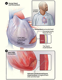
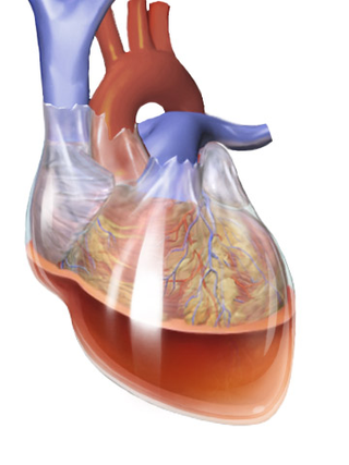
The pericardium, also called pericardial sac, is a double-walled sac containing the heart and the roots of the great vessels. It has two layers, an outer layer made of strong inelastic connective tissue, and an inner layer made of serous membrane. It encloses the pericardial cavity, which contains pericardial fluid, and defines the middle mediastinum. It separates the heart from interference of other structures, protects it against infection and blunt trauma, and lubricates the heart's movements.

<span class="mw-page-title-main">Cardiac tamponade</span> Buildup of fluid around the heart

Cardiac tamponade, also known as pericardial tamponade, is a compression of the heart due to pericardial effusion. Onset may be rapid or gradual. Symptoms typically include those of obstructive shock including shortness of breath, weakness, lightheadedness, and cough. Other symptoms may relate to the underlying cause.

<span class="mw-page-title-main">Chest pain</span> Discomfort or pain in the chest as a medical symptom

Chest pain is pain or discomfort in the chest, typically the front of the chest. It may be described as sharp, dull, pressure, heaviness or squeezing. Associated symptoms may include pain in the shoulder, arm, upper abdomen, or jaw, along with nausea, sweating, or shortness of breath. It can be divided into heart-related and non-heart-related pain. Pain due to insufficient blood flow to the heart is also called angina pectoris. Those with diabetes or the elderly may have less clear symptoms.

<span class="mw-page-title-main">Constrictive pericarditis</span> Medical condition

Constrictive pericarditis is a condition characterized by a thickened, fibrotic pericardium, limiting the heart's ability to function normally. In many cases, the condition continues to be difficult to diagnose and therefore benefits from a good understanding of the underlying cause.

<span class="mw-page-title-main">Pericardiocentesis</span> Procedure where fluid is aspirated from the pericardium

Pericardiocentesis (PCC), also called pericardial tap, is a medical procedure where fluid is aspirated from the pericardium.

Dressler syndrome is a secondary form of pericarditis that occurs in the setting of injury to the heart or the pericardium. It consists of fever, pleuritic pain, pericarditis and/or pericardial effusion.

A pericardial friction rub, also pericardial rub, is an audible medical sign used in the diagnosis of pericarditis. Upon auscultation, this sign is an extra heart sound of to-and-fro character, typically with three components, two systolic and one diastolic. It resembles the sound of squeaky leather and often is described as grating, scratching, or rasping. The sound seems very close to the ear and may seem louder than or may even mask the other heart sounds. The sound usually is best heard between the apex and sternum but may be widespread.

Pulsus paradoxus, also paradoxic pulse or paradoxical pulse, is an abnormally large decrease in stroke volume, systolic blood pressure and pulse wave amplitude during inspiration. Pulsus paradoxus is not related to pulse rate or heart rate, and it is not a paradoxical rise in systolic pressure. Normally, blood pressure drops less precipitously than 10 mmHg during inhalation. Pulsus paradoxus is a sign that is indicative of several conditions, most commonly pericardial effusion.

<span class="mw-page-title-main">Pericardial effusion</span> Medical condition

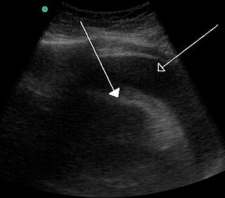
A pericardial effusion is an abnormal accumulation of fluid in the pericardial cavity. The pericardium is a two-part membrane surrounding the heart: the outer fibrous connective membrane and an inner two-layered serous membrane. The two layers of the serous membrane enclose the pericardial cavity between them. This pericardial space contains a small amount of pericardial fluid, normally 15-50 mL in volume. The pericardium, specifically the pericardial fluid provides lubrication, maintains the anatomic position of the heart in the chest, and also serves as a barrier to protect the heart from infection and inflammation in adjacent tissues and organs.

<span class="mw-page-title-main">Acute pericarditis</span> Medical condition

Acute pericarditis is a type of pericarditis usually lasting less than 6 weeks. It is the most common condition affecting the pericardium.

Pericardiectomy is the surgical removal of part or most of the pericardium. This operation is most commonly used to relieve constrictive pericarditis, or to remove a pericardium that is calcified and fibrous. It may also be used for severe or recurrent cases of pericardial effusion. Post-operative outcomes and mortality are significantly impacted by the disease it is used to treat.

<span class="mw-page-title-main">Myocardial rupture</span> Medical condition

Myocardial rupture is a laceration of the ventricles or atria of the heart, of the interatrial or interventricular septum, or of the papillary muscles. It is most commonly seen as a serious sequela of an acute myocardial infarction.

The following outline is provided as an overview of and topical guide to cardiology, the branch of medicine dealing with disorders of the human heart. The field includes medical diagnosis and treatment of congenital heart defects, coronary artery disease, heart failure, valvular heart disease and electrophysiology. Physicians who specialize in cardiology are called cardiologists.

Obstructive shock is one of the four types of shock, caused by a physical obstruction in the flow of blood. Obstruction can occur at the level of the great vessels or the heart itself. Causes include pulmonary embolism, cardiac tamponade, and tension pneumothorax. These are all life-threatening. Symptoms may include shortness of breath, weakness, or altered mental status. Low blood pressure and tachycardia are often seen in shock. Other symptoms depend on the underlying cause.

<span class="mw-page-title-main">Hemopericardium</span> Medical condition

Hemopericardium refers to blood in the pericardial sac of the heart. It is clinically similar to a pericardial effusion, and, depending on the volume and rapidity with which it develops, may cause cardiac tamponade.

Tuberculous pericarditis is a form of pericarditis. It is a condition in which the pericardium surrounding the heart is infected by the bacterial species Mycobacterium tuberculosis. Tuberculous pericarditis accounts for a significant percentage of presentations of tuberculosis worldwide. The condition has four stages of disease which manifests with clinical presentations ranging from acute pericarditis to overt heart failure. Tuberculous pericarditis is an under-diagnosed condition. Diagnosis often requires a range of diagnostic tools, including pericardiocentesis, biochemical tests, and imaging. Treatment of this disease is similar to treatment of pulmonary tuberculosis. Alternative treatment options to reduce cardiac complications are also available.

Myocardial infarction complications may occur immediately following a myocardial infarction, or may need time to develop. After an infarction, an obvious complication is a second infarction, which may occur in the domain of another atherosclerotic coronary artery, or in the same zone if there are any live cells left in the infarct.

<span class="mw-page-title-main">Postpericardiotomy syndrome</span> Medical condition

Postpericardiotomy syndrome (PPS) is a medical syndrome referring to an immune phenomenon that occurs days to months after surgical incision of the pericardium. PPS can also be caused after a trauma, a puncture of the cardiac or pleural structures, after percutaneous coronary intervention, or due to pacemaker or pacemaker wire placement.

<span class="mw-page-title-main">Purulent pericarditis</span> Inflammation of the sac surrounding the heart due to bacterial infection.

Purulent pericarditis refers to localized inflammation in the setting of infection of the pericardial sac surrounding the heart. In contrast to other causes of pericarditis which may have a viral etiology, purulent pericarditis refers specifically to bacterial or fungal infection of the pericardial sac. Clinical etiologies of purulent pericarditis may include recent surgery, adjacent infection, trauma, or even primary infection. The onset of purulent pericarditis is usually acute, with most individuals presenting to a medical facility approximately 3 days following the onset of symptoms.

References

  1. 1 2 3 4 5 6 7 8 9 "What Are the Signs and Symptoms of Pericarditis?". National Heart, Lung, and Blood Institute. September 26, 2012. Archived from the original on 2 October 2016. Retrieved 28 September 2016.
  2. 1 2 3 4 Imazio M, Gaita F (July 2015). "Diagnosis and treatment of pericarditis". Heart. 101 (14): 1159–68. doi:10.1136/heartjnl-2014-306362. PMID   25855795. S2CID   35310104.
  3. 1 2 "How Is Pericarditis Treated?". National Heart, Lung, and Blood Institute. September 26, 2012. Archived from the original on 2 October 2016. Retrieved 28 September 2016.
  4. 1 2 3 4 5 6 7 8 9 10 11 12 13 14 15 Tingle LE, Molina D, Calvert CW (November 2007). "Acute pericarditis". American Family Physician. 76 (10): 1509–14. PMID   18052017.
  5. 1 2 "What Causes Pericarditis?". National Heart, Lung, and Blood Institute. September 26, 2012. Archived from the original on 2 October 2016. Retrieved 28 September 2016.
  6. 1 2 3 4 5 6 7 8 Imazio M, Gaita F, LeWinter M (October 2015). "Evaluation and Treatment of Pericarditis: A Systematic Review". JAMA. 314 (14): 1498–506. doi:10.1001/jama.2015.12763. hdl: 2318/1576078 . PMID   26461998.
  7. Cunha BA (2010). Antibiotic Essentials. Jones & Bartlett Publishers. p. 71. ISBN   978-1-4496-1870-4.
  8. 1 2 3 "What Is Pericarditis?". National Heart, Lung, and Blood Institute. September 26, 2012. Archived from the original on October 2, 2016.
  9. McConaghy JR, Oza RS (February 2013). "Outpatient diagnosis of acute chest pain in adults". American Family Physician. 87 (3): 177–82. PMID   23418761.
  10. 1 2 "Who Is at Risk for Pericarditis?". National Heart, Lung, and Blood Institute. September 26, 2012. Archived from the original on 2 October 2016. Retrieved 28 September 2016.
  11. 1 2 3 4 5 6 American College of Physicians (ACP) (2009). "Pericardial disease". Medical Knowledge Self-Assessment Program (MKSAP-15): Cardiovascular Medicine. American College of Physicians. p. 64. ISBN   978-1-934465-28-8. Archived from the original on 2010-08-02.
  12. "Pericarditis". National Heart, Lung, and Blood Institute .nih.gov. Archived from the original on 8 August 2014. Retrieved 5 August 2014.
  13. Brook I (April 2009). "Pericarditis caused by anaerobic bacteria". International Journal of Antimicrobial Agents. 33 (4): 297–300. doi:10.1016/j.ijantimicag.2008.06.033. PMID   18789852.
  14. Ibe T, Nakamura T, Taniguchi Y, Momomura S (June 2016). "IgG4-related effusive constrictive pericarditis". European Heart Journal: Cardiovascular Imaging. 17 (6): 707. doi: 10.1093/ehjci/jew056 . PMID   27044912.
  15. Atallah PC, Kassier A, Powers S (September 2014). "IgG4-related disease with effusive-constrictive pericarditis, tamponade, and hepatopathy: a novel triad". International Journal of Cardiology. 176 (2): 516–8. doi:10.1016/j.ijcard.2014.07.046. PMID   25062559.
  16. Gharacholou S Michael; Vaca-Cartagena F Bryan; Parikh P Pragnesh; Pollak M Peter; Bruce J Charles. "Peri-Myocardial Infarction Pericarditis: Current Concepts". Edelweiss Publications Inc. Retrieved 15 February 2022.
  17. Kuntz, J.; Crane, B.; Weinmann, S.; Naleway, A. L.; Vaccine Safety Datalink Investigator Team (2018). "Myocarditis and pericarditis are rare following live viral vaccinations in adults". Vaccine. 36 (12): 1524–1527. doi:10.1016/j.vaccine.2018.02.030. PMC   6437672 . PMID   29456017.
  18. "Myocarditis, Pericarditis, and Dilated Cardiomyopathy after Smallpox Vaccination among Civilians in the United States, January–October 2003". 15 March 2008. Retrieved 22 January 2022.
  19. "Myocarditis and Pericarditis After mRNA COVID-19 Vaccination" . Retrieved 22 January 2022.
  20. Flint A (1862). "Lectures on the diagnosis of diseases of the heart: Lecture VIII". American Medical Times: Being a Weekly Series of the New York Journal of Medicine. 5 (July to December): 309–311.
  21. Ismail TF (January 2020). "Acute pericarditis: Update on diagnosis and management". Clinical Medicine. 20 (1): 48–51. doi:10.7861/clinmed.cme.20.1.4. PMC   6964178 . PMID   31941732.
  22. Klatt EC. "Cardiovascular Pathology Index Images". The Internet Pathology Laboratory for Medical Education. Florida State University College of Medicine. Archived from the original on 2007-05-24.
  23. Alabed S, Cabello JB, Irving GJ, Qintar M, Burls A (August 2014). "Colchicine for pericarditis" (PDF). The Cochrane Database of Systematic Reviews. 2014 (8): CD010652. doi:10.1002/14651858.CD010652.pub2. PMC   10645160 . PMID   25164988.
  24. Tombetti, Enrico; Mulè, Alice; Tamanini, Silvia; Matteucci, Luca; Negro, Enrica; Brucato, Antonio; Carnovale, Carla (August 2020). "Novel Pharmacotherapies for Recurrent Pericarditis: Current Options in 2020". Current Cardiology Reports. 22 (8): 59. doi:10.1007/s11886-020-01308-y. PMC   7303578 . PMID   32562029.
  25. 1 2 3 Andreis, Alessandro; Imazio, Massimo; Casula, Matteo; Avondo, Stefano; Brucato, Antonio (28 February 2021). "Recurrent pericarditis: an update on diagnosis and management". Internal and Emergency Medicine. 16 (3): 551–558. doi:10.1007/s11739-021-02639-6. PMC   7914388 . PMID   33641044.
  26. Center for Drug Evaluation and Research (18 March 2021). "FDA Approves First Treatment for Disease That Causes Recurrent Inflammation in Sac Surrounding Heart". FDA. U.S. Food and Drug Administration. Retrieved 19 March 2021.
  27. Marcolongo, Renzo; Russo, Rosario; Laveder, Francesco; Noventa, Franco; Agostini, Carlo (November 1995). "Immunosuppressive therapy prevents recurrent pericarditis". Journal of the American College of Cardiology. 26 (5): 1276–1279. doi:10.1016/0735-1097(95)00302-9. PMID   7594043. S2CID   8236269.
  28. del Fresno, M. Rosa; Peralta, Julio E.; Granados, Miguel Ángel; Enríquez, Eugenia; Domínguez-Pinilla, Nerea; de Inocencio, Jaime (2014-11-01). "Intravenous Immunoglobulin Therapy for Refractory Recurrent Pericarditis". Pediatrics. 134 (5): e1441–e1446. doi:10.1542/peds.2013-3900. ISSN   0031-4005. PMID   25287461. S2CID   12121925.
  29. Andreis, Alessandro; Imazio, Massimo; Casula, Matteo; Avondo, Stefano; Brucato, Antonio (April 2021). "Recurrent pericarditis: an update on diagnosis and management". Internal and Emergency Medicine. 16 (3): 551–558. doi:10.1007/s11739-021-02639-6. ISSN   1828-0447. PMC   7914388 . PMID   33641044.