Sinus node dysfunction

Last updated
Sinus node dysfunction
Other namesSick sinus syndrome or sinoatrial node disease
ECG Sinus Pause.jpg
Telemetry strip of a 44-year-old female with asymptomatic sinus pause found while admitted for mildly symptomatic COVID-19 pneumonia.
Complications Tachycardia-bradycardia syndrome
Diagnostic method electrocardiogram
Treatment Pacemaker implantation

Sinus node dysfunction (SND), also known as sick sinus syndrome (SSS), is a group of abnormal heart rhythms (arrhythmias) usually caused by a malfunction of the sinus node, the heart's primary pacemaker. [1] [2] Tachycardia-bradycardia syndrome is a variant of sick sinus syndrome in which the arrhythmia alternates between fast and slow heart rates. [3]

Contents

Signs and symptoms

Often sinus node dysfunction produces no symptoms, especially early in the disease course. Signs and symptoms usually appear in more advanced disease and more than 50% of patients will present with syncope or transient near-fainting spells as well as bradycardias that are accompanied by rapid heart rhythms, referred to as tachycardia-bradycardia syndrome [4] [5] Other presenting signs or symptoms can include confusion, fatigue, palpitations, chest pain, shortness of breath, headache, and nausea. Patients can also present with symptoms of congestive heart failure, stroke or transient ischemic attacks due to the abnormal rhythm. [5]

Complications

Electrocardiogram from a man with tachycardia-bradycardia syndrome following mitral valvuloplasty, resection of the left atrial appendage, and maze procedure. The ECG shows AV-junctional rhythm resulting in bradycardia at around 46 beats per minute. Brady-tachy syndrome AV-junctional rhythm.png
Electrocardiogram from a man with tachycardia-bradycardia syndrome following mitral valvuloplasty, resection of the left atrial appendage, and maze procedure. The ECG shows AV-junctional rhythm resulting in bradycardia at around 46 beats per minute.
This ECG from the same patient shows atrial fibrillation at around 126 beats per minute. Brady-tachy syndrome atrial fibrillation.png
This ECG from the same patient shows atrial fibrillation at around 126 beats per minute.

The most common complication of sinus node dysfunction is the development of tachycardia-bradycardia syndrome with abnormal atrial rhythms such as atrial tachycardia, atrial fibrillation, and flutter. [5] [3] These rhythms increases the risk of clot formation in the atrium, embolization, and stroke. [5]

Developing sinus arrest, sinus node exit block, sinus bradycardia, atrioventricular block, and other types of abnormal rhythms are also common complications. [5] [6] Sinus node dysfunction has a close association with the presence of atrial fibrillation due to their shared etiology of remodeling. [6]

Causes

Sinus node dysfunction can be caused by intrinsic and extrinsic factors that affect the normal functioning of the sinus node. Intrinsic causes can include degeneration, dysfunction, or remodeling of the sinus node while extrinsic causes can create or worsen underlying atrial arrhythmias. [5] Intrinsic causes tend to be responsible for permanent sinus node dysfunction while extrinsic causes are more commonly temporary. [5]

Intrinsic causes

Age-related degenerative fibrosis of the sinus node is often identified as the most common intrinsic cause. [5] [6] Other intrinsic causes include inherited ion channel dysfunctions, remodeling diseases such as heart failure and atrial fibrillation, infiltrative diseases such as sarcoidosis, amyloidosis, hemochromatosis, and connective tissue diseases, inflammatory etiology such as rheumatic fever, Chagas disease, and Lyme disease, as well as atherosclerotic and ischemic changes to the sinus node artery. [5] [6]

Inherited sinus node dysfunction has been associated with mutations of the gene responsible for the formation of the alpha subunit of the sodium channel (SCN5A). [5]

Extrinsic causes

Common cardiac pharmacology such as beta-blockers, calcium channel blockers, digoxin, sympatholytic medication, and other antiarrhythmics can alter sinus node function to create an arrhythmia such as sick sinus syndrome. Electrolyte abnormalities such as hyperkalemia, hypokalemia, and hypocalcemia can also alter normal sinus node functioning. Hypothyroidism, hypoxia, hypothermia, and various toxins have also been associated with sinus node dysfunctions. [5] [6]

Diagnosis

Electrocardiogram (ECG) of a 64-year-old female with sinus bradycardia. Heart rate 49 bpm. ECG Sinus Bradycardia 49 bpm.jpg
Electrocardiogram (ECG) of a 64-year-old female with sinus bradycardia. Heart rate 49 bpm.

Electrocardiogram

The primary 12-lead electrocardiogram (ECG) finding in sinus node dysfunction is inappropriate sinus bradycardia. [7] Sinus node dysfunction can also present with sudden sinus arrest with or without junctional escape, sinoatrial block, prolonged asystolic period followed by tachycardias, or tachycardia-bradycardia syndrome presenting as various atrial arrhythmias such as atrial fibrillation, flutter, tachycardia, or paroxysmal supraventricular tachycardia. [7] [5]

Clinical diagnosis

Diagnosing sinus node dysfunction requires clinical symptoms as well as ECG abnormalities. If ECG findings cannot be identified, prolonged cardiac monitoring should be pursued either with a Holter monitor in an outpatient setting or telemetry while inpatient, due to the transient nature of abnormal ECG findings. [5] If Holter or telemetry monitoring fails to identify ECG changes and suspicion of sinus node dysfunction remains high due to severe symptoms or episodes of syncope, implantable loop recorders should be considered for extended monitoring up to 24 months. [7]

Exercise stress tests can be utilized to identify intrinsic causes of sinus node dysfunction. Tilt table tests can be used to discriminate bradycardia caused by dysfunction of the autonomic nervous system. [5] [7]

Treatment

The primary reason for considering treatment is the presence of symptoms. [1] Pacemaker implantation is the primary treatment modality of symptomatic sinus node dysfunction. [5] [7] [8] The goal of this treatment modality is to relieve symptoms associated with sinus node dysfunction and improve quality of life. [5] Dual chamber pacemakers are preferred due to the possibility of developing atrioventricular block [5] as well as long term cost-effectiveness relative to single-chamber atrial pacemakers. [9]

In tachycardia-bradycardia syndrome, medication-based management can treat atrial tachyarrhythmias. However, these medications may exacerbate underlying bradyarrhythmia. Therefore, a dual-chamber pacemaker capable of managing atrial tachyarrhythmias as well as bradyarrhythmias is implanted before drug therapy is begun. [7]

Epidemiology

Overall incidence of sinus node dysfunction increases with age [10] with 1 in 1000 in adults over 45 years old [6] and 1 in 600 cardiac patients over 65 years old. [5] Sinus node dysfunction is the primary indication for approximately 30%-50% of all pacemaker implantation in the United States. [10] Sinus node dysfunction is a relatively uncommon syndrome in the young and middle-aged population.[ citation needed ]

Related Research Articles

<span class="mw-page-title-main">Bradycardia</span> Heart rate below the normal range

Bradycardia is a medical term used to describe a resting heart rate under 60 beats per minute (BPM). While bradycardia can result from a variety of pathologic processes, it is commonly a physiologic response to cardiovascular conditioning, or due to asymptomatic type 1 atrioventricular block. Resting heart rates less than 50 BPM are often normal during sleep in young and healthy adults, and in athletes. In large population studies of adults without underlying heart disease, resting heart rates of 45-50 BPM appear to be the lower limits of normal, dependent on age and sex. Bradycardia is most likely to be discovered in the elderly, as both age and underlying cardiac disease progression contribute to its development.

<span class="mw-page-title-main">Tachycardia</span> Heart rate that exceeds the normal resting rate

Tachycardia, also called tachyarrhythmia, is a heart rate that exceeds the normal resting rate. In general, a resting heart rate over 100 beats per minute is accepted as tachycardia in adults. Heart rates above the resting rate may be normal or abnormal.

<span class="mw-page-title-main">Wolff–Parkinson–White syndrome</span> Medical condition

Wolff–Parkinson–White syndrome (WPWS) is a disorder due to a specific type of problem with the electrical system of the heart involving an accessory pathway able to conduct electrical current between the atria and the ventricles, thus bypassing the atrioventricular node. About 60% of people with the electrical problem developed symptoms, which may include an abnormally fast heartbeat, palpitations, shortness of breath, lightheadedness, or syncope. Rarely, cardiac arrest may occur. The most common type of irregular heartbeat that occurs is known as paroxysmal supraventricular tachycardia.

<span class="mw-page-title-main">Palpitations</span> Perceived cardiac abnormality in which ones heartbeat can be felt

Palpitations are perceived abnormalities of the heartbeat characterized by awareness of cardiac muscle contractions in the chest, which is further characterized by the hard, fast and/or irregular beatings of the heart.

<span class="mw-page-title-main">Third-degree atrioventricular block</span> Medical condition

Third-degree atrioventricular block is a medical condition in which the electrical impulse generated in the sinoatrial node in the atrium of the heart can not propagate to the ventricles.

<span class="mw-page-title-main">Supraventricular tachycardia</span> Abnormally fast heart rhythm

Supraventricular tachycardia (SVT) is an umbrella term for fast heart rhythms arising from the upper part of the heart. This is in contrast to the other group of fast heart rhythms – ventricular tachycardia, which start within the lower chambers of the heart. There are four main types of SVT: atrial fibrillation, atrial flutter, paroxysmal supraventricular tachycardia (PSVT), and Wolff–Parkinson–White syndrome. The symptoms of SVT include palpitations, feeling of faintness, sweating, shortness of breath, and/or chest pain.

<span class="mw-page-title-main">AV nodal reentrant tachycardia</span> Medical condition

AV-nodal reentrant tachycardia (AVNRT) is a type of abnormal fast heart rhythm. It is a type of supraventricular tachycardia (SVT), meaning that it originates from a location within the heart above the bundle of His. AV nodal reentrant tachycardia is the most common regular supraventricular tachycardia. It is more common in women than men. The main symptom is palpitations. Treatment may be with specific physical maneuvers, medications, or, rarely, synchronized cardioversion. Frequent attacks may require radiofrequency ablation, in which the abnormally conducting tissue in the heart is destroyed.

<span class="mw-page-title-main">Sinus bradycardia</span> Sinus rhythm with a rate that is lower than normal

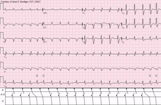
Sinus bradycardia is a sinus rhythm with a reduced rate of electrical discharge from the sinoatrial node, resulting in a bradycardia, a heart rate that is lower than the normal range.

Premature atrial contraction (PAC), also known as atrial premature complexes (APC) or atrial premature beats (APB), are a common cardiac dysrhythmia characterized by premature heartbeats originating in the atria. While the sinoatrial node typically regulates the heartbeat during normal sinus rhythm, PACs occur when another region of the atria depolarizes before the sinoatrial node and thus triggers a premature heartbeat, in contrast to escape beats, in which the normal sinoatrial node fails, leaving a non-nodal pacemaker to initiate a late beat.

Tachycardia-induced cardiomyopathy (TIC) is a disease where prolonged tachycardia or arrhythmia causes an impairment of the myocardium, which can result in heart failure. People with TIC may have symptoms associated with heart failure and/or symptoms related to the tachycardia or arrhythmia. Though atrial fibrillation is the most common cause of TIC, several tachycardias and arrhythmias have been associated with the disease.

Lown–Ganong–Levine syndrome (LGL) is a pre-excitation syndrome of the heart. Those with LGL syndrome have episodes of abnormal heart racing with a short PR interval and normal QRS complexes seen on their electrocardiogram when in a normal sinus rhythm. LGL syndrome was originally thought to be due to an abnormal electrical connection between the atria and the ventricles, but is now thought to be due to accelerated conduction through the atrioventricular node in the majority of cases. The syndrome is named after Bernard Lown, William Francis Ganong, Jr., and Samuel A. Levine.

<span class="mw-page-title-main">Lorcainide</span> Antiarrythmic agent

Lorcainide is a Class 1c antiarrhythmic agent that is used to help restore normal heart rhythm and conduction in patients with premature ventricular contractions, ventricular tachycardiac and Wolff–Parkinson–White syndrome. Lorcainide was developed by Janssen Pharmaceutica (Belgium) in 1968 under the commercial name Remivox and is designated by code numbers R-15889 or Ro 13-1042/001. It has a half-life of 8.9 +- 2.3 hrs which may be prolonged to 66 hrs in people with cardiac disease.

<span class="mw-page-title-main">Multifocal atrial tachycardia</span> Fast heart rhythm associated with exacerbations of COPD

Multifocal atrial tachycardia (MAT) is an abnormal heart rhythm, specifically a type of supraventricular tachycardia, that is particularly common in older people and is associated with exacerbations of chronic obstructive pulmonary disease (COPD). Normally, the heart rate is controlled by a cluster of cells called the sinoatrial node. When a number of different clusters of cells outside the SA node take over control of the heart rate, and the rate exceeds 100 beats per minute, this is called multifocal atrial tachycardia.

<span class="mw-page-title-main">Wandering atrial pacemaker</span> Medical condition

Wandering atrial pacemaker (WAP) is an atrial rhythm where the pacemaking activity of the heart originates from different locations within the atria. This is different from normal pacemaking activity, where the sinoatrial node is responsible for each heartbeat and keeps a steady rate and rhythm. Causes of wandering atrial pacemaker are unclear, but there may be factors leading to its development. It is often seen in the young, the old, and in athletes, and rarely causes symptoms or requires treatment. Diagnosis of wandering atrial pacemaker is made by an ECG.

<span class="mw-page-title-main">Accelerated idioventricular rhythm</span> Medical condition

Accelerated idioventricular rhythm is a ventricular rhythm with a rate of between 40 and 120 beats per minute. Idioventricular means “relating to or affecting the cardiac ventricle alone” and refers to any ectopic ventricular arrhythmia. Accelerated idioventricular arrhythmias are distinguished from ventricular rhythms with rates less than 40 and those faster than 120. Though some other references limit to between 60 and 100 beats per minute. It is also referred to as AIVR and "slow ventricular tachycardia."

<span class="mw-page-title-main">Junctional rhythm</span> Medical condition

Junctional rhythm describes an abnormal heart rhythm resulting from impulses coming from a locus of tissue in the area of the atrioventricular node(AV node), the "junction" between atria and ventricles.

<span class="mw-page-title-main">Ectopic pacemaker</span> Cardiac condition

An ectopic pacemaker, also known as ectopic focus or ectopic foci, is an excitable group of cells that causes a premature heart beat outside the normally functioning SA node of the heart. It is thus a cardiac pacemaker that is ectopic, producing an ectopic beat. Acute occurrence is usually non-life-threatening, but chronic occurrence can progress into tachycardia, bradycardia or ventricular fibrillation. In a normal heart beat rhythm, the SA node usually suppresses the ectopic pacemaker activity due to the higher impulse rate of the SA node. However, in the instance of either a malfunctioning SA node or an ectopic focus bearing an intrinsic rate superior to SA node rate, ectopic pacemaker activity may take over the natural heart rhythm. This phenomenon is called an escape rhythm, the lower rhythm having escaped from the dominance of the upper rhythm. As a rule, premature ectopic beats indicate increased myocyte or conducting tissue excitability, whereas late ectopic beats indicate proximal pacemaker or conduction failure with an escape 'ectopic' beat.

<span class="mw-page-title-main">Junctional ectopic tachycardia</span> Medical condition

Junctional ectopic tachycardia (JET) is a rare syndrome of the heart that manifests in patients recovering from heart surgery. It is characterized by cardiac arrhythmia, or irregular beating of the heart, caused by abnormal conduction from or through the atrioventricular node. In newborns and infants up to 6 weeks old, the disease may also be referred to as His bundle tachycardia or congenital JET.

<span class="mw-page-title-main">Arrhythmia</span> Group of medical conditions characterized by irregular heartbeat

Arrhythmias, also known as cardiac arrhythmias, heart arrhythmias, or dysrhythmias, are irregularities in the heartbeat, including when it is too fast or too slow. A resting heart rate that is too fast – above 100 beats per minute in adults – is called tachycardia, and a resting heart rate that is too slow – below 60 beats per minute – is called bradycardia. Some types of arrhythmias have no symptoms. Symptoms, when present, may include palpitations or feeling a pause between heartbeats. In more serious cases, there may be lightheadedness, passing out, shortness of breath, chest pain, or decreased level of consciousness. While most cases of arrhythmia are not serious, some predispose a person to complications such as stroke or heart failure. Others may result in sudden death.

<span class="mw-page-title-main">Automatic tachycardia</span> Medical condition

An automatic tachycardia is a cardiac arrhythmia which involves an area of the heart generating an abnormally fast rhythm, sometimes also called enhanced automaticity. These tachycardias, or fast heart rhythms, differ from reentrant tachycardias in which there is an abnormal electrical pathway which gives rise to the pathology. Most automatic tachycardias are supraventricular tachycardias (SVT). It is important to recognize an automatic tachycardia because the treatment will be different to that for a reentrant tachycardia. The most useful clue will be the presence of 'warm up' and 'cool down'. This means that whereas a reentrant tachycardia will both begin and end abruptly as cardiac conduction uses then ceases to use the accessory pathway, an automatic tachycardia will rise and fall gradually in rate as the automatic focus increases and decreases its automatic rate of electrical discharge.

References

  1. 1 2 Kusumoto FM, Schoenfeld MH, Barrett C, Edgerton JR, Ellenbogen KA, Gold MR, et al. (20 August 2019). "2018 ACC/AHA/HRS Guideline on the Evaluation and Management of Patients With Bradycardia and Cardiac Conduction Delay: A Report of the American College of Cardiology/American Heart Association Task Force on Clinical Practice Guidelines and the Heart Rhythm Society". Circulation. 140 (8): e382–e482. doi: 10.1161/CIR.0000000000000628 . PMID   30586772.
  2. Dobrzynski H, Boyett MR, Anderson RH (10 April 2007). "New Insights Into Pacemaker Activity: Promoting Understanding of Sick Sinus Syndrome". Circulation. 115 (14): 1921–1932. doi: 10.1161/CIRCULATIONAHA.106.616011 . PMID   17420362.
  3. 1 2 Tse G, Liu T, LiKH, Laxton V, Wong AO, Chan YW, et al. (March 2017). "Tachycardia-bradycardia syndrome: Electrophysiological mechanisms and future therapeutic approaches (Review)". International Journal of Molecular Medicine. 39 (3): 519–526. doi:10.3892/ijmm.2017.2877. PMC   5360359 . PMID   28204831.
  4. Hannibal GB, Walsh-Irwin C (1 October 2015). "Sick Sinus Syndrome". AACN Advanced Critical Care. 26 (4): 376–380. doi:10.1097/NCI.0000000000000099. PMID   26485000. S2CID   26255262.
  5. 1 2 3 4 5 6 7 8 9 10 11 12 13 14 15 16 17 18 Semelka M, Gera J, Usman S (15 May 2013). "Sick sinus syndrome: a review". American Family Physician. 87 (10): 691–6. PMID   23939447.
  6. 1 2 3 4 5 6 John RM, Kumar S (10 May 2016). "Sinus Node and Atrial Arrhythmias". Circulation. 133 (19): 1892–1900. doi: 10.1161/CIRCULATIONAHA.116.018011 . PMID   27166347.
  7. 1 2 3 4 5 6 De Ponti R, Marazzato J, Bagliani G, Leonelli FM, Padeletti L (June 2018). "Sick Sinus Syndrome". Cardiac Electrophysiology Clinics. 10 (2): 183–195. doi:10.1016/j.ccep.2018.02.002. PMID   29784479. S2CID   29154542.
  8. Drago F, Silvetti MS, Grutter G, De Santis A (1 July 2006). "Long term management of atrial arrhythmias in young patients with sick sinus syndrome undergoing early operation to correct congenital heart disease". EP Europace. 8 (7): 488–494. doi: 10.1093/europace/eul069 . PMID   16798761.
  9. Edwards SJ, Karner C, Trevor N, Wakefield V, Salih F (August 2015). "Dual-chamber pacemakers for treating symptomatic bradycardia due to sick sinus syndrome without atrioventricular block: a systematic review and economic evaluation". Health Technology Assessment . 19 (65): 1–210. doi:10.3310/hta19650. PMC   4781212 . PMID   26293406.
  10. 1 2 Jensen PN, Gronroos NN, Chen LY, Folsom AR, deFilippi C, Heckbert SR, et al. (August 2014). "Incidence of and Risk Factors for Sick Sinus Syndrome in the General Population". Journal of the American College of Cardiology. 64 (6): 531–538. doi:10.1016/j.jacc.2014.03.056. PMC   4139053 . PMID   25104519.