Labia majora

Last updated
Labia majora
Labium.JPG
Labia majora of a human
Details
Precursor Labioscrotal swellings
Part of Vulva
Artery Deep external pudendal artery, superficial external pudendal artery, posterior labial arteries
Vein Posterior labial veins, internal pudendal veins
Nerve Perineal branches of posterior femoral cutaneous nerve, genital branch of genitofemoral nerve, posterior labial nerves, anterior labial nerves, pudendal nerve, perineal nerve
Identifiers
Latin labium majus pudendi
TA98 A09.2.01.003
TA2 3549
FMA 20367
Anatomical terminology

In primates, and specifically in humans, the labia majora (sg.: labium majus), also known as the outer lips or outer labia, are two prominent longitudinal skin folds that extend downward and backward from the mons pubis to the perineum. [1] Together with the labia minora, they form the labia of the vulva.

Contents

The labia majora are homologous to the male scrotum. [2]

Etymology

Labia majora is the Latin plural for big ("major") lips. The Latin term labium/labia is used in anatomy for a number of usually paired parallel structures, but in English, it is mostly applied to two pairs of parts of the vulva—labia majora and labia minora. Traditionally, to avoid confusion with other lip-like structures of the body, the vulvar labia were termed by anatomists in Latin as labia majora (or minora) pudendi.

Embryology

Embryologically, they develop from labioscrotal folds. [3]

The labia majora after puberty may become of a darker color than the skin outside them and grow pubic hair on their external surface.

Function and structure

The main function of the labia majora is to cover and protect the other parts of the vulva. [4] The labia majora contain the labia minora, interlabial sulci, clitoral hood, clitoral glans, frenulum clitoridis, the Hart's Line, and the vulval vestibule, where the external openings of the urethra and the vagina are located. Each labium majus has two surfaces, an outer, pigmented and covered with strong, pubic hair; and an inner, smooth and beset with large sebaceous follicles. The labia majora are covered with squamous epithelium. Between the two, there is a considerable quantity of areolar tissue and fat, besides vessels, nerves, and glands. Below the skin of the labia majora, there is a tissue called the dartos muliebris, which gives them a wrinkled appearance. [5]

Pudendal cleft

Front view of the vulva. Pudendal cleft at 3. Vulva - front view.png
Front view of the vulva. Pudendal cleft at 3.

The labia majora constitute the lateral boundaries of the pudendal cleft. The pudendal cleft, also known as the vulvar cleft or cleft of Venus (rima vulvae or rima pudendi in Latin), [6] is a vertical fissure between the labia majora starting at the basal point of the mons pubis. After puberty, the clitoral hood and the labia minora can protrude into the pudendal cleft to a variable degree. [7]

Commissures

A) anterior commissure, B) fourchette, C) posterior commissure Labia commissures.jpg
A) anterior commissure, B) fourchette, C) posterior commissure

The labia majora are thicker in front, and form the anterior labial commissure where they meet below the mons pubis. Posteriorly, they are not really joined, but appear to become lost in the neighboring integument, ending close to, and nearly parallel to, each other. Together with the connecting skin between them, they form another commissure, the posterior labial commissure, which is also the posterior boundary of the vulva. The interval between the posterior commissure and the anus, from 2.5 to 3 cm in length, constitutes the perineum. [8] The anterior region of the perineum is known as the urogenital triangle, which separates it from the anal region. Between the labia majora and the inner thighs are the labiocrural folds. Between the labia majora and labia minora are the interlabial sulci. Labia majora atrophy after menopause.

In non-human primates

While the labia majora is present in all female primates, many have them until adulthood or become inconspicuous around that period. [9] Primates besides humans that always have visible labia majora are bonobos, strepsirrhines, tarsiers, cebid monkeys, and gibbons. [10] [11] [12]

In non-primate female mammals, the labia majora are absent since the labioscrotal swellings have disappeared as a fetus. [13]

Use in grafting

The fat pad of the labia majora can be used as a graft, often as a so-called "Martius labial fat pad graft", and can be used, for example, in urethrolysis. [14]

See also

Related Research Articles

<span class="mw-page-title-main">Labia minora</span> Flaps of skin on either side of the vaginal opening in the vulva

The labia minora, also known as the inner labia, inner lips, or nymphae, are two flaps of skin that are part of the primate vulva, extending outwards from the vaginal and urethral openings to encompass the vestibule. The labia minora are situated between the labia majora and together form the labia. They vary widely in size, color and shape from individual to individual.

<span class="mw-page-title-main">Pudendal nerve</span> Main nerve of the perineum

The pudendal nerve is the main nerve of the perineum. It is a mixed nerve and also conveys sympathetic autonomic fibers. It carries sensation from the external genitalia of both sexes and the skin around the anus and perineum, as well as the motor supply to various pelvic muscles, including the male or female external urethral sphincter and the external anal sphincter.

<span class="mw-page-title-main">Mons pubis</span> Rounded mass of fatty tissue found over the pubic symphysis

In human anatomy, and in mammals in general, the mons pubis or pubic mound is a rounded mass of fatty tissue found over the pubic symphysis of the pubic bones.

<span class="mw-page-title-main">Labia</span> Parts of the vulva

The labia are the major externally visible portions of the vulva. In humans and other primates, there are two pairs of labia: the labia majora are large and thick folds of skin that cover the vulva's other parts while the labia minora are the inner folds of skin between the outer labia that surround and protect the urethral and vaginal openings.

Vulvitis is inflammation of the vulva, the external female mammalian genitalia that include the labia majora, labia minora, clitoris, and introitus. It may co-occur as vulvovaginitis with vaginitis, inflammation of the vagina, and may have infectious or non-infectious causes. The warm and moist conditions of the vulva make it easily affected. Vulvitis is prone to occur in any female especially those who have certain sensitivities, infections, allergies, or diseases that make them likely to have vulvitis. Postmenopausal women and prepubescent girls are more prone to be affected by it, as compared to women in their menstruation period. It is so because they have low estrogen levels which makes their vulvar tissue thin and dry. Women having diabetes are also prone to be affected by vulvitis due to the high sugar content in their cells, increasing their vulnerability. Vulvitis is not a disease, it is just an inflammation caused by an infection, allergy or injury. Vulvitis may also be symptom of any sexually transmitted infection or a fungal infection.

<span class="mw-page-title-main">Labiaplasty</span> Plastic surgery procedure for altering the labia minora

Labiaplasty is a plastic surgery procedure for creating or altering the labia minora and the labia majora, the folds of skin of the human vulva. It is a type of vulvoplasty. There are two main categories of women seeking cosmetic genital surgery: those with conditions such as intersex, and those with no underlying condition who experience physical discomfort or wish to alter the appearance of their vulvas because they believe they do not fall within a normal range.

<span class="mw-page-title-main">Vulval vestibule</span> Part of the vulva that contains the openings

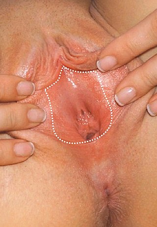
The vulval vestibule is the part of the vulva between the labia minora. On the inside, the urinary meatus and the vaginal opening open to the vestibule, while the outer edge is marked by Hart's line, named after David Berry Hart.

<span class="mw-page-title-main">Perineal nerve</span> Nerve of the perineum

The perineal nerve is a nerve of the pelvis. It arises from the pudendal nerve in the pudendal canal. It gives superficial branches to the skin, and a deep branch to muscles. It supplies the skin and muscles of the perineum. Its latency is tested with electrodes.

<span class="mw-page-title-main">Labioscrotal swelling</span> Embryonic structure of the reproductive system

The labioscrotal swellings are paired structures in the mammalian embryo that represent the final stage of development of the caudal end of the external genitals before sexual differentiation. In humans, the two swellings merge:

A commissure is the location at which two objects abut or are joined. The term is used especially in the fields of anatomy and biology.

<span class="mw-page-title-main">Posterior scrotal nerves</span>

The posterior scrotal branches are two in number, medial and lateral. They are branches of the perineal nerve, which is itself a branch of the pudendal nerve. The pudendal nerve arises from spinal roots S2 through S4, travels through the pudendal canal on the fascia of the obturator internus muscle, and gives off the perineal nerve in the perineum. The major branch of the perineal nerve is the posterior scrotal/posterior labial.

<span class="mw-page-title-main">Deep external pudendal artery</span> One of the pudendal arteries in human anatomy

The deep external pudendal artery is one of the pudendal arteries that is more deeply seated than the superficial external pudendal artery, passes medially across the pectineus and the adductor longus muscles; it is covered by the fascia lata, which it pierces at the medial side of the thigh, and is distributed, in the male, to the integument of the scrotum and perineum, in the female to the labia majora; its branches anastomose with the scrotal or labial branches of the perineal artery.

<span class="mw-page-title-main">Urogenital triangle</span> Anterior part of the perineum

The urogenital triangle is the anterior part of the perineum. In female mammals, it contains the vagina and associated parts of the internal genitalia.

<span class="mw-page-title-main">Posterior labial nerves</span>

The posterior labial nerves are branches of the pudendal nerve. They supply the female labia majora.

<span class="mw-page-title-main">Genital branch of genitofemoral nerve</span>

The genital branch of the genitofemoral nerve, also known as the external spermatic nerve in males, is a nerve in the abdomen that arises from the genitofemoral nerve. The genital branch supplies the cremaster muscle and anterior scrotal skin in males, and the skin of the mons pubis and labia majora in females.

The development of the reproductive system is the part of embryonic growth that results in the sex organs and contributes to sexual differentiation. Due to its large overlap with development of the urinary system, the two systems are typically described together as the genitourinary system.

<span class="mw-page-title-main">Clitoral hood reduction</span> Cosmetic surgical procedure

Clitoral hood reduction, also termed clitoral hoodectomy, clitoral unhooding, clitoridotomy, or (partial) hoodectomy, is a plastic surgery procedure for reducing the size and the area of the clitoral hood in order to further expose the glans of the clitoris.

<span class="mw-page-title-main">Vulva</span> External genitalia of the female mammal

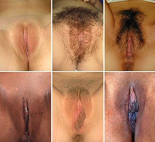
In mammals, the vulva consists of the external female genitalia. The human vulva includes the mons pubis, labia majora, labia minora, clitoris, vulval vestibule, urinary meatus, the vaginal opening, hymen, and Bartholin's and Skene's vestibular glands. The urinary meatus is also included as it opens into the vulval vestibule. The vulva includes the entrance to the vagina, which leads to the uterus, and provides a double layer of protection for this by the folds of the outer and inner labia. Pelvic floor muscles support the structures of the vulva. Other muscles of the urogenital triangle also give support.

Labial fusion is a medical condition of the vulva where the labia minora become fused together. It is generally a pediatric condition.

Crohn's disease (CD) of the vulva is a rare extra intestinal condition, with granulomatous cutaneous lesions affecting the female genitalia. Lesions connected to the affected gut via a healthy tissue are referred to as metastatic lesions.

References

  1. Blüm, Volker (2012). Vertebrate Reproduction: A Textbook. Springer Berlin Heidelberg. p. 74. ISBN   978-3-64271-074-2 . Retrieved November 19, 2023.
  2. "Sex Organ Homologies". www.meddean.luc.edu. Retrieved 2020-08-09.
  3. Manual of Obstetrics. (3rd ed.). Elsevier. pp. 1-16. ISBN   9788131225561.
  4. Kinsey, Brian (2011). Human Body From A to Z. Marshall Cavendish Reference. p. 465. ISBN   978-0-76149-976-3 . Retrieved September 29, 2023.
  5. Fahmy, Mohamed A. Baky (2021). Normal and Abnormal Scrotum. Springer International Publishing. p. 69. ISBN   978-3-03083-305-3 . Retrieved December 29, 2023.
  6. Gould, George M. (1936). Brownslow, C. V (ed.). Gould's Pocket Pronouncing Medical Dictionary (10 ed.). P. Blakinston's Son & C., Inc.
  7. Hennekam, RC; Allanson, JE; Biesecker, LG; Carey, JC; Opitz, JM; Vilain, E (June 2013). "Elements of morphology: standard terminology for the external genitalia". American Journal of Medical Genetics. Part A. 161A (6): 1238–63. doi:10.1002/ajmg.a.35934. PMC   4440541 . PMID   23650202.
  8. PD-icon.svgOne or more of the preceding sentences incorporates text in the public domain from page 1265 of  the 20th edition of Gray's Anatomy (1918)
  9. Sheets-Johnstone, Maxine (2010). The Roots of Thinking. Temple University Press. p. 184. ISBN   978-1-43990-365-0 . Retrieved November 19, 2023.
  10. Ankel-Simons, Friderun (2012). Primate Anatomy: An Introduction. Elsevier Science. p. 523. ISBN   978-0-08046-911-9 . Retrieved November 19, 2023.
  11. Rosenblum, Leonard A. (2013). Primate Behavior: Developments in Field and Laboratory Research · Volume 2. Elsevier Science. p. 251. ISBN   978-1-48327-182-8 . Retrieved November 19, 2023.
  12. Poiani, Aldo (2010). Animal Homosexuality: A Biosocial Perspective. Cambridge University Press. p. 156. ISBN   978-1-13949-038-2 . Retrieved November 19, 2023.
  13. Kumar, M.S.A. (2015). Clinically Oriented Anatomy of the Dog & Cat. Linus Learning. p. 1254. ISBN   978-1-60797-552-6.
  14. Carey, J. M.; Chon, J. K.; Leach, G. E. (2003). "Urethrolysis with martius labial fat pad graft for iatrogenic bladder outlet obstruction". Urology. 61 (4): 21–25. doi:10.1016/S0090-4295(03)00117-1. PMID   12657357.