
In amniotes, the clitoris is a female sex organ. In humans, it is the vulva's most erogenous area and generally the primary anatomical source of female sexual pleasure. The clitoris is a complex structure, and its size and sensitivity can vary. The visible portion, the glans, of the clitoris is typically roughly the size and shape of a pea and is estimated to have at least 8,000 nerve endings.
Clitoridectomy or clitorectomy is the surgical removal, reduction, or partial removal of the clitoris. It is rarely used as a therapeutic medical procedure, such as when cancer has developed in or spread to the clitoris. Commonly, non-medical removal of the clitoris is performed during female genital mutilation.

A clitoral hood piercing is a female genital piercing through the clitoral hood surrounding the clitoris. In addition to being an adornment, a clitoral hood piercing can enhance sexual pleasure during masturbation, foreplay and intercourse. In an empirical study at the University of South Alabama, the authors reported a positive relationship between vertical clitoral hood piercings and desire, frequency of intercourse, and sexual arousal. There are two main types of clitoral hood piercing: the vertical clitoral hood (VCH) piercing and the horizontal clitoral hood (HCH) piercing. As the names indicate, the difference is in the direction the piercing is oriented in the skin above the clitoris. Neither of these piercings penetrates the clitoris itself, although in common parlance they are sometimes called "clit" piercings. The deep hood piercing is a variation of the clitoral hood piercing that passes deeper through the clitoral hood.

In human anatomy, and in mammals in general, the mons pubis or pubic mound is a rounded mass of fatty tissue found over the pubic symphysis of the pubic bones.

The pubic symphysis is a secondary cartilaginous joint between the left and right superior rami of the pubis of the hip bones. It is in front of and below the urinary bladder. In males, the suspensory ligament of the penis attaches to the pubic symphysis. In females, the pubic symphysis is attached to the suspensory ligament of the clitoris. In most adults, it can be moved roughly 2 mm and with 1 degree rotation. This increases for women at the time of childbirth.

The bulbospongiosus muscles are a subgroup of the superficial muscles of the perineum. They have a slightly different origin, insertion and function in males and females. In males, these muscles cover the bulb of the penis, while in females, they cover the vestibular bulbs.

The female reproductive system is made up of the internal and external sex organs that function in the reproduction of new offspring. The human female reproductive system is immature at birth and develops to maturity at puberty to be able to produce gametes, and to carry a fetus to full term. The internal sex organs are the vagina, uterus, fallopian tubes, and ovaries. The female reproductive tract includes the vagina, uterus, and fallopian tubes and is prone to infections. The vagina allows for sexual intercourse and childbirth, and is connected to the uterus at the cervix. The uterus or womb accommodates the embryo, which develops into the fetus. The uterus also produces secretions, which help the transit of sperm to the fallopian tubes, where sperm fertilize ova produced by the ovaries. The external sex organs are also known as the genitals and these are the organs of the vulva including the labia, clitoris, and vaginal opening.
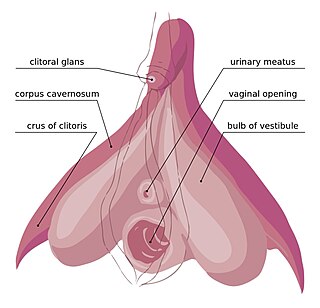
The clitoral crura are two erectile tissue structures, which together form a "V" shape. Crus is a Latin word that means "leg". Each "leg" of the V converges on the clitoral body. At each divergent point is a corpus cavernosum. Together with the vestibular bulbs, they form the clitoral root. The crura are attached to the pubic arch, and are adjacent to the vestibular bulbs. The crura flank the urethra, urethral sponge, and vagina and extend back toward the pubis. Each clitoral crus connects to the rami of the pubis and the ischium.

In vertebrates, the pubis or pubic bone forms the lower and anterior part of each side of the hip bone. The pubis is the most forward-facing of the three bones that make up the hip bone. The left and right pubic bones are each made up of three sections; a superior ramus, an inferior ramus, and a body.

The suspensory ligament of the penis is a triangular midline structure anchoring the penis to the pubic symphysis, holding the penis close to the pubic bone and supporting it during erection.

The fascia of Scarpa is the deep membranous layer (stratum membranosum) of the superficial fascia of the abdomen. It is a layer of the anterior abdominal wall. It is found deep to the fascia of Camper and superficial to the external oblique muscle.

The dorsal artery of the penis is a bilaterally paired terminal branch of the internal pudendal artery which passes upon the dorsum of the penis to the base of the glans penis, where it unites with its contralateral partner and supply the glans and foreskin.

The dorsal nerve of the penis is the deepest of three divisions of the pudendal nerve; it accompanies the internal pudendal artery along the ramus of the ischium; it then runs forward along the margin of the inferior ramus of the pubis, between the superior and inferior layers of the fascia of the urogenital diaphragm.

The corpus cavernosum of clitoris is one of a pair of sponge-like regions of erectile tissue of the clitoris. It is made of a sponge-like tissue that fills with blood during erection. This is homologous to the corpus cavernosum penis. The term corpora cavernosa literally means "cave-like bodies".

The perineal membrane is an anatomical term for a fibrous membrane in the perineum. The term "inferior fascia of urogenital diaphragm", used in older texts, is considered equivalent to the perineal membrane.
The dorsal nerve of the clitoris is a nerve in females that branches off the pudendal nerve to innervate the clitoris. The nerve is important for female sexual pleasure, and it may play a role in clitoral erections.

In human male anatomy, the dorsal veins of the penis are blood vessels that drain the shaft, the skin and the glans of the human penis. They are typically located in the midline on the dorsal aspect of the penis and they comprise the superficial dorsal veinof the penis, that lies in the subcutaneous tissue of the shaft, and the deep dorsal veinof the penis, that lies beneath the deep fascia.

The body or shaft of the penis is the free portion of the human penis that is located outside of the pelvic cavity. It is the continuation of the internal root, which is embedded in the pelvis and extends to the glans. It is made up of the two corpora cavernosa and the corpus spongiosum on the underside. The corpora cavernosa are intimately bound to one another with a dorsally fenestrated septum, which becomes a complete one before the penile crura. The body of the penis is homologous to the female clitoral body.

In human male anatomy, the radix or root of the penis is the internal and most proximal portion of the human penis that lies in the perineum. Unlike the pendulous body of the penis, which is suspended from the pubic symphysis, the root is attached to the pubic arch of the pelvis and is not visible externally. It is triradiate in form, consisting of three masses of erectile tissue; the two diverging crura, one on either side, and the median bulb of the penis or urethral bulb. Approximately one third to one half of the penis is embedded in the pelvis and can be felt through the scrotum and in the perineum.

Clitoral hood reduction, also termed clitoral hoodectomy, clitoral unhooding, clitoridotomy, or (partial) hoodectomy, is a plastic surgery procedure for reducing the size and the area of the clitoral hood in order to further expose the glans of the clitoris.
