Uterine cancer

Last updated
Uterine cancer
Other namesWomb cancer
Uterus with Cancer Origins.jpg
Diagram of uterus with labeled origins of cancer types.
Specialty Gynecology, oncology
Symptoms Endometrial cancer: vaginal bleeding, pelvic pain [1]
Uterine sarcoma: vaginal bleeding, mass in the vagina [2]
Types Endometrial cancer, uterine sarcoma [3]
Risk factors Endometrial cancer: obesity, metabolic syndrome, type 2 diabetes, tamoxifen use, unopposed estrogens, late menopause, family history of the condition [1]
Uterine sarcoma: radiation therapy to the pelvis [2]
Treatment Surgery, radiation therapy, chemotherapy, hormone therapy, targeted therapy [1] [2]
Frequency3.8 million (2015) [4]
Deaths90,000 (2015) [5]

Uterine cancer, also known as womb cancer, includes two types of cancer that develop from the tissues of the uterus. [3] Endometrial cancer forms from the lining of the uterus, and uterine sarcoma forms from the muscles or support tissue of the uterus. [1] [2] Endometrial cancer accounts for approximately 90% of all uterine cancers in the United States. [6] Symptoms of endometrial cancer include changes in vaginal bleeding or pain in the pelvis. [1] Symptoms of uterine sarcoma include unusual vaginal bleeding or a mass in the vagina. [2]

Contents

Risk factors for endometrial cancer include obesity, metabolic syndrome, type 2 diabetes, taking pills that contain estrogen without progesterone, a history of tamoxifen use, late menopause, and a family history of the condition. [7] [1] Risk factors for uterine sarcoma include prior radiation therapy to the pelvis. [2] Diagnosis of endometrial cancer is typically based on an endometrial biopsy. [1] A diagnosis of uterine sarcoma may be suspected based on symptoms, a pelvic exam, and medical imaging. [2]

Endometrial cancer can often be cured while uterine sarcoma typically is harder to treat. [3] Treatment may include a combination of surgery, radiation therapy, chemotherapy, hormone therapy, and targeted therapy. [1] [2] Just over 80% of women survive more than 5 years following diagnosis. [8]

In 2015 about 3.8 million women were affected globally and it resulted in 90,000 deaths. [4] [5] Endometrial cancer is relatively common while uterine sarcomas are rare. [3] In the United States, uterine cancers represent 3.5% of new cancer cases. [8] They most commonly occur in women between the ages of 45 and 74 with a median age of diagnosis of 63. [8]

Types

The terms uterine cancer and womb cancer may refer to several different types of cancer that occur in the uterus, namely:

Endometrial cancer

Uterine sarcomas

Signs and symptoms

Both types of uterine cancer can present with abnormal vaginal bleeding and discharge. [11] [12] Abnormalities can include change in duration or amount of menstrual bleeding as well as new bleeding between menses or after menopause. Sensations of new or increasing pelvic pressure or pain can also indicate tumor growth in the uterus. [12] Any of these findings warrant further workup by a doctor.

Causes and risk factors

It is not known with certainty what the causes for uterine cancer may be, though hormone imbalance is cited as a risk factor. Estrogen receptors, known to be present on the surfaces of cells of this type of cancer, are thought to interact with the hormone causing increased cell growth, which can then result in cancer. The exact mechanism of how this occurs is not understood. [13]

Risk factors for endometrial cancer include obesity, metabolic syndrome, type 2 diabetes, taking pills that contain estrogen without progesterone, a history of tamoxifen use, late menopause, and certain hereditary conditions (Lynch syndrome, Cowden syndrome). [7] [1] [11] Risk factors for uterine sarcoma include prior radiation therapy to the pelvis, a history of tamoxifen use, a history of childhood retinoblastoma, and hereditary leiomyomatosis and renal cell carcinoma (HLRCC) syndrome. [2] [12]

Diagnosis

To evaluate for uterine cancer, a clinician might perform a pelvic exam to visually inspect internal pelvic organs and to feel the size and position of the uterus and ovaries. A “pap smear” may also be done to brush the sides of the cervix to collect cells for testing and to look at under a microscope. A dilatation and curettage is often done to collect a sample of uterine lining tissue. An ultrasound is also often performed to look for tumors. [14]

Screening and Prevention

Screening for uterine cancers is not recommended except for in women with certain hereditary conditions that increase their risk (Lynch, Cowden, HLRCC). [11] [12]

In general, combined oral contraceptive pills and progestin-only pills are protective against uterine cancers. Weight loss and/or bariatric surgery reduces the risk for patients with obesity. [11]

Treatment

Treatment of uterine cancer may differ depending on the type of cancer and staging of the tumor. [15] In early stages, minimal invasive surgery is preferred. [16]

For endometrial cancer, five main types of treatments are used, including surgery, radiation therapy, chemotherapy, hormone therapy, and targeted therapy. The most common treatment modality for endometrial cancer is surgery, whereby the uterus is removed via a total hysterectomy. [15] Hysterectomies may also be accompanied by removal of ovaries and fallopian tubes, called a salpingo-oophorectomy. Additionally, hormone therapy which seeks to block the growth of cancer cells may also be used in the treatment of endometrial cancer. Targeted therapy may include monoclonal antibodies, mTOR inhibitors, and signal transduction inhibitors which all act to target cancer cells specifically. [15]

As of 2021, treatment options for uterine sarcoma include surgery, radiation therapy, chemotherapy, and hormone therapy. [15]

Prognosis

Prognosis varies for the different types of endometrial cancer. Factors that influence prognosis across types of uterine cancer are age at diagnosis, the stage of the cancer, the grade of the cancer, histology, depth of invasion into the myometrium, and the presence of spread to nearby lymph nodes or other regions. [17] Endometrial cancer typically has a good 5-year-survival when diagnosed early. [18] Generally, the prognosis is poorer for uterine sarcomas as compared to endometrial cancers. [19]

Epidemiology

Age-standardized death from cancer of the uterine body per 100,000 inhabitants in 2004.
.mw-parser-output .div-col{margin-top:0.3em;column-width:30em}.mw-parser-output .div-col-small{font-size:90%}.mw-parser-output .div-col-rules{column-rule:1px solid #aaa}.mw-parser-output .div-col dl,.mw-parser-output .div-col ol,.mw-parser-output .div-col ul{margin-top:0}.mw-parser-output .div-col li,.mw-parser-output .div-col dd{page-break-inside:avoid;break-inside:avoid-column}
.mw-parser-output .legend{page-break-inside:avoid;break-inside:avoid-column}.mw-parser-output .legend-color{display:inline-block;min-width:1.25em;height:1.25em;line-height:1.25;margin:1px 0;text-align:center;border:1px solid black;background-color:transparent;color:black}.mw-parser-output .legend-text{}
no data
less than 0.5
0.5-1
1-1.5
1.5-2
2-2.5
2.5-3
3-3.5
3.5-4
4-4.5
4.5-5
5-8
more than 8 Corpus uteri cancer world map - Death - WHO2004.svg
Age-standardized death from cancer of the uterine body per 100,000 inhabitants in 2004.
  no data
  less than 0.5
  0.5–1
  1–1.5
  1.5–2
  2–2.5
  2.5–3
  3–3.5
  3.5–4
  4–4.5
  4.5–5
  5–8
  more than 8

Uterine cancer effects approximately 3.1% of females during their lifetime. [8] Uterine cancer resulted in 45,000 deaths worldwide in 1990, with this number increasing to 58,000 deaths in 2010. [21] North America and Northern Europe have the highest rates of uterine cancer. Asia, Southern Europe, Australia and South America have moderate rates, with the lowest rates in Africa and Eastern Asia. [22] About 81% of women with uterine cancer survive for five years. This rate is higher with more localized cancer at 95% survival rate for five years and lower for a distant spread of the cancer, at a 16.8% survival rate for five years. [8]

United Kingdom

Uterine cancer is the fourth most common cancer in females in the UK (around 8,500 women were diagnosed with the disease in 2011), and it is the tenth most common cause of cancer death in females (around 2,000 women died in 2012). [23]

United States

Uterine cancer has a high prevalence in the United States, with approximately 772,247 women with the disease in 2016. [24] Of those uterine cancers, approximately 90% of the cases are endometrial cancers. [6] This is the fourth most commonly diagnosed type of cancer. [25]

In the United States, uterine cancer is the most common invasive gynecologic cancer. [22] The number of women diagnosed with uterine cancer has been steadily increasing, with 35,040 diagnosed in 1999 and 56,808 diagnosed in 2016. The age-adjusted rate of new cases in 1999 was 23.9 per 100,000 and has increased to 27.3 per 100,000 in 2016. [26] The incidence of uterine cancer increased even more in 2019, with an approximated 61,880 new cases. [24]

The rates of incidence and death for uterine cancer differ depending on race. The rate of diagnosis is highest for white females, with 28.1 new cases per 100,000 persons. Black females have a similar incidence with 27.4 new cases per 100,000 persons. Other ethnic groups had lower incidences: Hispanic females had 24.1 new cases per 100,000 persons, Asian/Pacific Islander females had 20.8 new cases per 100,000 persons, and American Indian/Alaska Native females had 19.7 new cases per 100,000 persons. For the death rates of uterine cancer, black females had the highest rates, 8.5 deaths per 100,000 persons. The death rates for the other ethnic groups were dramatically lower. White females had 4.4 deaths per 100,000 persons, Hispanic females had 3.9 deaths per 100,000 persons, American Indian/Alaska Native females had 3.5 deaths per 100,000 persons, and Asian/Pacific Islander females had 3.1 deaths per 100,000 persons. [8]

Research

As current diagnostic methods are invasive and inaccurate, researchers are looking into new ways to catch womb cancer, especially in its early stages. A study found that using a technique involving infrared light on simple blood test samples detected uterine cancer with high accuracy (87%), and could detect precancerous growths in all cases. [27] [28]

Related Research Articles

<span class="mw-page-title-main">Endometrium</span> Inner mucous membrane of the mammalian uterus

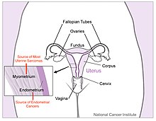
The endometrium is the inner epithelial layer, along with its mucous membrane, of the mammalian uterus. It has a basal layer and a functional layer: the basal layer contains stem cells which regenerate the functional layer. The functional layer thickens and then is shed during menstruation in humans and some other mammals, including apes, Old World monkeys, some species of bat, the elephant shrew and the Cairo spiny mouse. In most other mammals, the endometrium is reabsorbed in the estrous cycle. During pregnancy, the glands and blood vessels in the endometrium further increase in size and number. Vascular spaces fuse and become interconnected, forming the placenta, which supplies oxygen and nutrition to the embryo and fetus. The speculated presence of an endometrial microbiota has been argued against.

<span class="mw-page-title-main">Breast cancer</span> Cancer that originates in mammary glands

Breast cancer is a cancer that develops from breast tissue. Signs of breast cancer may include a lump in the breast, a change in breast shape, dimpling of the skin, milk rejection, fluid coming from the nipple, a newly inverted nipple, or a red or scaly patch of skin. In those with distant spread of the disease, there may be bone pain, swollen lymph nodes, shortness of breath, or yellow skin.

<span class="mw-page-title-main">Sarcoma</span> Type of malignant tumor (cancer)

A sarcoma is a malignant tumor, a type of cancer that arises from cells of mesenchymal origin. Connective tissue is a broad term that includes bone, cartilage, fat, vascular, or other structural tissues, and sarcomas can arise in any of these types of tissues. As a result, there are many subtypes of sarcoma, which are classified based on the specific tissue and type of cell from which the tumor originates. Sarcomas are primary connective tissue tumors, meaning that they arise in connective tissues. This is in contrast to secondary connective tissue tumors, which occur when a cancer from elsewhere in the body spreads to the connective tissue. Sarcomas are one of five different types of cancer, classified by the cell type from which they originate. The word sarcoma is derived from the Greek σάρκωμα sarkōma 'fleshy excrescence or substance', itself from σάρξsarx meaning 'flesh'.

<span class="mw-page-title-main">Endometrial cancer</span> Uterine cancer that is located in tissues lining the uterus

Endometrial cancer is a cancer that arises from the endometrium. It is the result of the abnormal growth of cells that have the ability to invade or spread to other parts of the body. The first sign is most often vaginal bleeding not associated with a menstrual period. Other symptoms include pain with urination, pain during sexual intercourse, or pelvic pain. Endometrial cancer occurs most commonly after menopause.

<span class="mw-page-title-main">Kidney cancer</span> Medical condition

Kidney cancer, also known as renal cancer, is a group of cancers that starts in the kidney. Symptoms may include blood in the urine, a lump in the abdomen, or back pain. Fever, weight loss, and tiredness may also occur. Complications can include spread to the lungs or brain.

<span class="mw-page-title-main">Oral cancer</span> Cancer of the lining of the lips, mouth, or upper throat

Oral cancer, also known as mouth cancer, is a cancer of the lining of the lips, mouth, or upper throat. In the mouth, it most commonly starts as a painless white patch, that thickens, develops red patches, an ulcer, and continues to grow. When on the lips, it commonly looks like a persistent crusting ulcer that does not heal, and slowly grows. Other symptoms may include difficult or painful swallowing, new lumps or bumps in the neck, a swelling in the mouth, or a feeling of numbness in the mouth or lips.

<span class="mw-page-title-main">Ovarian cancer</span> Cancer originating in or on the ovary

Ovarian cancer is a cancerous tumor of an ovary. It may originate from the ovary itself or more commonly from communicating nearby structures such as fallopian tubes or the inner lining of the abdomen. The ovary is made up of three different cell types including epithelial cells, germ cells, and stromal cells. When these cells become abnormal, they have the ability to divide and form tumors. These cells can also invade or spread to other parts of the body. When this process begins, there may be no or only vague symptoms. Symptoms become more noticeable as the cancer progresses. These symptoms may include bloating, vaginal bleeding, pelvic pain, abdominal swelling, constipation, and loss of appetite, among others. Common areas to which the cancer may spread include the lining of the abdomen, lymph nodes, lungs, and liver.

<span class="mw-page-title-main">Serous tumour</span> Medical condition

A serous tumour is a neoplasm that typically has papillary to solid formations of tumor cells with crowded nuclei, and which typically arises on the modified Müllerian-derived serous membranes that surround the ovaries in females. Such ovarian tumors are part of the surface epithelial-stromal tumour group of ovarian tumors. They are common neoplasms with a strong tendency to occur bilaterally, and they account for approximately a quarter of all ovarian tumors.

<span class="mw-page-title-main">Adenomyosis</span> Extension of endometrial tissue into the myometrium

Adenomyosis is a medical condition characterized by the growth of cells that proliferate on the inside of the uterus (endometrium) atypically located among the cells of the uterine wall (myometrium), as a result, thickening of the uterus occurs. As well as being misplaced in patients with this condition, endometrial tissue is completely functional. The tissue thickens, sheds and bleeds during every menstrual cycle.

<span class="mw-page-title-main">Uterine fibroid</span> Medical condition with benign tumors of uterus

Uterine fibroids, also known as uterine leiomyomas or fibroids, are benign smooth muscle tumors of the uterus. Most women with fibroids have no symptoms while others may have painful or heavy periods. If large enough, they may push on the bladder, causing a frequent need to urinate. They may also cause pain during penetrative sex or lower back pain. A woman can have one uterine fibroid or many. Occasionally, fibroids may make it difficult to become pregnant, although this is uncommon.

<span class="mw-page-title-main">Invasive carcinoma of no special type</span> Medical condition

Invasive carcinoma of no special type, invasive breast carcinoma of no special type (IBC-NST), invasive ductal carcinoma (IDC), infiltrating ductal carcinoma (IDC) or invasive ductal carcinoma, not otherwise specified (NOS) is a disease. For international audiences this article will use "invasive carcinoma NST" because it is the preferred term of the World Health Organization (WHO).

<span class="mw-page-title-main">Leiomyosarcoma</span> Medical condition

A leiomyosarcoma, also known as LMS, is a rare malignant (cancerous) smooth muscle tumor. The origin of the word is from leio- + myo- + sarcoma which means malignant smooth muscle tumor. The stomach, bladder, uterus, blood vessels, and intestines are examples of hollow organs made up of smooth muscles where LMS can be located, however the uterus or abdomen are the most common sites.

Adenomyoma is a tumor (-oma) including components derived from glands (adeno-) and muscle (-my-). It is a type of complex and mixed tumor, and several variants have been described in the medical literature. Uterine adenomyoma, the localized form of uterine adenomyosis, is a tumor composed of endometrial gland tissue and smooth muscle in the myometrium. Adenomyomas containing endometrial glands are also found outside of the uterus, most commonly on the uterine adnexa but can also develop at distant sites outside of the pelvis. Gallbladder adenomyoma, the localized form of adenomyomatosis, is a polypoid tumor in the gallbladder composed of hyperplastic mucosal epithelium and muscularis propria.

The uterine sarcomas form a group of malignant tumors that arises from the smooth muscle or connective tissue of the uterus.

<span class="mw-page-title-main">Endometrial intraepithelial neoplasia</span>

Endometrial intraepithelial neoplasia (EIN) is a premalignant lesion of the uterine lining that predisposes to endometrioid endometrial adenocarcinoma. It is composed of a collection of abnormal endometrial cells, arising from the glands that line the uterus, which have a tendency over time to progress to the most common form of uterine cancer—endometrial adenocarcinoma, endometrioid type.

<span class="mw-page-title-main">Mixed Müllerian tumor</span> Medical condition

A malignant mixed Müllerian tumor, also known as malignant mixed mesodermal tumor (MMMT) is a cancer found in the uterus, the ovaries, the fallopian tubes and other parts of the body that contains both carcinomatous and sarcomatous components. It is divided into two types, homologous and a heterologous type. MMMT account for between two and five percent of all tumors derived from the body of the uterus, and are found predominantly in postmenopausal women with an average age of 66 years. Risk factors are similar to those of adenocarcinomas and include obesity, exogenous estrogen therapies, and nulliparity. Less well-understood but potential risk factors include tamoxifen therapy and pelvic irradiation.

<span class="mw-page-title-main">Endometrial hyperplasia</span> Medical condition

Endometrial hyperplasia is a condition of excessive proliferation of the cells of the endometrium, or inner lining of the uterus.

<span class="mw-page-title-main">Uterine serous carcinoma</span> Type of cancer of the uterus

Uterine serous carcinoma is a malignant form of serous tumor that originates in the uterus. It is an uncommon form of endometrial cancer that typically arises in postmenopausal women. It is typically diagnosed on endometrial biopsy, prompted by post-menopausal bleeding.

Uterine clear-cell carcinoma (CC) is a rare form of endometrial cancer with distinct morphological features on pathology; it is aggressive and has high recurrence rate. Like uterine papillary serous carcinoma CC does not develop from endometrial hyperplasia and is not hormone sensitive, rather it arises from an atrophic endometrium. Such lesions belong to the type II endometrial cancers.

Neuroendocrine carcinoma of the cervix is best defined separately:Neuroendocrine: Of, relating to, or involving the interaction between the nervous system and the hormones of the endocrine glands.Carcinoma: An invasive malignant tumor derived from epithelial tissue that tends to metastasize to other areas of the body.

References

  1. 1 2 3 4 5 6 7 8 9 "Endometrial Cancer Treatment". National Cancer Institute. 26 April 2018. Retrieved 3 February 2019.
  2. 1 2 3 4 5 6 7 8 9 "Uterine Sarcoma Treatment". National Cancer Institute. 3 October 2018. Retrieved 3 February 2019.
  3. 1 2 3 4 "Uterine Cancer". National Cancer Institute. 1 January 1980. Retrieved 3 February 2019.
  4. 1 2 "Global, regional, and national incidence, prevalence, and years lived with disability for 310 diseases and injuries, 1990-2015: a systematic analysis for the Global Burden of Disease Study 2015". Lancet. 388 (10053): 1545–1602. October 2016. doi:10.1016/S0140-6736(16)31678-6. PMC   5055577 . PMID   27733282.
  5. 1 2 "Global, regional, and national life expectancy, all-cause mortality, and cause-specific mortality for 249 causes of death, 1980-2015: a systematic analysis for the Global Burden of Disease Study 2015". Lancet. 388 (10053): 1459–1544. October 2016. doi:10.1016/S0140-6736(16)31012-1. PMC   5388903 . PMID   27733281.
  6. 1 2 Felix AS, Brinton LA (September 2018). "Cancer Progress and Priorities: Uterine Cancer". Cancer Epidemiology, Biomarkers & Prevention. 27 (9): 985–994. doi:10.1158/1055-9965.EPI-18-0264. PMC   6504985 . PMID   30181320.
  7. 1 2 "Endometrial Cancer Risk Factors". www.cancer.org. Retrieved 2021-09-13.
  8. 1 2 3 4 5 6 "Uterine Cancer - Cancer Stat Facts". SEER. Retrieved 3 February 2019.
  9. 1 2 "What Is Endometrial Cancer?". www.cancer.org. Retrieved 2021-09-14.
  10. 1 2 "What Is Uterine Sarcoma?". www.cancer.org. Retrieved 2021-09-14.
  11. 1 2 3 4 "Endometrial carcinoma: Clinical features, diagnosis, prognosis, and screening". www.uptodate.com. Retrieved 2021-09-18.
  12. 1 2 3 4 "Uterine sarcoma: Classification, epidemiology, clinical manifestations, and diagnosis". www.uptodate.com. Retrieved 2021-09-18.
  13. Causes, Risk Factors, and Prevention TOPICS - Do we know what causes endometrial cancer? - cancer.org - American Cancer Society - Retrieved 5 January 2015.
  14. "Uterine Cancer - Diagnosis". Cancer.Net. 2012-06-25. Retrieved 2021-09-14.
  15. 1 2 3 4 "Uterine Cancer—Patient Version - National Cancer Institute". www.cancer.gov. Retrieved 2021-09-13.
  16. Filippova OT, Leitao MM (July 2020). "The current clinical approach to newly diagnosed uterine cancer". Expert Review of Anticancer Therapy. 20 (7): 581–590. doi:10.1080/14737140.2020.1782750. PMC   7416456 . PMID   32531179.
  17. Uharcek P (October 2008). "Prognostic factors in endometrial carcinoma". The Journal of Obstetrics and Gynaecology Research. 34 (5): 776–783. doi: 10.1111/j.1447-0756.2008.00796.x . PMID   18958927.
  18. "Survival Rates for Endometrial Cancer". www.cancer.org. Retrieved 2021-09-14.
  19. "Survival Rates for Uterine Sarcoma". www.cancer.org. Retrieved 2021-09-14.
  20. "WHO Disease and injury country estimates". World Health Organization. 2009. Retrieved Nov 11, 2009.
  21. Lozano R, Naghavi M, Foreman K, Lim S, Shibuya K, Aboyans V, et al. (December 2012). "Global and regional mortality from 235 causes of death for 20 age groups in 1990 and 2010: a systematic analysis for the Global Burden of Disease Study 2010". Lancet. 380 (9859): 2095–2128. doi:10.1016/S0140-6736(12)61728-0. hdl: 10536/DRO/DU:30050819 . PMC   10790329 . PMID   23245604. S2CID   1541253.
  22. 1 2 Felix AS, Brinton LA (September 2018). "Cancer Progress and Priorities: Uterine Cancer". Cancer Epidemiology, Biomarkers & Prevention. 27 (9): 985–994. doi:10.1158/1055-9965.EPI-18-0264. PMC   6504985 . PMID   30181320.
  23. "Uterine cancer statistics". Cancer Research UK. Retrieved 28 October 2014.
  24. 1 2 "Cancer of the Endometrium - Cancer Stat Facts". SEER. Retrieved 2019-11-12.
  25. Felix AS, Brinton LA (September 2018). "Cancer Progress and Priorities: Uterine Cancer". Cancer Epidemiology, Biomarkers & Prevention. 27 (9): 985–994. doi:10.1158/1055-9965.EPI-18-0264. PMC   6504985 . PMID   30181320.
  26. "USCS Data Visualizations". gis.cdc.gov. Archived from the original on 2019-01-25. Retrieved 2019-11-12.
  27. "Womb cancer could be detected early with an inexpensive new blood test". NIHR Evidence (Plain English summary). 2020-11-12. doi:10.3310/alert_42575. S2CID   241679195.
  28. Paraskevaidi M, Morais CL, Ashton KM, Stringfellow HF, McVey RJ, Ryan NA, et al. (May 2020). "Detecting Endometrial Cancer by Blood Spectroscopy: A Diagnostic Cross-Sectional Study". Cancers. 12 (5): 1256. doi: 10.3390/cancers12051256 . PMC   7281323 . PMID   32429365.