Lymphatic system

Last updated
Lymphatic system
Blausen 0623 LymphaticSystem Female.png
Human lymphatic system
Details
Identifiers
Latin systema lymphoideum
MeSH D008208
TA98 A13.0.00.000
TA2 5149
FMA 7162 74594, 7162
Anatomical terminology

The lymphatic system, or lymphoid system, is an organ system in vertebrates that is part of the immune system, and complementary to the circulatory system. It consists of a large network of lymphatic vessels, lymph nodes, lymphoid organs, lymphatic tissue and lymph. [1] [2] Lymph is a clear fluid carried by the lymphatic vessels back to the heart for re-circulation. The Latin word for lymph, lympha, refers to the deity of fresh water, "Lympha". [3]

Contents

Unlike the circulatory system that is a closed system, the lymphatic system is open. [4] [5] The human circulatory system processes an average of 20 litres of blood per day through capillary filtration, which removes plasma from the blood. Roughly 17 litres of the filtered blood is reabsorbed directly into the blood vessels, while the remaining three litres are left in the interstitial fluid. One of the main functions of the lymphatic system is to provide an accessory return route to the blood for the surplus three litres. [6]

The other main function is that of immune defense. Lymph is very similar to blood plasma, in that it contains waste products and cellular debris, together with bacteria and proteins. The cells of the lymph are mostly lymphocytes. Associated lymphoid organs are composed of lymphoid tissue, and are the sites either of lymphocyte production or of lymphocyte activation. These include the lymph nodes (where the highest lymphocyte concentration is found), the spleen, the thymus, and the tonsils. Lymphocytes are initially generated in the bone marrow. The lymphoid organs also contain other types of cells such as stromal cells for support. [7] Lymphoid tissue is also associated with mucosas such as mucosa-associated lymphoid tissue (MALT). [8]

Fluid from circulating blood leaks into the tissues of the body by capillary action, carrying nutrients to the cells. The fluid bathes the tissues as interstitial fluid, collecting waste products, bacteria, and damaged cells, and then drains as lymph into the lymphatic capillaries and lymphatic vessels. These vessels carry the lymph throughout the body, passing through numerous lymph nodes which filter out unwanted materials such as bacteria and damaged cells. Lymph then passes into much larger lymph vessels known as lymph ducts. The right lymphatic duct drains the right side of the region and the much larger left lymphatic duct, known as the thoracic duct, drains the left side of the body. The ducts empty into the subclavian veins to return to the blood circulation. Lymph is moved through the system by muscle contractions. [9] In some vertebrates, a lymph heart is present that pumps the lymph to the veins. [9] [10]

The lymphatic system was first described in the 17th century independently by Olaus Rudbeck and Thomas Bartholin. [11]

Structure

Anatomy of the lymphatic system showing primary and secondary lymphoid organs Anatomy of the lymphatic system.jpg
Anatomy of the lymphatic system showing primary and secondary lymphoid organs

The lymphatic system consists of a conducting network of lymphatic vessels, lymphoid organs, lymphoid tissues, and the circulating lymph. [1]

Primary lymphoid organs

The primary (or central) lymphoid organs generate lymphocytes from immature progenitor cells. The thymus and the bone marrow constitute the primary lymphoid organs involved in the production and early clonal selection of lymphocyte tissues.

Bone marrow

Bone marrow is responsible for both the creation of T cell precursors and the production and maturation of B cells, which are important cell types of the immune system. From the bone marrow, B cells immediately join the circulatory system and travel to secondary lymphoid organs in search of pathogens. T cells, on the other hand, travel from the bone marrow to the thymus, where they develop further and mature. Mature T cells then join B cells in search of pathogens. The other 95% of T cells begin a process of apoptosis, a form of programmed cell death.

Thymus

The thymus increases in size from birth in response to postnatal antigen stimulation. It is most active during the neonatal and pre-adolescent periods. The thymus is located between the inferior neck and the superior thorax. At puberty, by the early teens, the thymus begins to atrophy and regress, with adipose tissue mostly replacing the thymic stroma. However, residual T cell lymphopoiesis continues throughout adult life, providing some immune response. The thymus is where the T lymphocytes mature and become immunocompetent. The loss or lack of the thymus results in severe immunodeficiency and subsequent high susceptibility to infection. In most species, the thymus consists of lobules divided by septa which are made up of epithelium which is often considered an epithelial organ. T cells mature from thymocytes, proliferate, and undergo a selection process in the thymic cortex before entering the medulla to interact with epithelial cells.

Research on bony fish showed a buildup of T cells in the thymus and spleen of lymphoid tissues in salmon and showed that there are not many T cells in non-lymphoid tissues. [12]

The thymus provides an inductive environment for the development of T cells from hematopoietic progenitor cells. In addition, thymic stromal cells allow for the selection of a functional and self-tolerant T cell repertoire. Therefore, one of the most important roles of the thymus is the induction of central tolerance. However, the thymus is not where the infection is fought, as the T cells have yet to become immunocompetent.

Secondary lymphoid organs

The secondary (or peripheral) lymphoid organs, which include lymph nodes and the spleen, maintain mature naive lymphocytes and initiate an adaptive immune response. [13] The secondary lymphoid organs are the sites of lymphocyte activation by antigens. [14] Activation leads to clonal expansion, and affinity maturation. Mature lymphocytes recirculate between the blood and the secondary lymphoid organs until they encounter their specific antigen.

Spleen

The main functions of the spleen are:

  1. to produce immune cells to fight antigens
  2. to remove particulate matter and aged blood cells, mainly red blood cells
  3. to produce blood cells during fetal life.

The spleen synthesizes antibodies in its white pulp and removes antibody-coated bacteria and antibody-coated blood cells by way of blood and lymph node circulation. The white pulp of the spleen provides immune function due to the lymphocytes that are housed there. The spleen also consists of red pulp which is responsible for getting rid of aged red blood cells, as well as pathogens. This is carried out by macrophages present in the red pulp. A study published in 2009 using mice found that the spleen contains, in its reserve, half of the body's monocytes within the red pulp. [15] These monocytes, upon moving to injured tissue (such as the heart), turn into dendritic cells and macrophages while promoting tissue healing. [15] [16] [17] The spleen is a center of activity of the mononuclear phagocyte system and can be considered analogous to a large lymph node, as its absence causes a predisposition to certain infections. Notably, the spleen is important for a multitude of functions. The spleen removes pathogens and old erythrocytes from the blood (red pulp) and produces lymphocytes for immune response (white pulp). The spleen also is responsible for recycling some erythrocytes components and discarding others. For example, hemoglobin is broken down into amino acids that are reused.

Research on bony fish has shown that a high concentration of T cells are found in the white pulp of the spleen. [12]

Like the thymus, the spleen has only efferent lymphatic vessels. Both the short gastric arteries and the splenic artery supply it with blood. [18] The germinal centers are supplied by arterioles called penicilliary radicles. [19]

In the human until the fifth month of prenatal development, the spleen creates red blood cells; after birth, the bone marrow is solely responsible for hematopoiesis. As a major lymphoid organ and a central player in the reticuloendothelial system, the spleen retains the ability to produce lymphocytes. The spleen stores red blood cells and lymphocytes. It can store enough blood cells to help in an emergency. Up to 25% of lymphocytes can be stored at any one time. [20]

Lymph nodes

A lymph node showing afferent and efferent lymphatic vessels Illu lymph node structure.png
A lymph node showing afferent and efferent lymphatic vessels
Regional lymph nodes Lymph node regions.svg
Regional lymph nodes

A lymph node is an organized collection of lymphoid tissue, through which the lymph passes on its way back to the blood. Lymph nodes are located at intervals along the lymphatic system. Several afferent lymph vessels bring in lymph, which percolates through the substance of the lymph node, and is then drained out by an efferent lymph vessel. Of the nearly 800 lymph nodes in the human body, about 300 are located in the head and neck. [21] Many are grouped in clusters in different regions, as in the underarm and abdominal areas. Lymph node clusters are commonly found at the proximal ends of limbs (groin, armpits) and in the neck, where lymph is collected from regions of the body likely to sustain pathogen contamination from injuries. Lymph nodes are particularly numerous in the mediastinum in the chest, neck, pelvis, axilla, inguinal region, and in association with the blood vessels of the intestines. [8]

The substance of a lymph node consists of lymphoid follicles in an outer portion called the cortex. The inner portion of the node is called the medulla, which is surrounded by the cortex on all sides except for a portion known as the hilum. The hilum presents as a depression on the surface of the lymph node, causing the otherwise spherical lymph node to be bean-shaped or ovoid. The efferent lymph vessel directly emerges from the lymph node at the hilum. The arteries and veins supplying the lymph node with blood enter and exit through the hilum. The region of the lymph node called the paracortex immediately surrounds the medulla. Unlike the cortex, which has mostly immature T cells, or thymocytes, the paracortex has a mixture of immature and mature T cells. Lymphocytes enter the lymph nodes through specialised high endothelial venules found in the paracortex.

A lymph follicle is a dense collection of lymphocytes, the number, size, and configuration of which change in accordance with the functional state of the lymph node. For example, the follicles expand significantly when encountering a foreign antigen. The selection of B cells, or B lymphocytes, occurs in the germinal centre of the lymph nodes.

Secondary lymphoid tissue provides the environment for the foreign or altered native molecules (antigens) to interact with the lymphocytes. It is exemplified by the lymph nodes, and the lymphoid follicles in tonsils, Peyer's patches, spleen, adenoids, skin, etc. that are associated with the mucosa-associated lymphoid tissue (MALT).

In the gastrointestinal wall, the appendix has mucosa resembling that of the colon, but here it is heavily infiltrated with lymphocytes.

Tertiary lymphoid organs

Tertiary lymphoid organs (TLOs) are abnormal lymph node-like structures that form in peripheral tissues at sites of chronic inflammation, such as chronic infection, transplanted organs undergoing graft rejection, some cancers, and autoimmune and autoimmune-related diseases. [22] TLOs are regulated differently from the normal process whereby lymphoid tissues are formed during ontogeny, being dependent on cytokines and hematopoietic cells, but still drain interstitial fluid and transport lymphocytes in response to the same chemical messengers and gradients. [23] TLOs typically contain far fewer lymphocytes, and assume an immune role only when challenged with antigens that result in inflammation. They achieve this by importing the lymphocytes from blood and lymph. [24] TLOs often have an active germinal center, surrounded by a network of follicular dendritic cells (FDCs). [25]

TLOs are thought to play an important role in the immune response to cancer and to have possible implications in immunotherapy. They have been observed in a number of cancer types such as melanoma, non-small cell lung cancer and colorectal cancer (reviewed in [26] ) as well as glioma. [27] Patients with TLOs in the vicinity of their tumors tend to have a better prognosis, [28] [29] although the opposite is true for certain cancers. [30] TLOs that contain an active germinal center tend to have a better prognosis than those with TLOs without a germinal center. [28] [29] The reason that these patients tend to live longer is thought to be the immune response against the tumor, which is mediated by the TLOs. TLOs may also promote an anti-tumor response when patients are treated with immunotherapy. [31] TLOs have been referred to in many different ways, including as tertiary lymphoid structures (TLS) and ectopic lymphoid structures (ELS). When associated with colorectal cancer, they are often referred to as a Crohn's-like lymphoid reaction. [28]

Other lymphoid tissue

Lymphoid tissue associated with the lymphatic system is concerned with immune functions in defending the body against infections and the spread of tumours. It consists of connective tissue formed of reticular fibers, with various types of leukocytes (white blood cells), mostly lymphocytes enmeshed in it, through which the lymph passes. [32] Regions of the lymphoid tissue that are densely packed with lymphocytes are known as lymphoid follicles. Lymphoid tissue can either be structurally well organized as lymph nodes or may consist of loosely organized lymphoid follicles known as the mucosa-associated lymphoid tissue (MALT).

The central nervous system also has lymphatic vessels. The search for T cell gateways into and out of the meninges uncovered functional meningeal lymphatic vessels lining the dural sinuses, anatomically integrated into the membrane surrounding the brain. [33]

Lymphatic vessels

Lymph capillaries in the tissue spaces 2202 Lymphatic Capillaries big.png
Lymph capillaries in the tissue spaces

The lymphatic vessels, also called lymph vessels, are thin-walled vessels that conduct lymph between different parts of the body. [34] They include the tubular vessels of the lymph capillaries, and the larger collecting vessels–the right lymphatic duct and the thoracic duct (the left lymphatic duct). The lymph capillaries are mainly responsible for the absorption of interstitial fluid from the tissues, while lymph vessels propel the absorbed fluid forward into the larger collecting ducts, where it ultimately returns to the bloodstream via one of the subclavian veins.

The tissues of the lymphatic system are responsible for maintaining the balance of the body fluids. Its network of capillaries and collecting lymphatic vessels work to efficiently drain and transport extravasated fluid, along with proteins and antigens, back to the circulatory system. Numerous intraluminal valves in the vessels ensure a unidirectional flow of lymph without reflux. [35] Two valve systems, a primary and a secondary valve system, are used to achieve this unidirectional flow. [36] The capillaries are blind-ended, and the valves at the ends of capillaries use specialised junctions together with anchoring filaments to allow a unidirectional flow to the primary vessels. When interstitial fluid increases, it causes swelling that stretches collagen fibers anchored to adjacent connective tissue, in turn opening the unidirectional valves at the ends of these capillaries, facilitating the entry and subsequent drainage of excess lymph fluid. The collecting lymphatics, however, act to propel the lymph by the combined actions of the intraluminal valves and lymphatic muscle cells. [37]

Development

Lymphatic tissues begin to develop by the end of the fifth week of embryonic development.

Lymphatic vessels develop from lymph sacs that arise from developing veins, which are derived from mesoderm.

The first lymph sacs to appear are the paired jugular lymph sacs at the junction of the internal jugular and subclavian veins.

From the jugular lymph sacs, lymphatic capillary plexuses spread to the thorax, upper limbs, neck, and head.

Some of the plexuses enlarge and form lymphatic vessels in their respective regions. Each jugular lymph sac retains at least one connection with its jugular vein, the left one developing into the superior portion of the thoracic duct.

The spleen develops from mesenchymal cells between layers of the dorsal mesentery of the stomach.

The thymus arises as an outgrowth of the third pharyngeal pouch.

Function

The lymphatic system has multiple interrelated functions: [38] [39] [40] [41] [42] [43] [44]

Fat absorption

Nutrients in food are absorbed via intestinal vili (greatly enlarged in the picture) to blood and lymph. Long-chain fatty acids (and other lipids with similar fat solubility like some medicines) are absorbed to the lymph and move in it enveloped inside chylomicrons. They move via the thoracic duct of the lymphatic system and finally enter the blood via the left subclavian vein, thus bypassing the liver's first-pass metabolism completely. Nutrient absorbtion to blood and lymph.png
Nutrients in food are absorbed via intestinal vili (greatly enlarged in the picture) to blood and lymph. Long-chain fatty acids (and other lipids with similar fat solubility like some medicines) are absorbed to the lymph and move in it enveloped inside chylomicrons. They move via the thoracic duct of the lymphatic system and finally enter the blood via the left subclavian vein, thus bypassing the liver's first-pass metabolism completely.

Lymph vessels called lacteals are at the beginning of the gastrointestinal tract, predominantly in the small intestine. While most other nutrients absorbed by the small intestine are passed on to the portal venous system to drain via the portal vein into the liver for processing, fats (lipids) are passed on to the lymphatic system to be transported to the blood circulation via the thoracic duct. (There are exceptions, for example medium-chain triglycerides are fatty acid esters of glycerol that passively diffuse from the GI tract to the portal system.) The enriched lymph originating in the lymphatics of the small intestine is called chyle. The nutrients that are released into the circulatory system are processed by the liver, having passed through the systemic circulation.

Immune function

The lymphatic system plays a major role in the body's immune system, as the primary site for cells relating to adaptive immune system including T-cells and B-cells.

Cells in the lymphatic system react to antigens presented or found by the cells directly or by other dendritic cells.

When an antigen is recognized, an immunological cascade begins involving the activation and recruitment of more and more cells, the production of antibodies and cytokines and the recruitment of other immunological cells such as macrophages.

Clinical significance

The study of lymphatic drainage of various organs is important in the diagnosis, prognosis, and treatment of cancer. The lymphatic system, because of its closeness to many tissues of the body, is responsible for carrying cancerous cells between the various parts of the body in a process called metastasis. The intervening lymph nodes can trap the cancer cells. If they are not successful in destroying the cancer cells the nodes may become sites of secondary tumours.

Enlarged lymph nodes

Lymphadenopathy refers to one or more enlarged lymph nodes. Small groups or individually enlarged lymph nodes are generally reactive in response to infection or inflammation. This is called local lymphadenopathy. When many lymph nodes in different areas of the body are involved, this is called generalised lymphadenopathy. Generalised lymphadenopathy may be caused by infections such as infectious mononucleosis, tuberculosis and HIV, connective tissue diseases such as SLE and rheumatoid arthritis, and cancers, including both cancers of tissue within lymph nodes, discussed below, and metastasis of cancerous cells from other parts of the body, that have arrived via the lymphatic system. [45]

Lymphedema

Lymphedema is the swelling caused by the accumulation of lymph, which may occur if the lymphatic system is damaged or has malformations. It usually affects limbs, though the face, neck and abdomen may also be affected. In an extreme state, called elephantiasis, the edema progresses to the extent that the skin becomes thick with an appearance similar to the skin on elephant limbs. [46]

Causes are unknown in most cases, but sometimes there is a previous history of severe infection, usually caused by a parasitic disease, such as lymphatic filariasis.

Lymphangiomatosis is a disease involving multiple cysts or lesions formed from lymphatic vessels.[ relevant to this paragraph? ]

Lymphedema can also occur after surgical removal of lymph nodes in the armpit (causing the arm to swell due to poor lymphatic drainage) or groin (causing swelling of the leg). Conventional treatment is by manual lymphatic drainage and compression garments. Two drugs for the treatment of lymphedema are in clinical trials: Lymfactin [47] and Ubenimex/Bestatin. There is no evidence to suggest that the effects of manual lymphatic drainage are permanent. [48]

Cancer

Reed-Sternberg cells. Reed-Sternberg lymphocyte nci-vol-7172-300.jpg
Reed–Sternberg cells.

Cancer of the lymphatic system can be primary or secondary. Lymphoma refers to cancer that arises from lymphatic tissue. Lymphoid leukaemias and lymphomas are now considered to be tumours of the same type of cell lineage. They are called "leukaemia" when in the blood or marrow and "lymphoma" when in lymphatic tissue. They are grouped together under the name "lymphoid malignancy". [49]

Lymphoma is generally considered as either Hodgkin lymphoma or non-Hodgkin lymphoma. Hodgkin lymphoma is characterised by a particular type of cell, called a Reed–Sternberg cell, visible under microscope. It is associated with past infection with the Epstein–Barr virus, and generally causes a painless "rubbery" lymphadenopathy. It is staged, using Ann Arbor staging. Chemotherapy generally involves the ABVD and may also involve radiotherapy. [45] Non-Hodgkin lymphoma is a cancer characterised by increased proliferation of B-cells or T-cells, generally occurs in an older age group than Hodgkin lymphoma. It is treated according to whether it is high-grade or low-grade, and carries a poorer prognosis than Hodgkin lymphoma. [45]

Lymphangiosarcoma is a malignant soft tissue tumour, whereas lymphangioma is a benign tumour occurring frequently in association with Turner syndrome. Lymphangioleiomyomatosis is a benign tumour of the smooth muscles of the lymphatics that occurs in the lungs.

Lymphoid leukaemia is another form of cancer where the host is devoid of different lymphatic cells.

Other

History

Hippocrates, in the 5th century BC, was one of the first people to mention the lymphatic system. In his work On Joints, he briefly mentioned the lymph nodes in one sentence. Rufus of Ephesus, a Roman physician, identified the axillary, inguinal and mesenteric lymph nodes as well as the thymus during the 1st to 2nd century AD. [50] The first mention of lymphatic vessels was in the 3rd century BC by Herophilos, a Greek anatomist living in Alexandria, who incorrectly concluded that the "absorptive veins of the lymphatics," by which he meant the lacteals (lymph vessels of the intestines), drained into the hepatic portal veins, and thus into the liver. [50] The findings of Ruphus and Herophilos were further propagated by the Greek physician Galen, who described the lacteals and mesenteric lymph nodes which he observed in his dissection of apes and pigs in the 2nd century AD. [50]

In the mid 16th century, Gabriele Falloppio (discoverer of the fallopian tubes), described what is now known as the lacteals as "coursing over the intestines full of yellow matter." [50] In about 1563 Bartolomeo Eustachi, a professor of anatomy, described the thoracic duct in horses as vena alba thoracis. [50] The next breakthrough came when in 1622 a physician, Gaspare Aselli, identified lymphatic vessels of the intestines in dogs and termed them venae albae et lacteae, which are now known as simply the lacteals. The lacteals were termed the fourth kind of vessels (the other three being the artery, vein and nerve, which was then believed to be a type of vessel), and disproved Galen's assertion that chyle was carried by the veins. But, he still believed that the lacteals carried the chyle to the liver (as taught by Galen). [51] He also identified the thoracic duct but failed to notice its connection with the lacteals. [50] This connection was established by Jean Pecquet in 1651, who found a white fluid mixing with blood in a dog's heart. He suspected that fluid to be chyle as its flow increased when abdominal pressure was applied. He traced this fluid to the thoracic duct, which he then followed to a chyle-filled sac he called the chyli receptaculum, which is now known as the cisternae chyli; further investigations led him to find that lacteals' contents enter the venous system via the thoracic duct. [50] [51] Thus, it was proven convincingly that the lacteals did not terminate in the liver, thus disproving Galen's second idea: that the chyle flowed to the liver. [51] Johann Veslingius drew the earliest sketches of the lacteals in humans in 1641. [52]

The idea that blood recirculates through the body rather than being produced anew by the liver and the heart was first accepted as a result of works of William Harvey—a work he published in 1628. In 1652, Olaus Rudbeck (1630–1702) discovered certain transparent vessels in the liver that contained clear fluid (and not white), and thus named them hepatico-aqueous vessels. He also learned that they emptied into the thoracic duct and that they had valves. [51] He announced his findings in the court of Queen Christina of Sweden, but did not publish his findings for a year, [53] and in the interim similar findings were published by Thomas Bartholin, who additionally published that such vessels are present everywhere in the body, not just in the liver. He is also the one to have named them "lymphatic vessels." [51] This had resulted in a bitter dispute between one of Bartholin's pupils, Martin Bogdan, [54] and Rudbeck, whom he accused of plagiarism. [53]

Galen's ideas prevailed in medicine until the 17th century. It was thought that blood was produced by the liver from chyle contaminated with ailments by the intestine and stomach, to which various spirits were added by other organs, and that this blood was consumed by all the organs of the body. This theory required that the blood be consumed and produced many times over. Even in the 17th century, his ideas were defended by some physicians. [55] [56] [57]

Alexander Monro, of the University of Edinburgh Medical School, was the first to describe the function of the lymphatic system in detail. [58]

Etymology

Lymph originates in the Classical Latin word lympha "water", [59] which is also the source of the English word limpid. The spelling with y and ph was influenced by folk etymology with Greek νύμϕη (nýmphē) "nymph". [60]

The adjective used for the lymph-transporting system is lymphatic. The adjective used for the tissues where lymphocytes are formed is lymphoid. Lymphatic comes from the Latin word lymphaticus, meaning "connected to water."

See also

Related Research Articles

<span class="mw-page-title-main">Spleen</span> Organ recycling old red blood cells and also housing lymphocytes

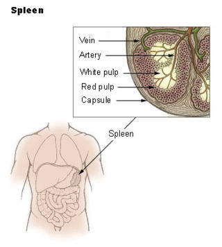
The spleen is an organ found in almost all vertebrates. Similar in structure to a large lymph node, it acts primarily as a blood filter. The word spleen comes from Ancient Greek σπλήν (splḗn).

<span class="mw-page-title-main">Lymph node</span> Organ of the lymphatic system

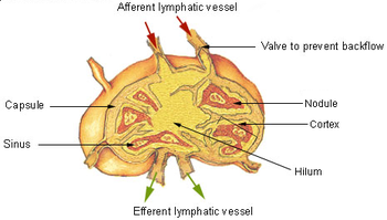
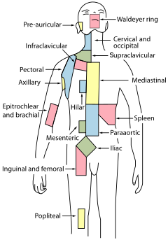
A lymph node, or lymph gland, is a kidney-shaped organ of the lymphatic system and the adaptive immune system. A large number of lymph nodes are linked throughout the body by the lymphatic vessels. They are major sites of lymphocytes that include B and T cells. Lymph nodes are important for the proper functioning of the immune system, acting as filters for foreign particles including cancer cells, but have no detoxification function.

<span class="mw-page-title-main">Lymph</span> Fluid that circulates throughout the lymphatic system

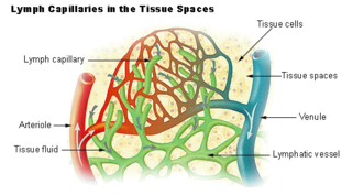
Lymph is the fluid that flows through the lymphatic system, a system composed of lymph vessels (channels) and intervening lymph nodes whose function, like the venous system, is to return fluid from the tissues to be recirculated. At the origin of the fluid-return process, interstitial fluid—the fluid between the cells in all body tissues—enters the lymph capillaries. This lymphatic fluid is then transported via progressively larger lymphatic vessels through lymph nodes, where substances are removed by tissue lymphocytes and circulating lymphocytes are added to the fluid, before emptying ultimately into the right or the left subclavian vein, where it mixes with central venous blood.

<span class="mw-page-title-main">Lymphatic vessel</span> Tubular vessels that are involved in the transport of lymph and lymphocytes

The lymphatic vessels are thin-walled vessels (tubes), structured like blood vessels, that carry lymph. As part of the lymphatic system, lymph vessels are complementary to the cardiovascular system. Lymph vessels are lined by endothelial cells, and have a thin layer of smooth muscle, and adventitia that binds the lymph vessels to the surrounding tissue. Lymph vessels are devoted to the propulsion of the lymph from the lymph capillaries, which are mainly concerned with the absorption of interstitial fluid from the tissues. Lymph capillaries are slightly bigger than their counterpart capillaries of the vascular system. Lymph vessels that carry lymph to a lymph node are called afferent lymph vessels, and those that carry it from a lymph node are called efferent lymph vessels, from where the lymph may travel to another lymph node, may be returned to a vein, or may travel to a larger lymph duct. Lymph ducts drain the lymph into one of the subclavian veins and thus return it to general circulation.

<span class="mw-page-title-main">Interstitium</span> Anatomical term

The interstitium is a contiguous fluid-filled space existing between a structural barrier, such as a cell membrane or the skin, and internal structures, such as organs, including muscles and the circulatory system. The fluid in this space is called interstitial fluid, comprises water and solutes, and drains into the lymph system. The interstitial compartment is composed of connective and supporting tissues within the body – called the extracellular matrix – that are situated outside the blood and lymphatic vessels and the parenchyma of organs. The role of the interstitium in solute concentration, protein transport and hydrostatic pressure impacts human pathology and physiological responses such as edema, inflammation and shock.

Gut-associated lymphoid tissue (GALT) is a component of the mucosa-associated lymphoid tissue (MALT) which works in the immune system to protect the body from invasion in the gut.

<span class="mw-page-title-main">Marginal zone</span> Part of the spleen

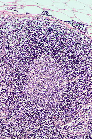
The marginal zone is the region at the interface between the non-lymphoid red pulp and the lymphoid white-pulp of the spleen.

<span class="mw-page-title-main">Myeloid tissue</span> Tissue of bone marrow

Myeloid tissue, in the bone marrow sense of the word myeloid, is tissue of bone marrow, of bone marrow cell lineage, or resembling bone marrow, and myelogenous tissue is any tissue of, or arising from, bone marrow; in these senses the terms are usually used synonymously, as for example with chronic myeloid/myelogenous leukemia.

<span class="mw-page-title-main">Kurloff cell</span> Cells found in the blood and organs of guinea pigs and capybara

Kurloff cells were described as mononuclear cells in the peripheral blood and organs of the guinea pig, capybara, paca, agouti and cavie. The Kurloff cell contains a characteristic proteoglycan-containing inclusion body. In the guinea pig, Kurloff cells are more numerous in the adult female than the adult male. A marked increase in the number of circulating Kurloff cells is present in the peripheral blood during pregnancy and after estrogen treatment in male and female animals. A relatively smaller number of cells take place in immature, non-pregnant, and non-estrogen-treated animals. The exact function of Kurloff cells remains unknown, but it has some of the characteristics of both monocytes and lymphocytes. In guinea-pigs, it has been proposed that Kurloff cells mainly involve in the function of the immune system, such as acting as a natural killer cell and preventing damage to the trophoblast by maternal defensive cells. Also, Kurloff cells present antibody-dependent cytotoxic activity in vitro.

<span class="mw-page-title-main">White pulp</span> Type of tissue in the spleen

White pulp is a histological designation for regions of the spleen, that encompasses approximately 25% of splenic tissue. White pulp consists entirely of lymphoid tissue.

Immune tolerance, also known as immunological tolerance or immunotolerance, refers to the immune system's state of unresponsiveness to substances or tissues that would otherwise trigger an immune response. It arises from prior exposure to a specific antigen and contrasts the immune system's conventional role in eliminating foreign antigens. Depending on the site of induction, tolerance is categorized as either central tolerance, occurring in the thymus and bone marrow, or peripheral tolerance, taking place in other tissues and lymph nodes. Although the mechanisms establishing central and peripheral tolerance differ, their outcomes are analogous, ensuring immune system modulation.

High endothelial venules (HEV) are specialized post-capillary venules characterized by plump endothelial cells as opposed to the usual flatter endothelial cells found in regular venules. HEVs enable lymphocytes circulating in the blood to directly enter a lymph node.

Lymphopoiesis (lĭm'fō-poi-ē'sĭs) is the generation of lymphocytes, one of the five types of white blood cells (WBCs). It is more formally known as lymphoid hematopoiesis.

<span class="mw-page-title-main">Tonsil</span> Lymphoid organs in the mouth and throat

The tonsils are a set of lymphoid organs facing into the aerodigestive tract, which is known as Waldeyer's tonsillar ring and consists of the adenoid tonsil, two tubal tonsils, two palatine tonsils, and the lingual tonsils. These organs play an important role in the immune system.

<span class="mw-page-title-main">Lymph capillary</span> Microvessel serving to drain and process extracellular fluid

Lymph capillaries or lymphatic capillaries are tiny, thin-walled microvessels located in the spaces between cells which serve to drain and process extracellular fluid. Upon entering the lumen of a lymphatic capillary, the collected fluid is known as lymph. Each lymphatic capillary carries lymph into a lymphatic vessel, which in turn connects to a lymph node, a small bean-shaped gland that filters and monitors the lymphatic fluid for infections. Lymph is ultimately returned to the venous circulation.

<span class="mw-page-title-main">CCL21</span> Mammalian protein found in Homo sapiens

Chemokine ligand 21 (CCL21) is a small cytokine belonging to the CC chemokine family. This chemokine is also known as 6Ckine, exodus-2, and secondary lymphoid-tissue chemokine (SLC). CCL21 elicits its effects by binding to a cell surface chemokine receptor known as CCR7. The main function of CCL21 is to guide CCR7 expressing leukocytes to the secondary lymphoid organs, such as lymph nodes and Peyer´s patches.

Lymphocyte homing receptors are cell adhesion molecules expressed on lymphocyte cell membranes that recognize addressins on target tissues. Lymphocyte homing refers to adhesion of the circulating lymphocytes in blood to specialized endothelial cells within lymphoid organs. These diverse tissue-specific adhesion molecules on lymphocytes and on endothelial cells contribute to the development of specialized immune responses.

<span class="mw-page-title-main">C-C chemokine receptor type 7</span> Protein-coding gene in the species Homo sapiens

C-C chemokine receptor type 7 is a protein that in humans is encoded by the CCR7 gene. Two ligands have been identified for this receptor: the chemokines ligand 19 (CCL19/ELC) and ligand 21 (CCL21). The ligands have similar affinity for the receptor, though CCL19 has been shown to induce internalisation of CCR7 and desensitisation of the cell to CCL19/CCL21 signals. CCR7 is a transmembrane protein with 7 transmembrane domains, which is coupled with heterotrimeric G proteins, which transduce the signal downstream through various signalling cascades. The main function of the receptor is to guide immune cells to immune organs by detecting specific chemokines, which these tissues secrete.

Lymph node stromal cells are essential to the structure and function of the lymph node whose functions include: creating an internal tissue scaffold for the support of hematopoietic cells; the release of small molecule chemical messengers that facilitate interactions between hematopoietic cells; the facilitation of the migration of hematopoietic cells; the presentation of antigens to immune cells at the initiation of the adaptive immune system; and the homeostasis of lymphocyte numbers. Stromal cells originate from multipotent mesenchymal stem cells.

<span class="mw-page-title-main">Follicular hyperplasia</span> Medical condition of lymphatic cells

Follicular hyperplasia (FH) is a type of lymphoid hyperplasia and is classified as a lymphadenopathy, which means a disease of the lymph nodes. It is caused by a stimulation of the B cell compartment and by abnormal cell growth of secondary follicles. This typically occurs in the cortex without disrupting the lymph node capsule. The follicles are pathologically polymorphous, are often contrasting and varying in size and shape. Follicular hyperplasia is distinguished from follicular lymphoma in its polyclonality and lack of bcl-2 protein expression, whereas follicular lymphoma is monoclonal, and expresses bcl-2.

References

  1. 1 2 Standring S (2016). Gray's anatomy: the anatomical basis of clinical practice (Forty-first ed.). Elsevier Limited. pp. 68–73. ISBN   9780702052309.
  2. Moore K (2018). Clinically oriented anatomy (Eighth ed.). Wolters Kluwer. pp. 43–45. ISBN   9781496347213.
  3. Natale G, Bocci G, Ribatti D (September 2017). "Scholars and scientists in the history of the lymphatic system". Journal of Anatomy. 231 (3): 417–429. doi:10.1111/joa.12644. PMC   5554832 . PMID   28614587.
  4. Zhang, Yufan; Zhang, Juxiang; Li, Xiaowei; Li, Jingru; Lu, Shuting; Li, Yuqiao; Ren, Panting; Zhang, Chunfu; Xiong, Liqin (2022-06-01). "Imaging of fluorescent polymer dots in relation to channels and immune cells in the lymphatic system". Materials Today Bio. 15: 100317. doi:10.1016/j.mtbio.2022.100317. ISSN   2590-0064. PMC   9213818 . PMID   35757035.
  5. Hu, Dan; Li, Long; Li, Sufang; Wu, Manyan; Ge, Nana; Cui, Yuxia; Lian, Zheng; Song, Junxian; Chen, Hong (2019-08-01). "Lymphatic system identification, pathophysiology and therapy in the cardiovascular diseases". Journal of Molecular and Cellular Cardiology. 133: 99–111. doi:10.1016/j.yjmcc.2019.06.002. ISSN   0022-2828. PMID   31181226. S2CID   184485255.
  6. Sherwood L (January 1, 2012). Human Physiology: From Cells to Systems. Cengage Learning. ISBN   9781111577438 via Google Books.
  7. Mak TW, Saunders ME, Saunders ME (2008). Primer to the immune response. Academic Press. pp. 28–. ISBN   978-0-12-374163-9 . Retrieved 12 November 2010.
  8. 1 2 Warwick R, Williams PL. "Angiology (Chapter 6)". Gray's anatomy (Thirty-fifth ed.). London: Longman. pp. 588–785.
  9. 1 2 Peyrot SM, Martin BL, Harland RM (March 2010). "Lymph heart musculature is under distinct developmental control from lymphatic endothelium". Developmental Biology. 339 (2): 429–38. doi:10.1016/j.ydbio.2010.01.002. PMC   2845526 . PMID   20067786.
  10. Jeltsch M, Tammela T, Alitalo K, Wilting J (October 2003). "Genesis and pathogenesis of lymphatic vessels". Cell and Tissue Research. 314 (1): 69–84. doi:10.1007/s00441-003-0777-2. PMID   12942362. S2CID   23318096.
  11. Eriksson G (2004). "[Olaus Rudbeck as scientist and professor of medicine]". Svensk Medicinhistorisk Tidskrift. 8 (1): 39–44. PMID   16025602.
  12. 1 2 Koppang EO, Fischer U, Moore L, Tranulis MA, Dijkstra JM, Köllner B, Aune L, Jirillo E, Hordvik I (December 2010). "Salmonid T cells assemble in the thymus, spleen and in novel interbranchial lymphoid tissue". J Anat. 217 (6): 728–39. doi:10.1111/j.1469-7580.2010.01305.x. PMC   3039185 . PMID   20880086.
  13. Ruddle NH, Akirav EM (August 2009). "Secondary lymphoid organs: responding to genetic and environmental cues in ontogeny and the immune response". Journal of Immunology. 183 (4): 2205–12. doi:10.4049/jimmunol.0804324. PMC   2766168 . PMID   19661265.
  14. Ruddle, N. H.; Akirav, E. M. (2009). "Secondary Lymphoid Organs: Responding to Genetic and Environmental Cues in Ontogeny and the Immune Response1". Journal of Immunology. 183 (4): 2205–2212. doi:10.4049/jimmunol.0804324. PMC   2766168 . PMID   19661265.
  15. 1 2 Swirski FK, Nahrendorf M, Etzrodt M, Wildgruber M, Cortez-Retamozo V, Panizzi P, et al. (July 2009). "Identification of splenic reservoir monocytes and their deployment to inflammatory sites". Science. 325 (5940): 612–6. Bibcode:2009Sci...325..612S. doi:10.1126/science.1175202. PMC   2803111 . PMID   19644120.
  16. Jia T, Pamer EG (July 2009). "Immunology. Dispensable but not irrelevant". Science. 325 (5940): 549–50. Bibcode:2009Sci...325..549J. doi:10.1126/science.1178329. PMC   2917045 . PMID   19644100.
  17. Angier N (August 3, 2009). "Finally, the Spleen Gets Some Respect". The New York Times. Archived from the original on 2018-01-27.
  18. Blackbourne LH (2008-04-01). Surgical recall. Lippincott Williams & Wilkins. p.  259. ISBN   978-0-7817-7076-7.
  19. "Penicilliary radicles". Saunders Comprehensive Veterinary Dictionary (3rd ed.). Elsevier, Inc. 2007. Archived from the original on 2016-03-04. Retrieved 2011-04-03 via The Free Dictionary by Farlex.
  20. "Spleen: Information, Surgery and Functions". Childrens Hospital of Pittsburgh - Chp.edu. 2010-11-17. Archived from the original on 2011-09-26. Retrieved 2011-04-03.
  21. Singh V (2017). Textbook of Anatomy Head, Neck, and Brain; Volume III (2nd ed.). Elsevier India. pp. 247–249. ISBN   9788131237274.
  22. Yin C, Mohanta S, Maffia P, Habenicht AJ (6 March 2017). "Editorial: Tertiary Lymphoid Organs (TLOs): Powerhouses of Disease Immunity". Frontiers in Immunology. 8: 228. doi: 10.3389/fimmu.2017.00228 . PMC   5337484 . PMID   28321222.
  23. Ruddle NH (March 2014). "Lymphatic vessels and tertiary lymphoid organs". The Journal of Clinical Investigation. 124 (3): 953–9. doi:10.1172/JCI71611. PMC   3934190 . PMID   24590281.
  24. Goldsby R, Kindt TJ, Osborne BA, Janis K (2003) [1992]. "Cells and Organs of the Immune System (Chapter 2)". Immunology (Fifth ed.). New York: W. H. Freeman and Company. pp.  24–56. ISBN   0-7167-4947-5.
  25. Hiraoka N, Ino Y, Yamazaki-Itoh R (2016-06-22). "Tertiary Lymphoid Organs in Cancer Tissues". Frontiers in Immunology. 7: 244. doi: 10.3389/fimmu.2016.00244 . PMC   4916185 . PMID   27446075.
  26. Sautès-Fridman, C; Petitprez, F; Calderaro, J; Fridman, WH (June 2019). "Tertiary lymphoid structures in the era of cancer immunotherapy" (PDF). Nature Reviews. Cancer. 19 (6): 307–325. doi:10.1038/s41568-019-0144-6. PMID   31092904. S2CID   155104003.
  27. van Hooren, L; Vaccaro, A; Ramachandran, M; Vazaios, K; Libard, S; van de Walle, T; Georganaki, M; Huang, H; Pietilä, I; Lau, J; Ulvmar, MH; Karlsson, MCI; Zetterling, M; Mangsbo, SM; Jakola, AS; Olsson Bontell, T; Smits, A; Essand, M; Dimberg, A (5 July 2021). "Agonistic CD40 therapy induces tertiary lymphoid structures but impairs responses to checkpoint blockade in glioma". Nature Communications. 12 (1): 4127. Bibcode:2021NatCo..12.4127V. doi:10.1038/s41467-021-24347-7. PMC   8257767 . PMID   34226552.
  28. 1 2 3 Maoz A, Dennis M, Greenson JK (2019). "The Crohn's-Like Lymphoid Reaction to Colorectal Cancer-Tertiary Lymphoid Structures With Immunologic and Potentially Therapeutic Relevance in Colorectal Cancer". Frontiers in Immunology. 10: 1884. doi: 10.3389/fimmu.2019.01884 . PMC   6714555 . PMID   31507584.
  29. 1 2 Sautès-Fridman C, Petitprez F, Calderaro J, Fridman WH (June 2019). "Tertiary lymphoid structures in the era of cancer immunotherapy" (PDF). Nature Reviews. Cancer. 19 (6): 307–325. doi:10.1038/s41568-019-0144-6. PMID   31092904. S2CID   155104003.
  30. Finkin S, Yuan D, Stein I, Taniguchi K, Weber A, Unger K, et al. (December 2015). "Ectopic lymphoid structures function as microniches for tumor progenitor cells in hepatocellular carcinoma". Nature Immunology. 16 (12): 1235–44. doi:10.1038/ni.3290. PMC   4653079 . PMID   26502405.
  31. Helmink BA, Reddy SM, Gao J, Zhang S, Basar R, Thakur R, et al. (January 2020). "B cells and tertiary lymphoid structures promote immunotherapy response" (PDF). Nature. 577 (7791): 549–555. Bibcode:2020Natur.577..549H. doi:10.1038/s41586-019-1922-8. PMC   8762581 . PMID   31942075. S2CID   210221106.
  32. "lymphoid tissue" at Dorland's Medical Dictionary
  33. Louveau A, Smirnov I, Keyes TJ, Eccles JD, Rouhani SJ, Peske JD, et al. (July 2015). "Structural and functional features of central nervous system lymphatic vessels". Nature. 523 (7560): 337–41. Bibcode:2015Natur.523..337L. doi:10.1038/nature14432. PMC   4506234 . PMID   26030524. we discovered functional lymphatic vessels lining the dural sinuses. These structures express all of the molecular hallmarks of lymphatic endothelial cells, are able to carry both fluid and immune cells from the cerebrospinal fluid, and are connected to the deep cervical lymph nodes. The unique location of these vessels may have impeded their discovery to date, thereby contributing to the long-held concept of the absence of lymphatic vasculature in the central nervous system. The discovery of the central nervous system lymphatic system may call for a reassessment of basic assumptions in neuroimmunology and sheds new light on the aetiology of neuroinflammatory and neurodegenerative diseases associated with immune system dysfunction.
  34. Kumar V (2018). Robbins basic pathology (Tenth ed.). Elsevier. p. 363. ISBN   9780323353175.
  35. Vittet D (November 2014). "Lymphatic collecting vessel maturation and valve morphogenesis". Microvascular Research. 96: 31–7. doi:10.1016/j.mvr.2014.07.001. PMID   25020266.
  36. Heppell C, Richardson G, Roose T (January 2013). "A model for fluid drainage by the lymphatic system". Bulletin of Mathematical Biology. 75 (1): 49–81. doi:10.1007/s11538-012-9793-2. PMID   23161129. S2CID   20438669.
  37. Bazigou E, Wilson JT, Moore JE (November 2014). "Primary and secondary lymphatic valve development: molecular, functional and mechanical insights". Microvascular Research. 96: 38–45. doi:10.1016/j.mvr.2014.07.008. PMC   4490164 . PMID   25086182.
  38. "The functions of the Lymphatic System". lymphnotes.com. Retrieved Feb 25, 2011.
  39. Hu, Dan; Li, Long; Li, Sufang; Wu, Manyan; Ge, Nana; Cui, Yuxia; Lian, Zheng; Song, Junxian; Chen, Hong (2019-08-01). "Lymphatic system identification, pathophysiology and therapy in the cardiovascular diseases". Journal of Molecular and Cellular Cardiology. 133: 99–111. doi:10.1016/j.yjmcc.2019.06.002. ISSN   0022-2828. PMID   31181226. S2CID   184485255.
  40. Munn, Lance L.; Padera, Timothy P. (2014-11-01). "Imaging the lymphatic system". Microvascular Research. SI: Lymphatics in Development and Pathology. 96: 55–63. doi:10.1016/j.mvr.2014.06.006. ISSN   0026-2862. PMC   4268344 . PMID   24956510.
  41. Chong, Chloé; Scholkmann, Felix; Bachmann, Samia B.; Luciani, Paola; Leroux, Jean-Christophe; Detmar, Michael; Proulx, Steven T. (2016-03-10). "In vivo visualization and quantification of collecting lymphatic vessel contractility using near-infrared imaging". Scientific Reports. 6 (1): 22930. Bibcode:2016NatSR...622930C. doi:10.1038/srep22930. ISSN   2045-2322. PMC   4785392 . PMID   26960708.
  42. Zhang, Yufan; Zhang, Juxiang; Li, Xiaowei; Li, Jingru; Lu, Shuting; Li, Yuqiao; Ren, Panting; Zhang, Chunfu; Xiong, Liqin (2022-06-01). "Imaging of fluorescent polymer dots in relation to channels and immune cells in the lymphatic system". Materials Today Bio. 15: 100317. doi:10.1016/j.mtbio.2022.100317. ISSN   2590-0064. PMC   9213818 . PMID   35757035.
  43. Schineis, Philipp; Runge, Peter; Halin, Cornelia (2019-01-01). "Cellular traffic through afferent lymphatic vessels". Vascular Pharmacology. Pioneering updates in vascular biology. 112: 31–41. doi:10.1016/j.vph.2018.08.001. ISSN   1537-1891. PMID   30092362. S2CID   51955021.
  44. Chavhan, Govind B.; Lam, Christopher Z.; Greer, Mary-Louise C.; Temple, Michael; Amaral, Joao; Grosse-Wortmann, Lars (2020-07-01). "Magnetic Resonance Lymphangiography". Radiologic Clinics of North America. 58 (4): 693–706. doi:10.1016/j.rcl.2020.02.002. ISSN   0033-8389. PMID   32471538. S2CID   218943574.
  45. 1 2 3 Colledge NR, Ralston SH, Walker BR, eds. (2011). Davidson's principles and practice of medicine (21st ed.). Edinburgh / New York: Churchill Livingstone / Elsevier. pp. 1001, 1037–1040. ISBN   978-0-7020-3085-7. OCLC   844959047.
  46. Douketis JD. "Lymphedema". Merck Manual.
  47. Herantis Pharma (2015-07-21). "Lymfactin® for lymphedema". Archived from the original on 2018-12-08. Retrieved 2018-12-28.
  48. Martín ML, Hernández MA, Avendaño C, Rodríguez F, Martínez H (March 2011). "Manual lymphatic drainage therapy in patients with breast cancer related lymphoedema". BMC Cancer. 11 (1): 94. doi: 10.1186/1471-2407-11-94 . PMC   3065438 . PMID   21392372.
  49. Fauci AS, Braunwald E, Kasper D, Hauser S, Longo DL (19 March 2009). Harrison's Manual of Medicine. McGraw Hill Professional. pp. 352–. ISBN   978-0-07-147743-7 . Retrieved 12 November 2010.
  50. 1 2 3 4 5 6 7 Ambrose CT (July 2006). "Immunology's first priority dispute--an account of the 17th-century Rudbeck-Bartholin feud". Cellular Immunology. 242 (1): 1–8. doi:10.1016/j.cellimm.2006.09.004. PMID   17083923.
  51. 1 2 3 4 5 Flourens P (1859). "Chapter 3: Aselli, Pecquet, Rudbeck, Bartholin". A History of the Discovery of the Circulation of the Blood. Rickey, Mallory & company. pp.  67–99. Retrieved 2008-07-11. william harvey.
  52. Natale, Gianfranco; Bocci, Guido; Ribatti, Domenico (September 2017). "Scholars and scientists in the history of the lymphatic system". Journal of Anatomy. 231 (3): 417–429. doi:10.1111/joa.12644. ISSN   0021-8782. PMC   5554832 . PMID   28614587.
  53. 1 2 Eriksson G (2004). "[Olaus Rudbeck as scientist and professor of medicine]". Svensk Medicinhistorisk Tidskrift (in Swedish). 8 (1): 39–44. PMID   16025602.
  54. "Disputatio anatomica, de circulatione sanguinis" [Account of Rudbeck's work on lymphatic system and dispute with Bartholin]. International League of Antiquarian Booksellers . Retrieved 2008-07-11.[ dead link ]
  55. "Chapter 25, "Galen in an Age of Change (1650–1820)", Maria Pia Donato". Brill's companion to the reception of Galen. Petros Bouras-Vallianatos, Barbara Zipser. Leiden. 2019. ISBN   978-90-04-39435-3. OCLC   1088603298.{{cite book}}: CS1 maint: location missing publisher (link) CS1 maint: others (link)
  56. Mitchell, Peter (2007). The Purple island and anatomy in early seventeenth-century literature, philosophy, and theology. Madison [NJ]: Fairleigh Dickinson University Press. p. 217. ISBN   978-0-8386-4018-0. OCLC   65207019.
  57. "Galen | Biography, Achievements, & Facts | Britannica". www.britannica.com. Retrieved 2022-12-07.
  58. Turner AL (1937). Story of a Great Hospital: The Royal Infirmary of Edinburgh 1729-1929. Oliver and Boyd. p.  360.
  59. lympha . Charlton T. Lewis and Charles Short. A Latin Dictionary on Perseus Project .
  60. "lymph" . Oxford English Dictionary (Online ed.). Oxford University Press.(Subscription or participating institution membership required.)