Starling equation

Last updated

The Starling principle holds that extracellular fluid movements between blood and tissues are determined by differences in hydrostatic pressure and colloid osmotic (oncotic) pressure between plasma inside microvessels and interstitial fluid outside them. The Starling Equation, proposed many years after the death of Starling, describes that relationship in mathematical form and can be applied to many biological and non-biological semipermeable membranes. The classic Starling principle and the equation that describes it have in recent years been revised and extended.

Contents

Every day around 8 litres of water (solvent) containing a variety of small molecules (solutes) leaves the blood stream of an adult human and perfuses the cells of the various body tissues. Interstitial fluid drains by afferent lymph vessels to one of the regional lymph node groups, where around 4 litres per day is reabsorbed to the blood stream. The remainder of the lymphatic fluid is rich in proteins and other large molecules and rejoins the blood stream via the thoracic duct which empties into the great veins close to the heart. [1] Filtration from plasma to interstitial (or tissue) fluid occurs in microvascular capillaries and post-capillary venules. In most tissues the micro vessels are invested with a continuous internal surface layer that includes a fibre matrix now known as the endothelial glycocalyx whose interpolymer spaces function as a system of small pores, radius circa 5 nm. Where the endothelial glycocalyx overlies a gap in the junction molecules that bind endothelial cells together (inter endothelial cell cleft), the plasma ultrafiltrate may pass to the interstitial space, leaving larger molecules reflected back into the plasma.

A small number of continuous capillaries are specialised to absorb solvent and solutes from interstitial fluid back into the blood stream through fenestrations in endothelial cells, but the volume of solvent absorbed every day is small.

Discontinuous capillaries as found in sinusoidal tissues of bone marrow, liver and spleen have little or no filter function.

The rate at which fluid is filtered across vascular endothelium (transendothelial filtration) is determined by the sum of two outward forces, capillary pressure () and interstitial protein osmotic pressure (), and two absorptive forces, plasma protein osmotic pressure () and interstitial pressure (). The Starling equation describes these forces in mathematical terms. It is one of the Kedem–Katchalski equations which bring nonsteady state thermodynamics to the theory of osmotic pressure across membranes that are at least partly permeable to the solute responsible for the osmotic pressure difference. [2] [3] The second Kedem–Katchalsky equation explains the trans endothelial transport of solutes, .

The equation

Diagram of the classic Starling model; the arteriole is shown in red on the left, and the venule in purple on the right. Note that the concentration of interstitial solutes (orange) increases proportionally to the distance from the arteriole. StarlingEquation.svg
Diagram of the classic Starling model; the arteriole is shown in red on the left, and the venule in purple on the right. Note that the concentration of interstitial solutes (orange) increases proportionally to the distance from the arteriole.

The classic Starling equation reads as follows: [4]

where:

By convention, outward force is defined as positive, and inward force is defined as negative. If Jv is positive, solvent is leaving the capillary (filtration). If negative, solvent is entering the capillary (absorption).

Applying the classic Starling equation, it had long been taught that continuous capillaries filter out fluid in their arteriolar section and reabsorb most of it in their venular section, as shown by the diagram. [4]

However, empirical evidence shows that, in most tissues, the flux of the intraluminal fluid of capillaries is continuous and, primarily, effluent. Efflux occurs along the whole length of a capillary. Fluid filtered to the space outside a capillary is mostly returned to the circulation via lymph nodes and the thoracic duct. [5]

A mechanism for this phenomenon is the Michel-Weinbaum model, in honour of two scientists who, independently, described the filtration function of the glycocalyx. Briefly, the colloid osmotic pressure πi of the interstitial fluid has been found to have no effect on Jv and the colloid osmotic pressure difference that opposes filtration is now known to be π'p minus the subglycocalyx π, which is close to zero while there is adequate filtration to flush interstitial proteins out of the interendothelial cleft. Consequently, Jv is much less than previously calculated, and the unopposed diffusion of interstitial proteins to the subglycocalyx space if and when filtration falls wipes out the colloid osmotic pressure difference necessary for reabsorption of fluid to the capillary. [4]

The revised Starling equation is compatible with the steady-state Starling principle:

where:

Pressures are often measured in millimetres of mercury (mmHg), and the filtration coefficient in millilitres per minute per millimetre of mercury (ml·min−1·mmHg−1).

Filtration coefficient

In some texts the product of hydraulic conductivity and surface area is called the filtration co-efficient Kfc.[ citation needed ]

Reflection coefficient

Staverman's reflection coefficient, σ, is a unitless constant that is specific to the permeability of a membrane to a given solute. [6]

The Starling equation, written without σ, describes the flow of a solvent across a membrane that is impermeable to the solutes contained within the solution. [7]

σn corrects for the partial permeability of a semipermeable membrane to a solute n. [7]

Where σ is close to 1, the plasma membrane is less permeable to the denotated species (for example, larger molecules such as albumin and other plasma proteins), which may flow across the endothelial lining, from higher to lower concentrations, more slowly, while allowing water and smaller solutes through the glycocalyx filter to the extravascular space. [7]

Approximated values

Following are typically quoted values for the variables in the classic Starling equation:

LocationPc (mmHg) [9] Pi (mmHg) [9] σπc (mmHg) [9] σπi (mmHg) [9]
arteriolar end of capillary +35−2+28+0.1
venular end of capillary+15−2+28+3

It is reasoned that some albumin escapes from the capillaries and enters the interstitial fluid where it would produce a flow of water equivalent to that produced by a hydrostatic pressure of +3 mmHg. Thus, the difference in protein concentration would produce a flow of fluid into the vessel at the venous end equivalent to 28  3 = 25 mmHg of hydrostatic pressure. The total oncotic pressure present at the venous end could be considered as +25 mmHg.[ citation needed ]

In the beginning (arteriolar end) of a capillary, there is a net driving force () outwards from the capillary of +9 mmHg. In the end (venular end), on the other hand, there is a net driving force of −8 mmHg.[ citation needed ]

Assuming that the net driving force declines linearly, then there is a mean net driving force outwards from the capillary as a whole, which also results in that more fluid exits a capillary than re-enters it. The lymphatic system drains this excess.[ citation needed ]

J. Rodney Levick argues in his textbook that the interstitial force is often underestimated, and measurements used to populate the revised Starling equation show the absorbing forces to be consistently less than capillary or venular pressures.

Specific organs

Kidneys

Glomerular capillaries have a continuous glycocalyx layer in health and the total transendothelial filtration rate of solvent () to the renal tubules is normally around 125 ml/ min (about 180 litres/ day). Glomerular capillary is more familiarly known as the glomerular filtration rate (GFR). In the rest of the body's capillaries, is typically 5 ml/ min (around 8 litres/ day), and the fluid is returned to the circulation via afferent and efferent lymphatics.[ citation needed ]

Lungs

The Starling equation can describe the movement of fluid from pulmonary capillaries to the alveolar air space. [10] [11]

Clinical significance

Woodcock and Woodcock showed in 2012 that the revised Starling equation (steady-state Starling principle) provides scientific explanations for clinical observations concerning intravenous fluid therapy. [12] Traditional teaching of both filtration and absorption of fluid occurring in a single capillary has been superseded by the concept of a vital circulation of extracellular fluid running parallel to the circulation of blood. New approaches to the treatment of oedema (tissue swelling) are suggested.

History

The Starling equation is named for the British physiologist Ernest Starling, who is also recognised for the Frank–Starling law of the heart. [13] Starling can be credited with identifying that the "absorption of isotonic salt solutions (from the extravascular space) by the blood vessels is determined by this osmotic pressure of the serum proteins" in 1896. [13]

See also

Related Research Articles

<span class="mw-page-title-main">Osmotic pressure</span> Measure of the tendency of a solution to take in pure solvent by osmosis

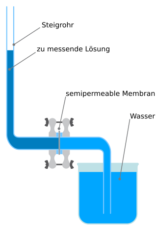
Osmotic pressure is the minimum pressure which needs to be applied to a solution to prevent the inward flow of its pure solvent across a semipermeable membrane. It is also defined as the measure of the tendency of a solution to take in its pure solvent by osmosis. Potential osmotic pressure is the maximum osmotic pressure that could develop in a solution if it were separated from its pure solvent by a semipermeable membrane

<span class="mw-page-title-main">Capillary</span> Smallest type of blood vessel

A capillary is a small blood vessel, from 5 to 10 micrometres in diameter, and is part of the microcirculation system. Capillaries are microvessels and the smallest blood vessels in the body. They are composed of only the tunica intima, consisting of a thin wall of simple squamous endothelial cells. They are the site of the exchange of many substances from the surrounding interstitial fluid, and they convey blood from the smallest branches of the arteries (arterioles) to those of the veins (venules). Other substances which cross capillaries include water, oxygen, carbon dioxide, urea, glucose, uric acid, lactic acid and creatinine. Lymph capillaries connect with larger lymph vessels to drain lymphatic fluid collected in microcirculation.

<span class="mw-page-title-main">Edema</span> Accumulation of excess fluid in body tissue

Edema, also spelled oedema, and also known as fluid retention, dropsy, hydropsy and swelling, is the build-up of fluid in the body's tissue. Most commonly, the legs or arms are affected. Symptoms may include skin which feels tight, the area may feel heavy, and joint stiffness. Other symptoms depend on the underlying cause.

<span class="mw-page-title-main">Oncotic pressure</span> Measure of pressure exerted by large dissolved molecules in biological fluids

Oncotic pressure, or colloid osmotic-pressure, is a type of osmotic pressure induced by the plasma proteins, notably albumin, in a blood vessel's plasma that causes a pull on fluid back into the capillary. Participating colloids displace water molecules, thus creating a relative water molecule deficit with water molecules moving back into the circulatory system within the lower venous pressure end of capillaries.

Hemodynamics or haemodynamics are the dynamics of blood flow. The circulatory system is controlled by homeostatic mechanisms of autoregulation, just as hydraulic circuits are controlled by control systems. The hemodynamic response continuously monitors and adjusts to conditions in the body and its environment. Hemodynamics explains the physical laws that govern the flow of blood in the blood vessels.

<span class="mw-page-title-main">Microcirculation</span> Circulation of the blood in the smallest blood vessels

The microcirculation is the circulation of the blood in the smallest blood vessels, the microvessels of the microvasculature present within organ tissues. The microvessels include terminal arterioles, metarterioles, capillaries, and venules. Arterioles carry oxygenated blood to the capillaries, and blood flows out of the capillaries through venules into veins.

In chemistry, colligative properties are those properties of solutions that depend on the ratio of the number of solute particles to the number of solvent particles in a solution, and not on the nature of the chemical species present. The number ratio can be related to the various units for concentration of a solution such as molarity, molality, normality (chemistry), etc. The assumption that solution properties are independent of nature of solute particles is exact only for ideal solutions, which are solutions that exhibit thermodynamic properties analogous to those of an ideal gas, and is approximate for dilute real solutions. In other words, colligative properties are a set of solution properties that can be reasonably approximated by the assumption that the solution is ideal.

<span class="mw-page-title-main">Extracellular fluid</span> Body fluid outside the cells of a multicellular organism

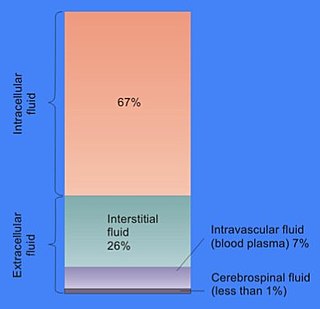
In cell biology, extracellular fluid (ECF) denotes all body fluid outside the cells of any multicellular organism. Total body water in healthy adults is about 50–60% of total body weight; women and the obese typically have a lower percentage than lean men. Extracellular fluid makes up about one-third of body fluid, the remaining two-thirds is intracellular fluid within cells. The main component of the extracellular fluid is the interstitial fluid that surrounds cells.

<span class="mw-page-title-main">Glomerular filtration rate</span> Renal function test

Renal functions include maintaining an acid–base balance; regulating fluid balance; regulating sodium, potassium, and other electrolytes; clearing toxins; absorption of glucose, amino acids, and other small molecules; regulation of blood pressure; production of various hormones, such as erythropoietin; and activation of vitamin D.

<span class="mw-page-title-main">Hydrostatics</span> Branch of fluid mechanics that studies fluids at rest

Fluid statics or hydrostatics is the branch of fluid mechanics that studies fluids at hydrostatic equilibrium and "the pressure in a fluid or exerted by a fluid on an immersed body".

<span class="mw-page-title-main">Glomerulus (kidney)</span> Functional unit of nephron

The glomerulus is a network of small blood vessels (capillaries) known as a tuft, located at the beginning of a nephron in the kidney. Each of the two kidneys contains about one million nephrons. The tuft is structurally supported by the mesangium, composed of intraglomerular mesangial cells. The blood is filtered across the capillary walls of this tuft through the glomerular filtration barrier, which yields its filtrate of water and soluble substances to a cup-like sac known as Bowman's capsule. The filtrate then enters the renal tubule of the nephron.

<span class="mw-page-title-main">Glycocalyx</span> Viscous, carbohydrate rich layer at the outermost periphery of a cell.

The glycocalyx, also known as the pericellular matrix and sometime cell coat, is a glycoprotein and glycolipid covering that surrounds the cell membranes of bacteria, epithelial cells, and other cells. It was described in a review article in 1970.

Water potential is the potential energy of water per unit volume relative to pure water in reference conditions. Water potential quantifies the tendency of water to move from one area to another due to osmosis, gravity, mechanical pressure and matrix effects such as capillary action. The concept of water potential has proved useful in understanding and computing water movement within plants, animals, and soil. Water potential is typically expressed in potential energy per unit volume and very often is represented by the Greek letter ψ.

<span class="mw-page-title-main">Osmotic concentration</span> Molarity of osmotically active particles

Osmotic concentration, formerly known as osmolarity, is the measure of solute concentration, defined as the number of osmoles (Osm) of solute per litre (L) of solution. The osmolarity of a solution is usually expressed as Osm/L, in the same way that the molarity of a solution is expressed as "M". Whereas molarity measures the number of moles of solute per unit volume of solution, osmolarity measures the number of osmoles of solute particles per unit volume of solution. This value allows the measurement of the osmotic pressure of a solution and the determination of how the solvent will diffuse across a semipermeable membrane (osmosis) separating two solutions of different osmotic concentration.

<span class="mw-page-title-main">Ultrafiltration (kidney)</span> Filtration by a semi-permeable membrane

In renal physiology, ultrafiltration occurs at the barrier between the blood and the filtrate in the glomerular capsule in the kidneys. As in nonbiological examples of ultrafiltration, pressure and concentration gradients lead to a separation through a semipermeable membrane. The Bowman's capsule contains a dense capillary network called the glomerulus. Blood flows into these capillaries through the afferent arterioles and leaves through the efferent arterioles.

The human body and even its individual body fluids may be conceptually divided into various fluid compartments, which, although not literally anatomic compartments, do represent a real division in terms of how portions of the body's water, solutes, and suspended elements are segregated. The two main fluid compartments are the intracellular and extracellular compartments. The intracellular compartment is the space within the organism's cells; it is separated from the extracellular compartment by cell membranes.

An intercellular cleft is a channel between two cells through which molecules may travel and gap junctions and tight junctions may be present. Most notably, intercellular clefts are often found between epithelial cells and the endothelium of blood vessels and lymphatic vessels, also helping to form the blood-nerve barrier surrounding nerves. Intercellular clefts are important for allowing the transportation of fluids and small solute matter through the endothelium.

Microvasculature comprises the microvessels – venules and capillaries of the microcirculation, with a maximum average diameter of 0.3 millimeters. As the vessels decrease in size, they increase their surface-area-to-volume ratio. This allows surface properties to play a significant role in the function of the vessel.

A depletion force is an effective attractive force that arises between large colloidal particles that are suspended in a dilute solution of depletants, which are smaller solutes that are preferentially excluded from the vicinity of the large particles. One of the earliest reports of depletion forces that lead to particle coagulation is that of Bondy, who observed the separation or "creaming" of rubber latex upon addition of polymer depletant molecules to solution. More generally, depletants can include polymers, micelles, osmolytes, ink, mud, or paint dispersed in a continuous phase.

<span class="mw-page-title-main">Membrane osmometer</span>

A membrane osmometer is a device used to indirectly measure the number average molecular weight of a polymer sample. One chamber contains pure solvent and the other chamber contains a solution in which the solute is a polymer with an unknown . The osmotic pressure of the solvent across the semipermeable membrane is measured by the membrane osmometer. This osmotic pressure measurement is used to calculate for the sample.

References

  1. Herring, Neil (2018). Levick's Introduction to Cardiovascular Physiology. 5th edition (6th ed.). London: CRC Press. pp. 149–213. ISBN   978-1498739849.
  2. Staverman, A. J. (1951). "The theory of measurement of osmotic pressure". Recueil des Travaux Chimiques des Pays-Bas. 70 (4): 344–352. doi:10.1002/recl.19510700409. ISSN   0165-0513.
  3. Kedem, O.; Katchalsky, A. (February 1958). "Thermodynamic analysis of the permeability of biological membranes to non-electrolytes". Biochimica et Biophysica Acta. 27 (2): 229–246. doi:10.1016/0006-3002(58)90330-5. ISSN   0006-3002. PMID   13522722.
  4. 1 2 3 Levick, J R (2004-06-15). "Revision of the Starling principle: new views of tissue fluid balance". The Journal of Physiology. 557 (Pt 3): 704. doi:10.1113/jphysiol.2004.066118. ISSN   0022-3751. PMC   1665155 . PMID   15131237.
  5. Levick, J.R.; Michel, C.C. (2010). "Microvascular fluid exchange and the revised Starling principle". Cardiovasc Res. 87 (2): 198–210. doi:10.1093/cvr/cvq062. PMID   20200043.
  6. Zelman, A. (1972-04-01). "Membrane Permeability: Generalization of the Reflection Coefficient Method of Describing Volume and Solute Flows". Biophysical Journal. 12 (4): 414–419. Bibcode:1972BpJ....12..414Z. doi:10.1016/S0006-3495(72)86093-4. ISSN   0006-3495. PMC   1484119 . PMID   5019478.
  7. 1 2 3 Michel, C. Charles; Woodcock, Thomas E.; Curry, Fitz-Roy E. (2020). "Understanding and extending the Starling principle". Acta Anaesthesiologica Scandinavica. 64 (8): 1032–1037. doi: 10.1111/aas.13603 . ISSN   1399-6576. PMID   32270491.
  8. Lautt, W. Wayne (April 7, 2009). Fluid Exchange. Morgan & Claypool Life Sciences via www.ncbi.nlm.nih.gov.
  9. 1 2 3 4 Boron, Walter F. (2005). Medical Physiology: A Cellular And Molecular Approaoch. Elsevier/Saunders. ISBN   978-1-4160-2328-9.
  10. Pal, Pramod K.; Chen, Robert (2014-01-01), Aminoff, Michael J.; Josephson, S. Andrew (eds.), "Chapter 1 - Breathing and the Nervous System", Aminoff's Neurology and General Medicine (Fifth Edition), Boston: Academic Press, pp. 3–23, doi:10.1016/b978-0-12-407710-2.00001-1, ISBN   978-0-12-407710-2, S2CID   56748572 , retrieved 2020-11-28
  11. Nadon, A. S.; Schmidt, E. P. (2014-01-01), McManus, Linda M.; Mitchell, Richard N. (eds.), "Pathobiology of the Acute Respiratory Distress Syndrome", Pathobiology of Human Disease, San Diego: Academic Press, pp. 2665–2676, doi:10.1016/b978-0-12-386456-7.05309-0, ISBN   978-0-12-386457-4 , retrieved 2020-11-28
  12. Woodcock, T. E.; Woodcock, T. M. (29 January 2012). "Revised Starling equation and the glycocalyx model of transvascular fluid exchange: an improved paradigm for prescribing intravenous fluid therapy". British Journal of Anaesthesia. 108 (3): 384–394. doi: 10.1093/bja/aer515 . PMID   22290457.
  13. 1 2 Starling, Ernest H. (1896-05-05). "On the Absorption of Fluids from the Connective Tissue Spaces". The Journal of Physiology. 19 (4): 312–326. doi:10.1113/jphysiol.1896.sp000596. PMC   1512609 . PMID   16992325.