Kikuchi disease

Last updated
Kikuchi disease
Other namesHistiocytic necrotizing lymphadenitis, Kikuchi-Fujimoto disease
Histiocytic necrotizing lymphadenitis - intermed mag.jpg
Micrograph of a lymph node with Kikuchi disease showing the characteristic features (abundant histiocytes, necrosis without neutrophils). H&E stain.
Specialty Angiology   OOjs UI icon edit-ltr-progressive.svg

Kikuchi disease was described in 1972 in Japan. It is also known as histiocytic necrotizing lymphadenitis, Kikuchi necrotizing lymphadenitis, phagocytic necrotizing lymphadenitis, subacute necrotizing lymphadenitis, and necrotizing lymphadenitis. [1] [2] [3] [4] Kikuchi disease occurs sporadically in people with no family history of the condition. [5]

Contents

It was first described by Dr Masahiro Kikuchi (1935–2012) in 1972 [6] and independently by Y. Fujimoto.

Signs and symptoms

The signs and symptoms of Kikuchi disease are fever, enlargement of the lymph nodes (lymphadenopathy), skin rashes, and headache. [7] In sixty to ninety percent of cases, lymphadenopathy presents in the posterior cervical lymph nodes with diameter enlargement typically being between one and two centimeters, but up to seven centimeters has been reported in literature. Occasionally, the supraclavicular and axillary lymph nodes become swollen as well. [8] Rarely, enlargement of the liver and spleen and nervous system involvement resembling meningitis are seen. Often a bout of extreme fatigue can occur - often taking hold during latter parts of the day and the affected person can be more prone to fatigue from exercise.[ citation needed ]

Pathophysiology

Some studies have suggested a genetic predisposition to the proposed autoimmune response. Several infectious candidates have been associated with Kikuchi disease. [9]

Many theories exist about the cause of KFD. Microbial/viral or autoimmune causes have been suggested. Mycobacterium szulgai and Yersinia and Toxoplasma species have been implicated. More recently, growing evidence suggests a role for Epstein-Barr virus, as well as other viruses (HHV6, HHV8, parvovirus B19, HIV and HTLV-1) in the pathogenesis of KFD. [1] However, many independent studies have failed to identify the presence of these infectious agents in cases of Kikuchi lymphadenopathy. [10] In addition, serologic tests including antibodies to a host of viruses have consistently proven noncontributory and no viral particles have been identified ultrastructurally. KFD is now proposed to be a nonspecific hyperimmune reaction to a variety of infectious, chemical, physical, and neoplastic agents. Other autoimmune conditions and manifestations such as antiphospholipid syndrome, polymyositis, systemic juvenile idiopathic arthritis, bilateral uveitis, arthritis and cutaneous necrotizing vasculitis have been linked to KFD. KFD may represent an exuberant T-cell-mediated immune response in a genetically susceptible individual to a variety of nonspecific stimuli. [1]

Certain Human leukocyte antigen class II genes appear more frequently in patients with Kikuchi disease, suggesting that there may be a genetic predisposition to the proposed autoimmune response. [11]

Diagnosis

It is diagnosed by lymph node excision biopsy. Kikuchi disease is a self-limiting illness which has symptoms which may overlap with Hodgkin's lymphoma leading to misdiagnosis in some patients. Antinuclear antibodies, antiphospholipid antibodies, anti-dsDNA, and rheumatoid factor are usually negative, and may help in differentiation from systemic lupus erythematosus. [12]

Differential diagnosis

The differential diagnosis of Kikuchi disease includes systemic lupus erythematosus (SLE), disseminated tuberculosis, lymphoma, sarcoidosis, and viral lymphadenitis. Clinical findings sometimes may include positive results for IgM/IgG/IgA antibodies. For other causes of lymph node enlargement, see lymphadenopathy. [13]

Management

No specific cure is known. Treatment is largely supportive. Nonsteroidal anti-inflammatory drugs (NSAIDs) are indicated for tender lymph nodes and fever, and corticosteroids are useful in severe extranodal or generalized disease. [12]

Symptomatic measures aimed at relieving the distressing local and systemic complaints have been described as the main line of management of KFD. Analgesics, antipyretics, NSAIDs, and corticosteroids have been used. If the clinical course is more severe, with multiple flares of bulky enlarged cervical lymph nodes and fever, then a low-dose corticosteroid treatment has been suggested. [12]

Epidemiology

Kikuchi-Fujimoto disease (KFD) is a rare, self-limiting disorder that typically affects the cervical lymph nodes. Recognition of this condition is crucial, especially because it can easily be mistaken for tuberculosis, lymphoma, or even adenocarcinoma. Awareness of this disorder helps prevent misdiagnosis and inappropriate treatment. [1]

Kikuchi's disease is a very rare disease mainly seen in Japan. Isolated cases are reported in North America, Europe, Asia, England, and at least two cases in New Zealand. It is possible that the prevalence of KFD is greater than is reported given lymphadenopathy can be overlooked and the disease's self-limiting nature. That a definite identification of KFD can only be done via a biopsy of affected tissues further suggests that cases go unrecognized or undiagnosed. [14] It is mainly a disease of young adults (20–30 years), with a slight bias towards females. The cause of this disease is not known, although infectious and autoimmune causes have been proposed. The course of the disease is generally benign and self-limiting. Lymph node enlargement usually resolves over several weeks to six months. The recurrence rate is about 3%. Death from Kikuchi disease is extremely rare and usually occurs due to liver, respiratory, or heart failure.[ citation needed ]

See also

Related Research Articles

<span class="mw-page-title-main">Lymph node</span> Organ of the lymphatic system

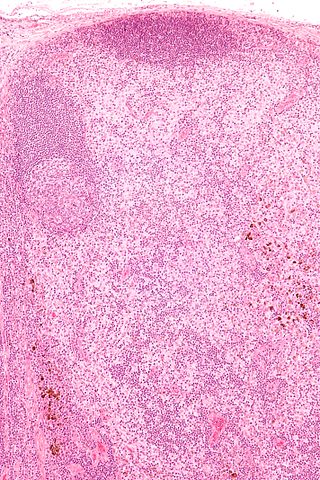
A lymph node, or lymph gland, is a kidney-shaped organ of the lymphatic system and the adaptive immune system. A large number of lymph nodes are linked throughout the body by the lymphatic vessels. They are major sites of lymphocytes that include B and T cells. Lymph nodes are important for the proper functioning of the immune system, acting as filters for foreign particles including cancer cells, but have no detoxification function.

<span class="mw-page-title-main">Infectious mononucleosis</span> Common viral infectious disease

Infectious mononucleosis, also known as glandular fever, is an infection usually caused by the Epstein–Barr virus (EBV). Most people are infected by the virus as children, when the disease produces few or no symptoms. In young adults, the disease often results in fever, sore throat, enlarged lymph nodes in the neck, and fatigue. Most people recover in two to four weeks; however, feeling tired may last for months. The liver or spleen may also become swollen, and in less than one percent of cases splenic rupture may occur.

<span class="mw-page-title-main">Lymphogranuloma venereum</span> Sexually transmitted infection

Lymphogranuloma venereum is a sexually transmitted infection caused by the invasive serovars L1, L2, L2a, L2b, or L3 of Chlamydia trachomatis.

<span class="mw-page-title-main">Mycobacterial cervical lymphadenitis</span> Human medical condition

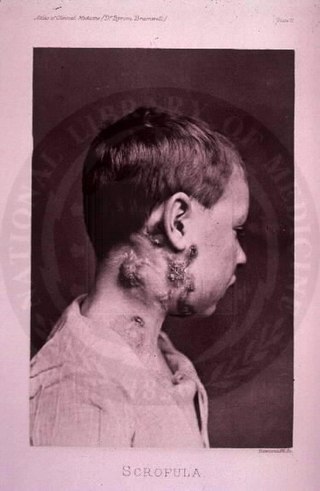
The disease mycobacterial cervical lymphadenitis, also known as scrofula and historically as king's evil, involves a lymphadenitis of the cervical lymph nodes associated with tuberculosis as well as nontuberculous (atypical) mycobacteria.

<span class="mw-page-title-main">Castleman disease</span> Group of lymphoproliferative disorders

Castlemandisease (CD) describes a group of rare lymphoproliferative disorders that involve enlarged lymph nodes, and a broad range of inflammatory symptoms and laboratory abnormalities. Whether Castleman disease should be considered an autoimmune disease, cancer, or infectious disease is currently unknown.

<span class="mw-page-title-main">Lymphadenopathy</span> Disease of lymph nodes

Lymphadenopathy or adenopathy is a disease of the lymph nodes, in which they are abnormal in size or consistency. Lymphadenopathy of an inflammatory type is lymphadenitis, producing swollen or enlarged lymph nodes. In clinical practice, the distinction between lymphadenopathy and lymphadenitis is rarely made and the words are usually treated as synonymous. Inflammation of the lymphatic vessels is known as lymphangitis. Infectious lymphadenitis affecting lymph nodes in the neck is often called scrofula.

<span class="mw-page-title-main">Kawasaki disease</span> Disease found in young children

Kawasaki disease is a syndrome of unknown cause that results in a fever and mainly affects children under 5 years of age. It is a form of vasculitis, where medium-sized blood vessels become inflamed throughout the body. The fever typically lasts for more than five days and is not affected by usual medications. Other common symptoms include large lymph nodes in the neck, a rash in the genital area, lips, palms, or soles of the feet, and red eyes. Within three weeks of the onset, the skin from the hands and feet may peel, after which recovery typically occurs. The disease is the leading cause of acquired heart disease in children in developed countries, which include the formation of coronary artery aneurysms and myocarditis.

Talaromycosis is a fungal infection that presents with painless skin lesions of the face and neck, as well as an associated fever, anaemia, and enlargement of the lymph glands and liver.

Persistent generalized lymphadenopathy (PGL) is enlarged, painless, non-tender lymph nodes occurring in a couple of different areas for more than three to six months for which no other reason can be found. To expand, the common site where PGL occurs is within the head and neck region; parotid gland alterations and nasopharyngeal lymphatic tissue enlargement are often frequent comorbidities of Persistent generalized lymphadenopathy. Due to lymphoproliferation in the intraglandular lymphoid tissue, obstruction within the epithelium results in cystic expansion, which is the cause of cystic parotid lesions found in PGL. This condition frequently occurs in people in the latency period of HIV/AIDS.

Lymphoid hyperplasia is the rapid proliferation of normal lymphocytic cells that resemble lymph tissue which may occur with bacterial or viral infections. The growth is termed hyperplasia which may result in enlargement of various tissue including an organ, or cause a cutaneous lesion.

<span class="mw-page-title-main">Rosai–Dorfman disease</span> Medical condition

Rosai–Dorfman disease, also known as sinus histiocytosis with massive lymphadenopathy or sometimes as Destombes–Rosai–Dorfman disease, is a rare disorder of unknown cause that is characterized by abundant histiocytes in the lymph nodes or other locations throughout the body.

Diffuse infiltrative lymphocytosis syndrome (DILS) is a rare multi-system complication of HIV believed to occur secondary to an abnormal persistence of the initial CD8+ T cell expansion that regularly occurs in an HIV infection. This persistent CD8+ T cell expansion occurs in the setting of a low CD4+/CD8+ T cell ratio and ultimately invades and destroys tissues and organs resulting in the various complications of DILS. DILS classically presents with bilateral salivary gland enlargement (parotitis), cervical lymphadenopathy, and sicca symptoms such as xerophthalmia and xerostomia, but it may also involve the lungs, nervous system, kidneys, liver, digestive tract, and muscles. Once suspected, current diagnostic workups include (1) confirming HIV infection, (2) confirming six or greater months of characteristic signs and symptoms, (3) confirming organ infiltration by CD8+ T cells, and (4) exclusion of other autoimmune conditions. Once the diagnosis of DILS is confirmed, management includes highly active antiretroviral therapy (HAART) and as-needed steroids. With proper treatment, the overall prognosis of DILS is favorable.

Necrotizing vasculitis, also called systemic necrotizing vasculitus, is a category of vasculitis, comprising vasculitides that present with necrosis. Examples include giant cell arteritis, microscopic polyangiitis, and granulomatosis with polyangiitis. ICD-10 uses the variant "necrotizing vasculopathy". ICD-9, while classifying these conditions together, does not use a dedicated phrase, instead calling them "polyarteritis nodosa and allied conditions".

<span class="mw-page-title-main">Cervical lymphadenopathy</span> Disease of the lymph nodes

Cervical lymphadenopathy refers to lymphadenopathy of the cervical lymph nodes. The term lymphadenopathy strictly speaking refers to disease of the lymph nodes, though it is often used to describe the enlargement of the lymph nodes. Similarly, the term lymphadenitis refers to inflammation of a lymph node, but often it is used as a synonym of lymphadenopathy.

<span class="mw-page-title-main">Dermatopathic lymphadenopathy</span> Medical condition

In pathology, dermatopathic lymphadenopathy, is lymph node pathology due to skin disease.

<span class="mw-page-title-main">Cat-scratch disease</span> Human disease

Cat-scratch disease (CSD) or felinosis is an infectious disease that most often results from a scratch or bite of a cat. Symptoms typically include a non-painful bump or blister at the site of injury and painful and swollen lymph nodes. People may feel tired, have a headache, or a fever. Symptoms typically begin within 3–14 days following infection.

<span class="mw-page-title-main">Follicular hyperplasia</span> Medical condition of lymphatic cells

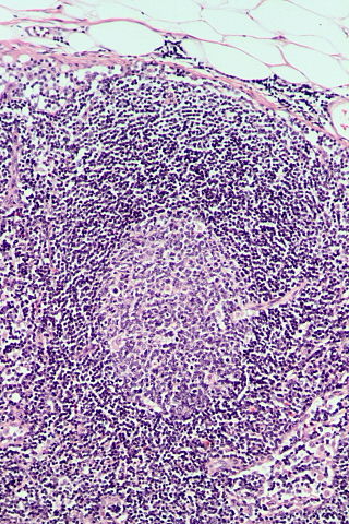
Follicular hyperplasia (FH) is a type of lymphoid hyperplasia and is classified as a lymphadenopathy, which means a disease of the lymph nodes. It is caused by a stimulation of the B cell compartment and by abnormal cell growth of secondary follicles. This typically occurs in the cortex without disrupting the lymph node capsule. The follicles are pathologically polymorphous, are often contrasting and varying in size and shape. Follicular hyperplasia is distinguished from follicular lymphoma in its polyclonality and lack of bcl-2 protein expression, whereas follicular lymphoma is monoclonal, and expresses bcl-2.

<span class="mw-page-title-main">Unicentric Castleman disease</span> Medical condition

Unicentric Castleman disease is a subtype of Castleman disease, a group of lymphoproliferative disorders characterized by lymph node enlargement, characteristic features on microscopic analysis of enlarged lymph node tissue, and a range of symptoms and clinical findings.

<span class="mw-page-title-main">Idiopathic multicentric Castleman disease</span> Medical condition

Idiopathic multicentric Castleman disease (iMCD) is a subtype of Castleman disease (also known as giant lymph node hyperplasia, lymphoid hamartoma, or angiofollicular lymph node hyperplasia), a group of lymphoproliferative disorders characterized by lymph node enlargement, characteristic features on microscopic analysis of enlarged lymph node tissue, and a range of symptoms and clinical findings.

Epstein–Barr virus–associated lymphoproliferative diseases are a group of disorders in which one or more types of lymphoid cells, i.e. B cells, T cells, NK cells, and histiocytic-dendritic cells, are infected with the Epstein–Barr virus (EBV). This causes the infected cells to divide excessively, and is associated with the development of various non-cancerous, pre-cancerous, and cancerous lymphoproliferative disorders (LPDs). These LPDs include the well-known disorder occurring during the initial infection with the EBV, infectious mononucleosis, and the large number of subsequent disorders that may occur thereafter. The virus is usually involved in the development and/or progression of these LPDs although in some cases it may be an "innocent" bystander, i.e. present in, but not contributing to, the disease.

References

  1. 1 2 3 4 Rammohan A, Cherukuri SD, Manimaran AB, Manohar RR, Naidu RM (June 2012). "Kikuchi-Fujimoto Disease: A Sheep in Wolf's Clothing". J Otolaryngol Head Neck Surg. 41 (3): 222–26. PMID   22762705.
  2. Kaushik V, Malik TH, Bishop PW, Jones PH (June 2004). "Histiocytic necrotising lymphadenitis (Kikuchi's disease): a rare cause of cervical lymphadenopathy". Surgeon. 2 (3): 179–82. doi:10.1016/s1479-666x(04)80084-2. PMID   15570824.
  3. Bosch X, Guilabert A (2006). "Kikuchi-Fujimoto disease". Orphanet J Rare Dis. 1: 18. doi: 10.1186/1750-1172-1-18 . PMC   1481509 . PMID   16722618.
  4. Bosch X, Guilabert A, Miquel R, Campo E (July 2004). "Enigmatic Kikuchi-Fujimoto disease: a comprehensive review". Am. J. Clin. Pathol. 122 (1): 141–52. doi: 10.1309/YF08-1L4T-KYWV-YVPQ . PMID   15272543.
  5. "kikuchi disease". Archived from the original on 8 May 2019. Retrieved 17 April 2018.
  6. Kikuchi M. Lymphadenitis showing focal reticulum cell hyperplasia with nuclear debris and phagocytes. Acta Hematol Jpn 1972;35:379–80.
  7. "Kikuchi disease". Genetic and Rare Diseases Information Center (GARD). Archived from the original on 8 May 2019. Retrieved 17 April 2018.
  8. Muhammad, Masab (2023). "Kikuchi Disease". National Library of Medicine. PMID   28613580 . Retrieved 17 November 2022.
  9. Atwater AR, Longley BJ, Aughenbaugh WD (July 2008). "Kikuchi's disease: case report and systematic review of cutaneous and histopathologic presentations". J. Am. Acad. Dermatol. 59 (1): 130–36. doi:10.1016/j.jaad.2008.03.012. PMID   18462833.
  10. Rosado, F. G.; Tang, Y. W.; Hasserjian, R. P.; McClain, C. M.; Wang, B; Mosse, C. A. (2013). "Kikuchi-Fujimoto lymphadenitis: Role of parvovirus B-19, Epstein-Barr virus, human herpesvirus 6, and human herpesvirus 8". Human Pathology. 44 (2): 255–59. doi:10.1016/j.humpath.2012.05.016. PMID   22939574.
  11. Tanaka, T.; Ohmori, M.; Yasunaga, S.; Ohshima, K.; Kikuchi, M.; Sasazuki, T. (1999). "DNA typing of HLA class II genes (HLA-DR, -DQ and -DP) in Japanese patients with histiocytic necrotizing lymphadentis (Kikuchi's disease) 8". Tissue Antigens. 54 (3): 246–253. doi:10.1034/j.1399-0039.1999.540305.x. PMID   10519361.
  12. 1 2 3 Bosch X, Guilabert A (23 May 2006). "Kikuchi-Fujimoto disease". Orphanet Journal of Rare Diseases. 1 (18): 18. doi: 10.1186/1750-1172-1-18 . PMC   1481509 . PMID   16722618.
  13. "Lymphadenopathy". The Lecturio Medical Concept Library. Retrieved 7 August 2021.
  14. Ulug, Mehmet; Aslan, Vahap (March 1, 2012). "An unusual cause of cervical lymphadenopathy: Kikuchi-Fujimoto disease". Journal of Microbiology and Infectious Diseases. 2 (1): 21–25. doi: 10.5799/ahinjs.02.2012.01.0036 .