Thymoma

Last updated
Thymoma
Encapsulated thymoma.jpg
An encapsulated thymoma (mixed lymphocytic and epithelial type)
Specialty Oncology, cardiothoracic surgery
Usual onset Adulthood
Treatmentsurgical removal, chemotherapy (in malignant cases).

A thymoma is a tumor originating from the epithelial cells of the thymus that is considered a rare malignancy. Thymomas are frequently associated with neuromuscular disorders such as myasthenia gravis; [1] thymoma is found in 20% of patients with myasthenia gravis. [2] Once diagnosed, thymomas may be removed surgically. In the rare case of a malignant tumor, chemotherapy may be used.

Contents

Signs and symptoms

A third of all people with a thymoma have symptoms caused by compression of the surrounding organs by an expansive mass. These problems may take the form of superior vena cava syndrome, dysphagia (difficulty swallowing), cough, or chest pain. [1]

One-third of patients have their tumors discovered because they have an associated autoimmune disorder. As mentioned earlier, the most common of those conditions is myasthenia gravis (MG); 10–15% of patients with MG have a thymoma and, conversely, 30–45% of patients with thymomas have MG. Additional associated autoimmune conditions include thymoma-associated multiorgan autoimmunity, pure red cell aplasia and Good syndrome (thymoma with combined immunodeficiency and hypogammaglobulinemia). Other reported disease associations are with acute pericarditis, agranulocytosis, alopecia areata, ulcerative colitis, Cushing's disease, hemolytic anemia, limbic encephalopathy, myocarditis, nephrotic syndrome, panhypopituitarism, pernicious anemia, polymyositis, rheumatoid arthritis, sarcoidosis, scleroderma, sensorimotor radiculopathy, stiff person syndrome , systemic lupus erythematosus and thyroiditis. [1] [3]

One-third to one-half of all persons with thymoma have no symptoms at all, and the mass is identified on a chest X-ray or CT/CAT scan performed for an unrelated problem. [1]

Pathology

Thymoma originates from the epithelial cell population in the thymus, and several microscopic subtypes are now recognized. [1] There are three principal histological types of thymoma, depending on the appearance of the cells by microscopy:

Thymic cortical epithelial cells have abundant cytoplasm, vesicular nucleus with finely divided chromatin and small nucleoli and cytoplasmic filaments contact adjacent cells. Thymic medullary epithelial cells in contrast are spindle shaped with oval dense nucleus and scant cytoplasm thymoma if recapitulates cortical cell features more, is thought to be less benign.

Diagnosis

CT scan of the chest revealing a large necrotic mass in the left anterior mediastinum (indicated by the red line). Histology later established the diagnosis of a thymoma. Anterior mediastinal mass thymoma diagram.jpg
CT scan of the chest revealing a large necrotic mass in the left anterior mediastinum (indicated by the red line). Histology later established the diagnosis of a thymoma.
Another axial slice of a CT scan of the chest showing a small thymoma anterior to the heart (marked with the red line). Tumor Thymoma1.JPG
Another axial slice of a CT scan of the chest showing a small thymoma anterior to the heart (marked with the red line).

When a thymoma is suspected, a CT/CAT scan is generally performed to estimate the size and extent of the tumor, and the lesion is sampled with a CT-guided needle biopsy. Increased vascular enhancement on CT scans can be indicative of malignancy, as can be pleural deposits. [1] Limited[ clarification needed ] biopsies are associated with a very small risk of pneumomediastinum or mediastinitis and an even-lower risk of damaging the heart or large blood vessels. Sometimes thymoma metastasize for instance to the abdomen. [5]

The diagnosis is made via histologic examination by a pathologist, after obtaining a tissue sample of the mass. Final tumor classification and staging is accomplished pathologically after formal[ clarification needed ] surgical removal of the thymic tumor.

Selected laboratory tests can be used to look for associated problems or possible tumor spread. These include: full blood count, protein electrophoresis, antibodies to the acetylcholine receptor (indicative of myasthenia), electrolytes, liver enzymes and renal function. [1]

Staging

The Masaoka Staging System is used widely and is based on the anatomic extent of disease at the time of surgery: [6]

Treatment

Surgery is the mainstay of treatment for thymoma. If the tumor is apparently invasive and large, preoperative (neoadjuvant) chemotherapy and/or radiotherapy may be used to decrease the size and improve resectability, before surgery is attempted. When the tumor is an early stage (Masaoka I through IIB), no further therapy is necessary. Removal of the thymus in adults does not appear to induce immune deficiency. In children, however, postoperative immunity may be abnormal and vaccinations for several infectious agents are recommended. Invasive thymomas may require additional treatment with radiotherapy and chemotherapy (cyclophosphamide, doxorubicin and cisplatin). [1] [ citation needed ]. [7] Recurrences of thymoma are described in 10-30% of cases up to 10 years after surgical resection, and in the majority of cases also pleural recurrences can be removed. Recently, surgical removal of pleural recurrences can be followed by hyperthermic intrathoracic perfusion chemotherapy or intrathoracic hyperthermic perfused chemotherapy (ITH). [8]

Prognosis

Prognosis is much worse for stage III or IV thymomas as compared with stage I and II tumors. Invasive thymomas uncommonly can also metastasize, generally to pleura, bones, liver or brain in approximately 7% of cases. [1] A study found that slightly over 40% of observed patients with stage III and IV tumors survived for at least 10 years after diagnosis. The median age of these patients at the time of thymoma diagnosis was 57 years. [9]

Patients who have undergone thymectomy for thymoma should be warned of possible severe side effects after yellow fever vaccination. This is probably caused by inadequate T-cell response to live attenuated yellow fever vaccine. Deaths have been reported.[ citation needed ]

Epidemiology

The incidence of thymomas is around 0.13-0.26 per 100,000 people per year. [10] Males are affected slightly less frequently than females. [10] The typical age at diagnosis is in the 40s and 50s, though the age may range from six years to 83 years. [10]

See also

Related Research Articles

<span class="mw-page-title-main">Myasthenia gravis</span> Autoimmune disease resulting in skeletal muscle weakness

Myasthenia gravis (MG) is a long-term neuromuscular junction disease that leads to varying degrees of skeletal muscle weakness. The most commonly affected muscles are those of the eyes, face, and swallowing. It can result in double vision, drooping eyelids, and difficulties in talking and walking. Onset can be sudden. Those affected often have a large thymus or develop a thymoma.

<span class="mw-page-title-main">Thymus</span> Endocrine gland

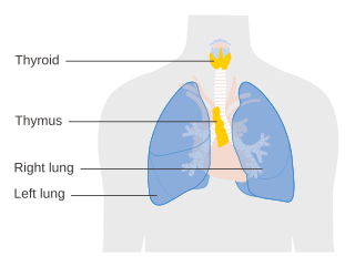
The thymus is a specialized primary lymphoid organ of the immune system. Within the thymus, thymus cell lymphocytes or T cells mature. T cells are critical to the adaptive immune system, where the body adapts to specific foreign invaders. The thymus is located in the upper front part of the chest, in the anterior superior mediastinum, behind the sternum, and in front of the heart. It is made up of two lobes, each consisting of a central medulla and an outer cortex, surrounded by a capsule.

<span class="mw-page-title-main">Mesothelioma</span> Cancer associated with asbestos

Mesothelioma is a type of cancer that develops from the thin layer of tissue that covers many of the internal organs. The area most commonly affected is the lining of the lungs and chest wall. Less commonly the lining of the abdomen and rarely the sac surrounding the heart, or the sac surrounding the testis may be affected. Signs and symptoms of mesothelioma may include shortness of breath due to fluid around the lung, a swollen abdomen, chest wall pain, cough, feeling tired, and weight loss. These symptoms typically come on slowly.

A thymectomy is an operation to remove the thymus. It usually results in remission of myasthenia gravis with the help of medication including steroids. However, this remission may not be permanent. Thymectomy is indicated when thymoma are present in the thymus. Anecdotal evidence suggests MG patients with no evidence of thymoma may still benefit from thymectomy.

<span class="mw-page-title-main">Surface epithelial-stromal tumor</span> Medical condition

Surface epithelial-stromal tumors are a class of ovarian neoplasms that may be benign or malignant. Neoplasms in this group are thought to be derived from the ovarian surface epithelium or from ectopic endometrial or fallopian tube (tubal) tissue. Tumors of this type are also called ovarian adenocarcinoma. This group of tumors accounts for 90% to 95% of all cases of ovarian cancer; however is mainly only found in postmenopausal women with the exception of the United States where 7% of cases occur in women under the age of 40. Serum CA-125 is often elevated but is only 50% accurate so it is not a useful tumor marker to assess the progress of treatment. 75% of women with epithelial ovarian cancer are found within the advanced-stages; however younger patients are more likely to have better prognoses than older patients.

<span class="mw-page-title-main">Pseudomyxoma peritonei</span> Medical condition

Pseudomyxoma peritonei (PMP) is a clinical condition caused by cancerous cells that produce abundant mucin or gelatinous ascites. The tumors cause fibrosis of tissues and impede digestion or organ function, and if left untreated, the tumors and mucin they produce will fill the abdominal cavity. This will result in compression of organs and will destroy the function of the colon, small intestine, stomach, or other organs. Prognosis with treatment in many cases is optimistic, but the disease is lethal if untreated, with death occurring via cachexia, bowel obstruction, or other types of complications.

<span class="mw-page-title-main">Invasive carcinoma of no special type</span> Medical condition

Invasive carcinoma of no special type, invasive breast carcinoma of no special type (IBC-NST), invasive ductal carcinoma (IDC), infiltrating ductal carcinoma (IDC) or invasive ductal carcinoma, not otherwise specified (NOS) is a disease. For international audiences this article will use "invasive carcinoma NST" because it is the preferred term of the World Health Organization (WHO).

<span class="mw-page-title-main">Oligoastrocytoma</span> Medical condition

Oligoastrocytomas are a subset of brain tumors that present with an appearance of mixed glial cell origin, astrocytoma and oligodendroglioma. However, the term "Oligoastrocytoma" is now considered obsolete by the National Comprehensive Cancer Network stating "the term should no longer be used as such morphologically ambiguous tumors can be reliably resolved into astrocytomas and oligodendrogliomas with molecular testing."

<span class="mw-page-title-main">Hemangiopericytoma</span> Medical condition

A hemangiopericytoma is a type of soft-tissue sarcoma that originates in the pericytes in the walls of capillaries. When inside the nervous system, although not strictly a meningioma tumor, it is a meningeal tumor with a special aggressive behavior. It was first characterized in 1942.

<span class="mw-page-title-main">Mediastinal tumors</span> Medical condition

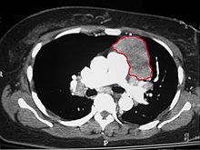
A mediastinal tumor is a tumor in the mediastinum, the cavity that separates the lungs from the rest of the chest. It contains the heart, esophagus, trachea, thymus, and aorta. The most common mediastinal masses are neurogenic tumors, usually found in the posterior mediastinum, followed by thymoma (15–20%) located in the anterior mediastinum. Lung cancer typically spreads to the lymph nodes in the mediastinum.

<span class="mw-page-title-main">Mucoepidermoid carcinoma</span> Medical condition

Mucoepidermoid carcinoma (MEC) is the most common type of minor salivary gland malignancy in adults. Mucoepidermoid carcinoma can also be found in other organs, such as bronchi, lacrimal sac, and thyroid gland.

<span class="mw-page-title-main">Thymic carcinoma</span> Medical condition

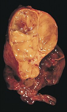
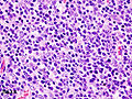
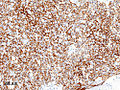
Thymic carcinoma, or type C thymoma, is a malignancy of the thymus. It is a rare cancer that is often diagnosed at advanced stages. Recurrence following treatment is common, and thymic carcinoma is associated with a poor prognosis.

Lung cancer staging is the assessment of the extent to which a lung cancer has spread from its original source. As with most cancers, staging is an important determinant of treatment and prognosis. In general, more advanced stages of cancer are less amenable to treatment and have a worse prognosis.

<span class="mw-page-title-main">Esthesioneuroblastoma</span> Medical condition

Esthesioneuroblastoma is a rare cancer of the nasal cavity. Arising from the upper nasal tract, esthesioneuroblastoma is believed to originate from sensory neuroepithelial cells, also known as neuroectodermal olfactory cells.

Thymus hyperplasia refers to an enlargement ("hyperplasia") of the thymus.

<span class="mw-page-title-main">Philip Bernatz</span>

Philip Eugene Bernatz was an eminent American physician and thoracic surgeon at the Mayo Clinic, and an international authority on the diagnosis and treatment of thymoma and other tumors of the chest.

<span class="mw-page-title-main">Hyperthermic intraperitoneal chemotherapy</span>

Intraperitoneal hyperthermic chemoperfusion is a type of hyperthermia therapy used in combination with surgery in the treatment of advanced abdominal cancers. In this procedure, warmed anti-cancer medications are infused and circulated in the peritoneal cavity (abdomen) for a short period of time. The chemotherapeutic agents generally infused during IPHC are mitomycin-C and cisplatin.

Ectopic thymus is a condition where thymus tissue is found in an abnormal location. It usually does not cause symptoms, but may leads to a mass in the neck that may compress the trachea and the esophagus. It is thought to be the result of either a failure of descent or a failure of involution of normal thymus tissue. It may be diagnosed with radiology, such as an ultrasound or magnetic resonance imaging. If it causes illness, surgery can be used to remove it. Recurrence after surgery is very unlikely.

Hyperthermic intrathoracic chemotherapy (HITOC) is part of a surgical strategy employed in the treatment of various pleural malignancies. The pleura in this situation could be considered to include the surface linings of the chest wall, lungs, mediastinum, and diaphragm. HITOC is the chest counterpart of HIPEC. Traditionally used in the treatment of malignant mesothelioma, a primary malignancy of the pleura, this modality has recently been evaluated in the treatment of secondary pleural malignancies.

Cytoreductive surgery (CRS) is a surgical procedure that aims to reduce the amount of cancer cells in the abdominal cavity for patients with tumors that have spread intraabdominally. It is often used to treat ovarian cancer but can also be used for other abdominal malignancies.

References

  1. 1 2 3 4 5 6 7 8 9 Thomas CR, Wright CD, Loehrer PJ (July 1999). "Thymoma: state of the art". Journal of Clinical Oncology. 17 (7): 2280–9. doi:10.1200/JCO.1999.17.7.2280. PMID   10561285. S2CID   12621667.
  2. Mitchell, Richard Sheppard; Kumar, Vinay; Robbins, Stanley L.; Abbas, Abul K.; Fausto, Nelson (2007). Robbins basic pathology. Saunders/Elsevier. ISBN   978-1-4160-2973-1.[ page needed ]
  3. Bernard C, Frih H, Pasquet F, Kerever S, Jamilloux Y, Tronc F, Guibert B, Isaac S, Devouassoux M, Chalabreysse L, Broussolle C, Petiot P, Girard N, Sève P (January 2016). "Thymoma associated with autoimmune diseases: 85 cases and literature review". Autoimmunity Reviews. 15 (1): 82–92. doi:10.1016/j.autrev.2015.09.005. PMID   26408958.
  4. Dadmanesh F, Sekihara T, Rosai J (May 2001). "Histologic typing of thymoma according to the new World Health Organization classification". Chest Surgery Clinics of North America. 11 (2): 407–20. PMID   11413764.
  5. van Geffen WH, Sietsma J, Roelofs PM, Hiltermann TJ (December 2011). "A malignant retroperitoneal mass – a rare presentation of recurrent thymoma". BMJ Case Reports. 2011: bcr0920114737. doi:10.1136/bcr.09.2011.4737. PMC   3229325 . PMID   22674945.
  6. Masaoka A, Monden Y, Nakahara K, Tanioka T (December 1981). "Follow-up study of thymomas with special reference to their clinical stages". Cancer. 48 (11): 2485–92. doi: 10.1002/1097-0142(19811201)48:11<2485::AID-CNCR2820481123>3.0.CO;2-R . PMID   7296496.
  7. NCCN Thymoma, Guidelines (2016). "NCCN Thymoma Guidelines" (PDF). NCCN Guidelines.
  8. Ambrogi, Marcello (2016). "Pleural recurrence of thymoma: surgical resection followed by hyperthermic intrathoracic perfusion chemotherapy". Eur J Cardiothorac Surg. 49 (1): 321–6. doi:10.1093/ejcts/ezv039.
  9. Wilkins KB, Sheikh E, Green R, Patel M, George S, Takano M, Diener-West M, Welsh J, Howard S, Askin F, Bulkley GB (October 1999). "Clinical and pathologic predictors of survival in patients with thymoma". Annals of Surgery. 230 (4): 562–72, discussion 572–4. doi:10.1097/00000658-199910000-00012. PMC   1420905 . PMID   10522726.
  10. 1 2 3 WHO Classification of Tumours Editorial Board (2021). "5. Tumors of the thymus". Thoracic Tumours. Vol. 5 (5th ed.). Lyon (France): World Health Organization. pp. 320–325. ISBN   978-92-832-4506-3.