A lymph node, or lymph gland, is a kidney-shaped organ of the lymphatic system and the adaptive immune system. A large number of lymph nodes are linked throughout the body by the lymphatic vessels. They are major sites of lymphocytes that include B and T cells. Lymph nodes are important for the proper functioning of the immune system, acting as filters for foreign particles including cancer cells, but have no detoxification function.
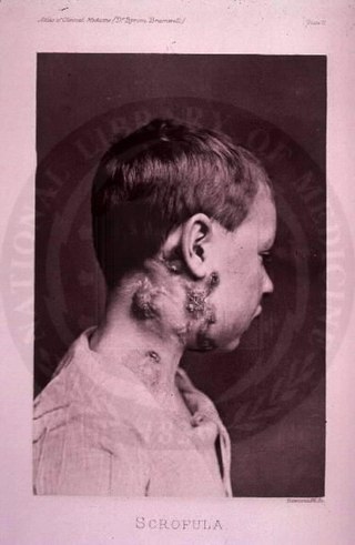
The disease mycobacterial cervical lymphadenitis, also known as scrofula and historically as king's evil, involves a lymphadenitis of the cervical lymph nodes associated with tuberculosis as well as nontuberculous (atypical) mycobacteria.

Chancroid is a bacterial sexually transmitted infection characterized by painful sores on the genitalia. Chancroid is known to spread from one individual to another solely through sexual contact. However, there have been reports of accidental infection through the hand.

Gastrointestinal stromal tumors (GISTs) are the most common mesenchymal neoplasms of the gastrointestinal tract. GISTs arise in the smooth muscle pacemaker interstitial cell of Cajal, or similar cells. They are defined as tumors whose behavior is driven by mutations in the KIT gene (85%), PDGFRA gene (10%), or BRAF kinase (rare). 95% of GISTs stain positively for KIT (CD117). Most (66%) occur in the stomach and gastric GISTs have a lower malignant potential than tumors found elsewhere in the GI tract.
Peripheral tuberculous lymphadenitis is a form of tuberculosis infection occurring outside of the lungs. In general, it describes tuberculosis infection of the lymph nodes, leading to lymphadenopathy. When cervical lymph nodes are affected, it is commonly referred to as "Scrofula." A majority of tuberculosis infections affect the lungs, and extra-pulmonary tuberculosis infections account for the remainder; these most commonly involve the lymphatic system. Although the cervical region is most commonly affected, tuberculous lymphadenitis can occur all around the body, including the axillary and inguinal regions.

A mediastinal tumor is a tumor in the mediastinum, the cavity that separates the lungs from the rest of the chest. It contains the heart, esophagus, trachea, thymus, and aorta. The most common mediastinal masses are neurogenic tumors, usually found in the posterior mediastinum, followed by thymoma (15–20%) located in the anterior mediastinum. Lung cancer typically spreads to the lymph nodes in the mediastinum.
Splenic marginal zone lymphoma (SMZL) is a type of marginal zone lymphoma, a cancer made up of B-cells that replace the normal architecture of the white pulp of the spleen. The neoplastic cells are both small lymphocytes and larger, transformed lymphoblasts, and they invade the mantle zone of splenic follicles and erode the marginal zone, ultimately invading the red pulp of the spleen. Frequently, the bone marrow and splenic hilar lymph nodes are involved along with the peripheral blood. The neoplastic cells circulating in the peripheral blood are termed villous lymphocytes due to their characteristic appearance.

A schwannoma is a usually benign nerve sheath tumor composed of Schwann cells, which normally produce the insulating myelin sheath covering peripheral nerves.

Extramammary Paget's disease (EMPD) is a rare and slow-growing malignancy which occurs within the epithelium and accounts for 6.5% of all Paget's disease. The clinical presentation of this disease is similar to the characteristics of mammary Paget's disease (MPD). However, unlike MPD, which occurs in large lactiferous ducts and then extends into the epidermis, EMPD originates in glandular regions rich in apocrine secretions outside the mammary glands. EMPD incidence is increasing by 3.2% every year, affecting hormonally-targeted tissues such as the vulva and scrotum. In women, 81.3% of EMPD cases are related to the vulva, while for men, 43.2% of the manifestations present at the scrotum.

Rosai–Dorfman disease, also known as sinus histiocytosis with massive lymphadenopathy or sometimes as Destombes–Rosai–Dorfman disease, is a rare disorder of unknown cause that is characterized by abundant histiocytes in the lymph nodes or other locations throughout the body.
Cancer of unknown primary origin (CUP) is a cancer that is determined to be at the metastatic stage at the time of diagnosis, but a primary tumor cannot be identified. A diagnosis of CUP requires a clinical picture consistent with metastatic disease and one or more biopsy results inconsistent with a tumor cancer
Malignant triton tumor (MTT) is a relatively rare, aggressive tumor made up of both malignant schwannoma cells and malignant rhabdomyoblasts. It is classified as a malignant peripheral nerve sheath tumor with rhabdomyosarcomatous differentiation.
A Warthin–Finkeldey cell is a type of giant multinucleate cell found in hyperplastic lymph nodes early in the course of measles and also in HIV-infected individuals, as well as in Kimura disease, and more rarely in a number of neoplastic and non-neoplastic lymph node disorders. Their origin is uncertain, but they have previously been shown to stain with markers similar to those of follicular dendritic cells, including CD21. Under the light microscope, these cells consist of a large, grape-like cluster of nuclei.

High-grade prostatic intraepithelial neoplasia (HGPIN) is an abnormality of prostatic glands and believed to precede the development of prostate adenocarcinoma.

Metanephric adenoma (MA) is a rare, benign tumour of the kidney, that can have a microscopic appearance similar to a nephroblastoma, or a papillary renal cell carcinoma.

Endosalpingiosis is a condition in which fallopian tube-like epithelium is found outside the fallopian tube. It is unknown what causes this condition. It is generally accepted that the condition develops from transformation of coelomic tissue. It is often an incidental finding and is not usually associated with any pathology.

Mammary-type myofibroblastoma (MFB), also named mammary and extramammary myofibroblastoma, was first termed myofibrolastoma of the breast, or, more simply, either mammary myofibroblastoma (MMFB) or just myofibroblastoma. The change in this terminology occurred because the initial 1987 study and many subsequent studies found this tumor only in breast tissue. However, a 2001 study followed by numerous reports found tumors with the microscopic histopathology and other key features of mammary MFB in a wide range of organs and tissues. Further complicating the issue, early studies on MFB classified it as one of various types of spindle cell tumors that, except for MFB, were ill-defined. These other tumors, which have often been named interchangeably in different reports, are: myelofibroblastoma, benign spindle cell tumor, fibroma, spindle cell lipoma, myogenic stromal tumor, and solitary stromal tumor. Finally, studies suggest that spindle cell lipoma and cellular angiofibroma are variants of MFB. Here, the latter two tumors are tentatively classified as MFB variants but otherwise MFB is described as it is more strictly defined in most recent publications. The World Health Organization in 2020 classified mammary type myofibroblastoma tumors and myofibroblastoma tumors as separate tumor forms within the category of fibroblastic and myofibroblastic tumors.

Unicentric Castleman disease is a subtype of Castleman disease, a group of lymphoproliferative disorders characterized by lymph node enlargement, characteristic features on microscopic analysis of enlarged lymph node tissue, and a range of symptoms and clinical findings.
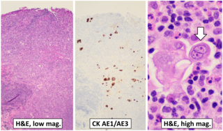
AE1/AE3 is an antibody cocktail that is used in immunohistochemistry, being generally positive in the cytoplasm of carcinomas.
Mature T-cell lymphoma, also called peripheral T-cell lymphoma, is a group of rare, aggressive lymphomas that develop from mature white blood cells and originate from lymphoid tissues outside of the bone marrow. Mature T-cell lymphoma is under the category of non-Hodgkin lymphoma. Mature T-cell lymphomas account for 10% to 15% of all lymphomas and is more common in Asia than in Europe and America. Its common subtypes include angioimmunoblastic T-cell lymphoma, anaplastic large cell lymphoma and peripheral T-cell lymphoma not otherwise specified. While different subtypes have variable symptoms, common symptoms include enlarged painless lymph nodes, fever, weight loss, rash and night sweats.