Neuroendocrine tumor

Last updated
Neuroendocrine tumor
Small intestine neuroendocrine tumour high mag.jpg
Micrograph of a neuroendocrine tumor. H&E stain
Specialty Endocrine oncology   OOjs UI icon edit-ltr-progressive.svg

Neuroendocrine tumors (NETs) are neoplasms that arise from cells of the endocrine (hormonal) and nervous systems. They most commonly occur in the intestine, where they are often called carcinoid tumors, but they are also found in the pancreas, lung, and the rest of the body.

Contents

Although there are many kinds of NETs, they are treated as a group of tissue because the cells of these neoplasms share common features, including a similar histological appearance, having special secretory granules, and often producing biogenic amines and polypeptide hormones. [1]

The term "neuro" refers to the dense core granules (DCGs), similar to the DCGs in the serotonergic neurons storing monoamines. The term "endocrine" refers to the synthesis and secretion of these monoamines. The neuroendocrine system includes endocrine glands such as the pituitary, the parathyroids and the neuroendocrine adrenals, as well as endocrine islet tissue embedded within glandular tissue such as in the pancreas, and scattered cells in the exocrine parenchyma. The latter is known as the diffuse endocrine system. [2] [3]

Classification

WHO

The World Health Organization (WHO) classification scheme places neuroendocrine tumors into three main categories, which emphasize the tumor grade rather than the anatomical origin: [4] [5]

Additionally, the WHO scheme recognizes mixed tumors with both neuroendocrine and epithelial carcinoma features, such as goblet cell cancer, a rare gastrointestinal tract tumor. [6]

Placing a given tumor into one of these categories depends on well-defined histological features: size, lymphovascular invasion, mitotic count, Ki-67 labelling index, invasion of adjacent organs, presence of metastases and whether they produce hormones. [4] [5]

The WHO grading from 2022 endorses a three-tiered grading system for most NETs, in particular NETs of the gastrointestinal or pancreaticobiliary tract, as well as NETs of the upper aerodigestive tract and the salivary glands. The grading system is based on proliferation assessed by mitotic rate and Ki-67 index and stratifies NETs into grade 1 (G1, low-grade), grade 2 (G2, intermediate-grade) and grade 3 (G3, high-grade). Tumor necrosis, although recognized as a factor associated with a potentially worse prognosis, is not included in the grading of NETs of the gastrointestinal or pancreaticobiliary tract. However, the absence or presence of tumor necrosis is a component of the grading of NETs of many other origins, such as the upper aerodigestive tract, the lung and the thymus. [7]

Neuroendocrine carcinomas are poorly differentiated high-grade neuroendocrine neoplasms and a designation of tumor grade is therefore redundant. [7] Lung and thymic neuroendocrine neoplasms are classified in a similar manner, including typical and atypical carcinoids, small cell and large cell neuroendocrine carincomas. [7]

Furthermore, the 2022 WHO classification introduces a two-tiered grading system for medullary thyroid carcinomas based on mitotic count, Ki-67 index and the absence or presence of tumor necrosis. Here, it may be noted that different cut-offs than with tumors of gastrointestinal, aerodigestive and lung origin are applied. [7]

Anatomic distribution

Traditionally, neuroendocrine tumors have been classified by their anatomic site of origin. NETs can arise in many different areas of the body, and are most often located in the intestine, pancreas or the lungs. The various kinds of cells that can give rise to NETs are present in endocrine glands and are also diffusely distributed throughout the body, most commonly Kulchitsky cells or similar enterochromaffin-like cells, that are relatively more common in the gastrointestinal and pulmonary systems. [8]

NETs include certain tumors of the gastrointestinal tract and of the pancreatic islet cells, [1] certain thymus and lung tumors, and medullary carcinoma of the parafollicular cells of the thyroid. [1] Tumors with similar cellular characteristics in the pituitary, parathyroid, and adrenomedullary glands are sometimes included [9] or excluded. [1]

Within the broad category of neuroendocrine tumors there are many different tumor types, [10] representing only a small proportion of the tumors or cancers in most of these tissues[ citation needed ]:

Grading

Neuroendocrine lesions are graded histologically according to markers of cellular proliferation, rather than cellular polymorphism. The following grading scheme is currently recommended for all gastroenteropancreatic neuroendocrine neoplasms by the World Health Organization: [38]

Mitoses in a neuroendocrine tumor. Mitoses in neuroendocrine tumor.jpg
Mitoses in a neuroendocrine tumor.
GMitotic count (per 10 HPF)Ki-67 index (%)
GXGrade cannot be assessed
G1< 2< 3%
G22 to 203–20%
G3> 20> 20%

If mitotic count and Ki-67 are discordant, the figure which gives the highest grade is used.

G1 and G2 neuroendocrine neoplasms are called neuroendocrine tumors (NETs) – formerly called carcinoid tumours. G3 neoplasms are called neuroendocrine carcinomas (NECs).[ citation needed ]

It has been proposed that the current G3 category be further separated into histologically well-differentiated and poorly-differentiated neoplasms to better reflect prognosis. [39]

Staging

Lymph node metastasis of a neuroendocrine tumor. Lymph node metastasis from neuroendocrine tumor.jpg
Lymph node metastasis of a neuroendocrine tumor.

Currently there is no one staging system for all neuroendocrine neoplasms. Well-differentiated lesions generally have their own staging system based on anatomical location, whereas poorly differentiated and mixed lesions are staged as carcinomas of that location. For example, gastric NEC and mixed adenoneuroendocrine cancers are staged as primary carcinoma of the stomach. [40]

TNM staging of gastroenteropancreatic Grade 1 and Grade 2 neuroendocrine tumors are as follows:

Stomach [41]
Primary Tumor (T)
T CategoryTumor Criteria
TXPrimary tumour cannot be assessed
T0No evidence of primary tumour
T1Invades the lamina propria or submucosa, and less than or equal to 1 cm in size
T2Invades the muscularis propria, or greater than 1 cm in size
T3Invades through the muscularis propria into subserosal tissue without penetration of overlying serosa
T4Invades visceral peritoneum (serosal) or other organs or adjacent structures
Regional Lymph Node (N)
N CategoryN Criteria
NXRegional lymph nodes cannot be assessed
N0No regional lymph node metastasis
N1Regional lymph node metastasis
Distant Metastasis (M)
M CategoryM Criteria
M0No distant metastasis
M1Distant metastasis
   M1aMetastasis confined to liver
   M1bMetastasis in at least one extra-hepatic site
   M1cBoth hepatic and extra-hepatic metastases
AJCC Prognostic Stage Groups
StageCriteria
IT1, N0, M0
IIT2 or T3, N0, M0
IIIAny T, N1, M0; T4, N0, M0
IVAny T, any N, M1
Duodenum / Ampulla of Vater [42]
Primary Tumor (T)
T CategoryTumor Criteria
TXPrimary tumour cannot be assessed
T1Invades the mucosa or submucosa only, and less than or equal to 1 cm in size (duodenal tumors)
Confined within the sphincter of Oddi, and less than or equal to 1 cm in size (ampullary tumors)
T2Invades the muscularis propria, or is > 1 cm (duodenal)
Invades through sphincter into duodenal submucosa or muscularis propria, or is > 1 cm (ampullary)
T3Invades the pancreas or peripancreatic adipose tissue
T4Invades visceral peritoneum (serosal) or other organs
Regional Lymph Node (N)
N CategoryN Criteria
NXRegional lymph nodes cannot be assessed
N0No regional lymph node metastasis
N1Regional lymph node metastasis
Distant Metastasis (M)
M CategoryM Criteria
M0No distant metastasis
M1Distant metastasis
   M1aMetastasis confined to liver
   M1bMetastasis in at least one extra-hepatic site
   M1cBoth hepatic and extra-hepatic metastases
AJCC Prognostic Stage Groups
StageCriteria
IT1, N0, M0
IIT2 or T3, N0, M0
IIIT4, N0, M0; Any T, N1, M0
IVAny T, any N, M1
Jejunum and Ileum [43]
Primary Tumor (T)
T CategoryTumor Criteria
TXPrimary tumour cannot be assessed
T0No evidence of primary tumour
T1Invades the lamina propria or submucosa, and less than or equal to 1 cm in size
T2Invades the muscularis propria, or greater than 1 cm in size
T3Invades through the muscularis propria into subserosal tissue without penetration of overlying serosa
T4Invades visceral peritoneum (serosal) or other organs or adjacent structures
Regional Lymph Node (N)
N CategoryN Criteria
NXRegional lymph nodes cannot be assessed
N0No regional lymph node metastasis
N1Regional lymph node metastasis less than 12 nodes
N2Large mesenteric masses (> 2 cm) and / or extensive nodal deposits (12 or greater), especially those that encase the superior mesenteric vessels
Distant Metastasis (M)
M CategoryM Criteria
M0No distant metastasis
M1Distant metastasis
   M1aMetastasis confined to liver
   M1bMetastasis in at least one extra-hepatic site
   M1cBoth hepatic and extra-hepatic metastases
AJCC Prognostic Stage Groups
StageCriteria
IT1, N0, M0
IIT2 or T3, N0, M0
IIIAny T, N1 or N2, M0; T4, N0, M0;
IVAny T, any N, M1
Appendix [44]
Primary Tumor (T)
T CategoryTumor Criteria
TXPrimary tumour cannot be assessed
T0No evidence of primary tumour
T12 cm or less in greatest dimension
T2Tumor more than 2 cm but less than or equal to 4 cm
T3Tumor more than 4 cm or with subserosal invasion or involvement of the mesoappendix
T4Perforates the peritoneum or directly invades other organs or structures (excluding direct mural extension to adjacent subserosa of adjacent bowel)
Regional Lymph Node (N)
N CategoryN Criteria
NXRegional lymph nodes cannot be assessed
N0No regional lymph node metastasis
N1Regional lymph node metastasis
Distant Metastasis (M)
M CategoryM Criteria
M0No distant metastasis
M1Distant metastasis
   M1aMetastasis confined to liver
   M1bMetastasis in at least one extra-hepatic site
   M1cBoth hepatic and extra-hepatic metastases
AJCC Prognostic Stage Groups
StageCriteria
IT1, N0, M0
IIT2 or T3, N0, M0
IIIAny T, N1, M0; T4, N1, M0
IVAny T, any N, M1
Colon and Rectum [45]
Primary Tumor (T)
T CategoryTumor Criteria
TXPrimary tumour cannot be assessed
T0No evidence of primary tumour
T1Invades the lamina propria or submucosa, and less than or equal to 2 cm
   T1aLess than 1 cm in greatest dimension
   T1b1–2 cm in greatest dimension
T2Invades the muscularis propria, or greater than 2 cm in size with invasion of the lamina propria or submucosa
T3Invades through the muscularis propria into subserosal tissue without penetration of overlying serosa
T4Invades visceral peritoneum (serosal) or other organs or adjacent structures
Regional Lymph Node (N)
N CategoryN Criteria
NXRegional lymph nodes cannot be assessed
N0No regional lymph node metastasis
N1Regional lymph node metastasis
Distant Metastasis (M)
M CategoryM Criteria
M0No distant metastasis
M1Distant metastasis
   M1aMetastasis confined to liver
   M1bMetastasis in at least one extra-hepatic site
   M1cBoth hepatic and extra-hepatic metastases
AJCC Prognostic Stage Groups
StageCriteria
IT1, N0, M0
IIAT2, N0, M0
IIBT3, N0, M0
IIIAT4, N0, M0
IIIBAny T, N1, M0
IVAny T, any N, M1
Pancreas [46]
Primary Tumor (T)
T CategoryTumor Criteria
TXPrimary tumour cannot be assessed
T1Limited to the pancreas, less than or equal to 2 cm in size
T2Limited to the pancreas, 2–4 cm in size
T3Limited to the pancreas, > 4 cm; or invading the duodenum or bile duct
T4Invading adjacent organs or the wall of large vessels
Regional Lymph Node (N)
N CategoryN Criteria
NXRegional lymph nodes cannot be assessed
N0No regional lymph node involvement
N1Regional lymph node involvement
Distant Metastasis (M)
M CategoryM Criteria
M0No distant metastasis
M1Distant metastasis
   M1aMetastasis confined to liver
   M1bMetastasis in at least one extra-hepatic site
   M1cBoth hepatic and extra-hepatic metastases
AJCC Prognostic Stage Groups
StageCriteria
IT1, N0, M0
IIT2 or T3, N0, M0
IIIAny T, N1, M0; T4, N0, M0
IVAny T, any N, M1

Signs and symptoms

Gastroenteropancreatic

Conceptually, there are two main types of NET within the gastroenteropancreatic neuroendocrine tumors (GEP-NET) category: those which arise from the gastrointestinal (GI) system and those that arise from the pancreas. In usage, the term "carcinoid" has often been applied to both, although sometimes it is restrictively applied to NETs of GI origin (as herein), or alternatively to those tumors which secrete functional hormones or polypeptides associated with clinical symptoms, as discussed.[ citation needed ]

Carcinoid tumors

Carcinoids most commonly affect the small bowel, particularly the ileum, and are the most common malignancy of the appendix. Many carcinoids are asymptomatic and are discovered only upon surgery for unrelated causes. These coincidental carcinoids are common; one study found that one person in ten has them. [47] Many tumors do not cause symptoms even when they have metastasized. [48] Other tumors even if very small can produce adverse effects by secreting hormones. [49]

Ten per cent (10%) [50] or less of carcinoids, primarily some midgut carcinoids, secrete excessive levels of a range of hormones, most notably serotonin (5-HT) or substance P, [51] causing a constellation of symptoms called carcinoid syndrome:[ citation needed ]

A carcinoid crisis with profound flushing, bronchospasm, tachycardia, and widely and rapidly fluctuating blood pressure [1] can occur if large amounts of hormone are acutely secreted, [51] which is occasionally triggered by factors such as diet, [51] alcohol, [51] surgery [1] [51] chemotherapy, [51] embolization therapy or radiofrequency ablation. [1]

Chronic exposure to high levels of serotonin causes thickening of the heart valves, particularly the tricuspid and the pulmonic valves, and over a long period can lead to congestive heart failure. [51] However, valve replacement is rarely needed. [52] The excessive outflow of serotonin can cause a depletion of tryptophan leading to niacin deficiency, and thus pellagra, [1] which is associated with dermatitis, dementia, and diarrhea. Many other hormones can be secreted by some of these tumors, most commonly growth hormone that can cause acromegaly, or cortisol, that can cause Cushing's syndrome. [53]

Occasionally, haemorrhage or the effects of tumor bulk are the presenting symptoms. Bowel obstruction can occur, sometimes due to fibrosing effects of NET secretory products [49] with an intense desmoplastic reaction at the tumor site, or of the mesentery.

Pancreatic neuroendocrine tumors

Pancreatic neuroendocrine tumors (PanNETs) are often referred to as "islet cell tumors", [54] [55] or "pancreatic endocrine tumors" [4]

The PanNET denomination is in line with current WHO guidelines. Historically, PanNETs have also been referred to by a variety of terms, and are still often called "islet cell tumors" or "pancreatic endocrine tumors". [4] originate within the pancreas. PanNETs are quite distinct from the usual form of pancreatic cancer, adenocarcinoma, which arises in the exocrine pancreas. About 95 percent of pancreatic tumors are adenocarcinoma; only 1 or 2% of clinically significant pancreas neoplasms are GEP-NETs.[ citation needed ]

Well or intermediately differentiated PanNETs are sometimes called islet cell tumors; neuroendocrine cancer (NEC) (synonymous with islet cell carcinoma) is more aggressive. Up to 60% of PanNETs are nonsecretory or nonfunctional, which either don't secrete, or the quantity or type of products such as pancreatic polypeptide (PPoma), chromogranin A, and neurotensin do not cause a clinical syndrome, although blood levels may be elevated. [32] Functional tumors are often classified by the hormone most strongly secreted by the pancreatic neuroendocrine tumor, as discussed in that main article.[ citation needed ]

Other

In addition to the two main categories of GEP-NET, there are rarer forms of neuroendocrine tumors that arise anywhere in the body, including within the lung, thymus and parathyroid. Bronchial carcinoid can cause airway obstruction, pneumonia, pleurisy, difficulty with breathing, cough, and hemoptysis, or may be associated with weakness, nausea, weight loss, night sweats, neuralgia, and Cushing's syndrome. Some are asymptomatic.[ citation needed ]Animal neuroendocrine tumors include neuroendocrine cancer of the liver in dogs, and devil facial tumor disease in Tasmanian devils. [56] [57] [58]

Familial syndromes

Most pancreatic NETs are sporadic. [54] However, neuroendocrine tumors can be seen in several inherited familial syndromes, including: [32]

Given these associations, recommendations in NET include family history evaluation, evaluation for second tumors, and in selected circumstances testing for germline mutations such as for MEN1. [1]

Pathophysiology

NETs are believed to arise from various neuroendocrine cells whose normal function is to serve at the neuroendocrine interface. Neuroendocrine cells are present not only in endocrine glands throughout the body that produce hormones, but are found in all body tissues. [59]

Diagnosis

Markers

Symptoms from secreted hormones may prompt measurement of the corresponding hormones in the blood or their associated urinary products, for initial diagnosis or to assess the interval change in the tumor. Secretory activity of the tumor cells is sometimes dissimilar to the tissue immunoreactivity to particular hormones. [60]

Synaptophysin immunohistochemistry of neuroendocrine tumor, staining positively. Synaptophysin immunohistochemistry of neuroendocrine tumor.jpg
Synaptophysin immunohistochemistry of neuroendocrine tumor, staining positively.

Given the diverse secretory activity of NETs there are many other potential markers, but a limited panel is usually sufficient for clinical purposes. [1] Aside from the hormones of secretory tumors, the most important markers are:

Newer markers include N-terminally truncated variant of Hsp70 is present in NETs but absent in normal pancreatic islets. [62] High levels of CDX2, a homeobox gene product essential for intestinal development and differentiation, are seen in intestinal NETs. Neuroendocrine secretory protein-55, a member of the chromogranin family, is seen in pancreatic endocrine tumors but not intestinal NETs. [62]

Imaging

For morphological imaging, CT-scans, MRIs, sonography (ultrasound), and endoscopy (including endoscopic ultrasound) are commonly used. Multiphase CT and MRI are typically used both for diagnostics and for evaluation of therapy. The multiphase CT should be performed before and after an intravenous injection of an iodine-based contrast agent, both in the late arterial phase and in the portal venous phase (triple-phase study). While MRI is generally superior to CT, both for detection of the primary tumor and for evaluation of metastases, CECT is more widely available, even at academic institutions. Therefore, multiphase CT is often the modality of choice. [5] [63]

Advances in nuclear medicine imaging, also known as molecular imaging, have improved diagnostic and treatment paradigms in patients with neuroendocrine tumors. This is because of its ability to not only identify sites of disease but also characterize them. Neuroendocrine tumours express somatostatin receptors providing a unique target for imaging. Octreotide is a synthetic modification of somatostatin with a longer half-life.[ citation needed ] OctreoScan, also called somatostatin receptor scintigraphy (SRS or SSRS), utilizes intravenously administered octreotide that is chemically bound to a radioactive substance, often indium-111, to detect larger lesions with tumor cells that are avid for octreotide.[ citation needed ]

Somatostatin receptor imaging can now be performed with positron emission tomography (PET) which offers higher resolution, three-dimensional and more rapid imaging. Gallium-68 receptor PET-CT is much more accurate than an Octreotide scan. [64] Thus, octreotide scanning for NET tumors is being increasingly replaced by gallium-68 DOTATOC scan. [65]

Imaging with fluorine-18 fluorodeoxyglucose (FDG) PET may be valuable to image some neuroendocrine tumors. [66] This scan is performed by injected radioactive sugar intravenously. Tumors that grow more quickly use more sugar. Using this scan, the aggressiveness of the tumor can be assessed.[ citation needed ] However, neuroendocrine tumors are often slow growing and indolent, and these do not show well on FDG-PET.

Functional imaging with gallium-labelled somatostatin analog and 18F-FDG PET tracers ensures better staging and prognostication of neuroendocrine neoplasms. [67]

The combination of somatostatin receptor and FDG PET imaging is able to quantify somatostatin receptor cell surface (SSTR) expression and glycolytic metabolism, respectively. [66] The ability to perform this as a whole body study is highlighting the limitations of relying on histopathology obtained from a single site. This is enabling better selection of the most appropriate therapy for an individual patient. [68]

Histopathology

Nuclei of neuroendocrine tumors often show granular "salt-and-pepper" chromatin, as seen here on H&E stain and Pap stain. Well-differentiated neuroendocrine tumor with salt-and-pepper chromatin.png
Nuclei of neuroendocrine tumors often show granular "salt-and-pepper" chromatin, as seen here on H&E stain and Pap stain.
Small intestinal neuroendocrine tumor at bottom third of image, showing the typical intramural (within the wall) location, and overlying intact epithelium. H&E stain Small intestine neuroendocrine tumour low mag.jpg
Small intestinal neuroendocrine tumor at bottom third of image, showing the typical intramural (within the wall) location, and overlying intact epithelium. H&E stain

Features in common

Neuroendocrine tumors, despite differing embryological origin, have common phenotypic characteristics. NETs show tissue immunoreactivity for markers of neuroendocrine differentiation (pan-neuroendocrine tissue markers) and may secrete various peptides and hormones. There is a lengthy list of potential markers in neuroendocrine tumors; several reviews provide assistance in understanding these markers. [70] [60] Widely used neuroendocrine tissue markers are various chromogranins, synaptophysin and PGP9.5. Neuron-specific enolase (NSE) is less specific. [1] [8] The nuclear neuroendocrine marker insulinoma-associated protein-1 (INSM1) has proven to be sensitive as well as highly specific for neuroendocrine differentiation. [71]

NETs are often small, yellow or tan masses, often located in the submucosa or more deeply intramurally, and they can be very firm due to an accompanying intense desmoplastic reaction. The overlying mucosa may be either intact or ulcerated. Some GEP-NETs invade deeply to involve the mesentery. [72] Histologically, NETs are an example of "small blue cell tumors," showing uniform cells which have a round to oval stippled nucleus and scant, pink granular cytoplasm. The cells may align variously in islands, glands or sheets. High power examination shows bland cytopathology. Electron microscopy can identify secretory granules. There is usually minimal pleomorphism but less commonly there can be anaplasia, mitotic activity, and necrosis.[ citation needed ]

Some neuroendocrine tumor cells possess especially strong hormone receptors, such as somatostatin receptors and uptake hormones strongly. This avidity can assist in diagnosis and may make some tumors vulnerable to hormone targeted therapies.[ citation needed ]

Argentaffin and hormone secretion

NETs from a particular anatomical origin often show similar behavior as a group, such as the foregut (which conceptually includes pancreas, and even thymus, airway and lung NETs), midgut and hindgut; individual tumors within these sites can differ from these group benchmarks:[ citation needed ]

  • Foregut NETs are argentaffin negative. Despite low serotonin content, they often secrete 5-hydroxytryptophan (5-HTP), histamine, and several polypeptide hormones. There may be associated atypical carcinoid syndrome, acromegaly, Cushing disease, other endocrine disorders, telangiectasia, or hypertrophy of the skin in the face and upper neck. [73]   These tumors can metastasize to bone.
  • Midgut NETs are argentaffin positive, can produce high levels of serotonin 5-hydroxytryptamine (5-HT), kinins, prostaglandins, substance P (SP), and other vasoactive peptides, and sometimes produce corticotropic hormone (previously adrenocorticotropic hormone [ACTH]). Bone metastasis is uncommon.
  • Hindgut NETs are argentaffin negative and rarely secrete 5-HT, 5-HTP, or any other vasoactive peptides. Bone metastases are not uncommon.

Treatment

Several issues help define appropriate treatment of a neuroendocrine tumor, including its location, invasiveness, hormone secretion, and metastasis. Treatments may be aimed at curing the disease or at relieving symptoms (palliation). Observation may be feasible for non-functioning low-grade neuroendocrine tumors. If the tumor is locally advanced or has metastasized, but is nonetheless slowly growing, treatment that relieves symptoms may often be preferred over immediate challenging surgeries.[ citation needed ]

Intermediate and high grade tumors (noncarcinoids) are usually best treated by various early interventions (active therapy) rather than observation (wait-and-see approach). [74]

Treatments have improved over the past several decades, and outcomes are improving. [49] In malignant carcinoid tumors with carcinoid syndrome, the median survival has improved from two years to more than eight years. [75]

Detailed guidelines for managing neuroendocrine tumors are available from ESMO, [76] NCCN [77] and a UK panel. [1] The NCI has guidelines for several categories of NET: islet cell tumors of the pancreas, [78] gastrointestinal carcinoids, [79] Merkel cell tumors [80] and pheochromocytoma/paraganglioma. [81] However, effective predictive biomarkers are yet to be discovered. Similarly, recent advances in understanding neuroendocrine tumor's molecular and genomic alterations still have to find their ways into a definitive management strategy. [82]

Surgery

Even if the tumor has advanced and metastasized, making curative surgery infeasible, surgery often has a role in neuroendocrine cancers for palliation of symptoms and possibly increased lifespan. [74]

Cholecystectomy is recommended if there is a consideration of long-term treatment with somatostatin analogs. [83] :46

Symptomatic relief

In secretory tumors, somatostatin analogs given subcutaneously or intramuscularly alleviate symptoms by blocking hormone release. A consensus review has reported on the use of somatostatin analogs for GEP-NETs. [84]

These medications may also anatomically stabilize or shrink tumors, as suggested by the PROMID study (Placebo-controlled prospective randomized study on the antiproliferative efficacy of Octreotide LAR in patients with metastatic neuroendocrine MIDgut tumors): at least in this subset of NETs, average tumor stabilization was 14.3 months compared to 6 months for placebo. [85]

The CLARINET study (a randomized, double-blind, placebo-controlled study on the antiproliferative effects of lanreotide in patients with enteropancreatic neuroendocrine tumors) further demonstrated the antiproliferative potential of lanreotide, a somatostatin analog and recently approved FDA treatment for GEP-NETS. In this study, lanreotide showed a statistically significant improvement in progression-free survival, meeting its primary endpoint. The disease in sixty-five percent of patients treated with lanreotide in the study had not progressed or caused death at 96 weeks, the same was true of 33% of patients on placebo. This represented a 53% reduction in risk of disease progression or death with lanreotide based on a hazard ratio of .47. [86]

Lanreotide is the first and only FDA approved antitumor therapy demonstrating a statistically significant progression-free survival benefit in a combined population of patients with GEP-NETS.[ citation needed ]

Other medications that block particular secretory effects can sometimes relieve symptoms. [52]

Chemotherapy

Interferon is sometimes used to treat GEP-NETs. [87] Its effectiveness is somewhat uncertain, but low doses can be titrated within each person, often considering the effect on the blood leukocyte count; [87] Interferon is often used in combination with other agents, especially somatostatin analogs such as octreotide. [88]

Gastrointestinal neuroendocrine tumors

Most gastrointestinal carcinoid tumors tend not to respond to chemotherapy agents, [52] showing 10 to 20% response rates that are typically less than 6 months. Combining chemotherapy medications has not usually been of significant improvement [52] showing 25 to 35% response rates that are typically less than 9 months.

The exceptions are poorly differentiated (high-grade or anaplastic) metastatic disease, where cisplatin with etoposide may be used [52] and Somatostatin Receptor Scintigraphy (SSRS) negative tumors which had a response rate in excess of 70% compared to 10% in strongly positive SRSS carcinoid tumors. [1]

PanNETs

Targeted therapy with everolimus (Afinitor) and sunitinib (Sutent) is approved by the FDA in unresectable, locally advanced or metastatic PanNETs. Some PanNETs are more responsive to chemotherapy than gastroenteric carcinoid tumors. Several agents have shown activity [52] and combining several medicines, particularly doxorubicin with streptozocin and fluorouracil (5-FU or f5U), is often more effective. Although marginally effective in well-differentiated PETs, cisplatin with etoposide is active in poorly differentiated neuroendocrine cancers (PDNECs). [52]

Radionuclide therapy

Peptide receptor radionuclide therapy (PRRT) is a type of radioisotope therapy (RIT) [9] in which a peptide or hormone conjugated to a radionuclide or radioligand is given intravenously, the peptide or neuroamine hormone previously having shown good uptake of a tracer dose, using Somatostatin receptor imaging as detailed above. This type of radiotherapy is a systemic therapy and will impact somatostatin positive disease. [89] The peptide receptor may be bound to lutetium-177, yttrium-90, indium-111 and other isotopes including alpha emitters. [90] This is a highly targeted and effective therapy with minimal side effects in tumors with high levels of somatostatin cell surface expression, because the radiation is absorbed at the sites of the tumor, or excreted in the urine. The radioactively labelled hormones enter the tumor cells which, together with nearby cells, are damaged by the attached radiation. Not all cells are immediately killed; cell death can go on for up to two years.[ citation needed ]

PRRT was initially used for low grade NETs. It is also very useful in more aggressive NETs such as Grade 2 and 3 NETs [91] [92] provided they demonstrate high uptake on SSTR imaging to suggest benefit.

Hepatic artery

Metastases to the liver can be treated by several types of hepatic artery treatments based on the observation that tumor cells get nearly all their nutrients from the hepatic artery, while the normal cells of the liver get about 70–80 percent of their nutrients and 50% their oxygen supply from the portal vein, and thus can survive with the hepatic artery effectively blocked. [49] [93]

Other therapies

AdVince, a type of gene therapy using a genetically modified oncolytic adenovirus [97] and supported by the crowdfunding campaign iCancer [98] was used in a Phase 1 trial against NET in 2016. [99]

Further efforts towards more personalized therapies in neuroendocrine tumors are undertaken [100] i.a. combining drug screening platforms and patient-derived ex vivo cell cultures that mimic relevant aspects of the original tumors. [101]

Epidemiology

Although estimates vary, the annual incidence of clinically significant neuroendocrine tumors is approximately 2.5–5 per 100,000; [102] two thirds are carcinoid tumors and one third are other NETs.

The prevalence has been estimated as 35 per 100,000, [102] and may be considerably higher if clinically silent tumors are included. An autopsy study of the pancreas in people who died from unrelated causes discovered a remarkably high incidence of tiny asymptomatic NETs. Routine microscopic study of three random sections of the pancreas found NETs in 1.6%, and multiple sections identified NETs in 10%. [103] As diagnostic imaging increases in sensitivity, such as endoscopic ultrasonography, very small, clinically insignificant NETs may be coincidentally discovered; being unrelated to symptoms, such neoplasms may not require surgical excision.[ citation needed ]

History

Small intestinal neuroendocrine tumors were first distinguished from other tumors in 1907. [104] [48] They were named carcinoid tumors because their slow growth was considered to be "cancer-like" rather than truly cancerous. [48]

However, in 1938 it was recognized that some of these small bowel tumors could be malignant. [104] [48] Despite the differences between these two original categories, and further complexities due to subsequent inclusion of other NETs of pancreas and pulmonary origin, all NETs are sometimes (incorrectly) subsumed into the term "carcinoid".[ citation needed ]

Enterochromaffin cells, which give rise to carcinoid tumors, were identified in 1897 by Nikolai Kulchitsky and their secretion of serotonin was established in 1953 [104] when the "flushing" effect of serotonin had become clinically recognized. Carcinoid heart disease was identified in 1952, and carcinoid fibrosis in 1961. [104]

Neuroendocrine tumors were sometimes called APUDomas because these cells often show amine precursor (L-DOPA and 5-hydroxytryptophan) uptake and decarboxylation to produce biogenic amines such as catecholamines and serotonin. Although this behavior was also part of the disproven hypothesis that these cells might all embryologically arise from the neural crest, [59] [74] [75] neuroendocrine cells sometimes produce various types of hormones and amines, [75] and they can also have strong receptors for other hormones to which they respond.

There have been multiple nomenclature systems for these tumors, [4] and the differences between these schema have often been confusing. Nonetheless, these systems all distinguish between well-differentiated (low and intermediate-grade) and poorly differentiated (high-grade) NETs. Cellular proliferative rate is of considerable significance in this prognostic assessment. [4]

Related Research Articles

<span class="mw-page-title-main">Pancreatic cancer</span> Type of endocrine gland cancer

Pancreatic cancer arises when cells in the pancreas, a glandular organ behind the stomach, begin to multiply out of control and form a mass. These cancerous cells have the ability to invade other parts of the body. A number of types of pancreatic cancer are known.

<span class="mw-page-title-main">Insulinoma</span> Medical condition

An insulinoma is a tumour of the pancreas that is derived from beta cells and secretes insulin. It is a rare form of a neuroendocrine tumour. Most insulinomas are benign in that they grow exclusively at their origin within the pancreas, but a minority metastasize. Insulinomas are one of the functional pancreatic neuroendocrine tumour (PNET) group. In the Medical Subject Headings classification, insulinoma is the only subtype of "islet cell adenoma".

<span class="mw-page-title-main">Carcinoid</span> A slow-growing type of neuroendocrine tumor

A carcinoid is a slow-growing type of neuroendocrine tumor originating in the cells of the neuroendocrine system. In some cases, metastasis may occur. Carcinoid tumors of the midgut are associated with carcinoid syndrome.

A VIPoma or vipoma is a rare endocrine tumor that overproduces vasoactive intestinal peptide. The incidence is about 1 per 10,000,000 per year. VIPomas usually originate from the non-β islet cells of the pancreas. They are sometimes associated with multiple endocrine neoplasia type 1. Roughly 50–75% of VIPomas are malignant, but even when they are benign, they are problematic because they tend to cause a specific syndrome: the massive amounts of VIP cause a syndrome of profound and chronic watery diarrhea and resultant dehydration, hypokalemia, achlorhydria, acidosis, flushing and hypotension, hypercalcemia, and hyperglycemia. This syndrome is called Verner–Morrison syndrome (VMS), WDHA syndrome, or pancreatic cholera syndrome (PCS). The eponym reflects the physicians who first described the syndrome.

<span class="mw-page-title-main">Gastrinoma</span> Medical condition

Gastrinomas are neuroendocrine tumors (NETs), usually located in the duodenum or pancreas, that secrete gastrin and cause a clinical syndrome known as Zollinger–Ellison syndrome (ZES). A large number of gastrinomas develop in the pancreas or duodenum, with near-equal frequency, and approximately 10% arise as primary neoplasms in lymph nodes of the pancreaticoduodenal region.

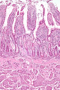
Somatostatinomas are a tumor of the delta cells of the endocrine pancreas that produces somatostatin. Increased levels of somatostatin inhibit pancreatic hormones and gastrointestinal hormones. Thus, somatostatinomas are associated with mild diabetes mellitus, steatorrhoea and gallstones, and achlorhydria. Somatostatinomas are commonly found in the head of pancreas. Only ten percent of somatostatinomas are functional tumours [9], and 60–70% of tumours are malignant. Nearly two-thirds of patients with malignant somatostatinomas will present with metastatic disease.

<span class="mw-page-title-main">Enteroendocrine cell</span>

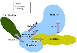
Enteroendocrine cells are specialized cells of the gastrointestinal tract and pancreas with endocrine function. They produce gastrointestinal hormones or peptides in response to various stimuli and release them into the bloodstream for systemic effect, diffuse them as local messengers, or transmit them to the enteric nervous system to activate nervous responses. Enteroendocrine cells of the intestine are the most numerous endocrine cells of the body. They constitute an enteric endocrine system as a subset of the endocrine system just as the enteric nervous system is a subset of the nervous system. In a sense they are known to act as chemoreceptors, initiating digestive actions and detecting harmful substances and initiating protective responses. Enteroendocrine cells are located in the stomach, in the intestine and in the pancreas. Microbiota play key roles in the intestinal immune and metabolic responses in these enteroendocrine cells via their fermentation product, acetate.

<span class="mw-page-title-main">Lanreotide</span> Pharmaceutical drug

Lanreotide, sold under the brand name Somatuline among others, is a medication used in the management of acromegaly and symptoms caused by neuroendocrine tumors, most notably carcinoid syndrome. It is a long-acting analogue of somatostatin, like octreotide.

<span class="mw-page-title-main">Somatostatin receptor 2</span> Protein-coding gene in the species Homo sapiens

Somatostatin receptor type 2 is a protein that in humans is encoded by the SSTR2 gene.

<span class="mw-page-title-main">Edotreotide</span> Chemical compound

Edotreotide (USAN, also known as (DOTA0-Phe1-Tyr3) octreotide, DOTA-TOC, DOTATOC) is a substance which, when bound to various radionuclides, is used in the treatment and diagnosis of certain types of cancer. When used therapeutically it is an example of peptide receptor radionuclide therapy.

<span class="mw-page-title-main">Pancreatic tumor</span> Medical condition

A pancreatic tumor is an abnormal growth in the pancreas. In adults, almost 90% are pancreatic cancer and a few are benign. Pancreatic tumors are rare in children.

Advanced Accelerator Applications is a France-based pharmaceutical group, specialized in the field of nuclear medicine. The group operates in all three segments of nuclear medicine to diagnose and treat serious conditions in the fields of oncology, neurology, cardiology, infectious and inflammatory diseases.

Pulmonary neuroendocrine tumors are neuroendocrine tumors localized to the lung: bronchus or pulmonary parenchyma.

Neuroendocrine differentiation is a term primarily used in relation to prostate cancers that display a significant neuroendocrine cell population on histopathological examination. These types of prostate cancer comprise true neuroendocrine cancers, such as small cell carcinoma, carcinoid and carcinoid-like tumors, as well as prostatic adenocarcinoma exhibiting focal neuroendocrine phenotype.

<span class="mw-page-title-main">Pancreatic neuroendocrine tumor</span> Medical condition

Pancreatic neuroendocrine tumours, often referred to as "islet cell tumours", or "pancreatic endocrine tumours" are neuroendocrine neoplasms that arise from cells of the endocrine (hormonal) and nervous system within the pancreas.

Hepatic artery embolization, also known as trans-arterial embolization (TAE), is one of the several therapeutic methods to treat primary liver tumors or metastases to the liver. The embolization therapy can reduce the size of the tumor, and decrease the tumor's impact such its hormone production, effectively decreasing symptoms. The treatment was initially developed in the early 1970s. The several types of hepatic artery treatments are based on the observation that tumor cells get nearly all their nutrients from the hepatic artery, while the normal cells of the liver get about 70-80 percent of their nutrients and 50% their oxygen supply from the portal vein, and thus can survive with the hepatic artery effectively blocked. In practice, hepatic artery embolization occludes the blood flow to the tumors, achieving significant tumor shrinkage in over 80% of people. Shrinkage rates vary.

<span class="mw-page-title-main">Peptide receptor radionuclide therapy</span> Type of radiotherapy

Peptide receptor radionuclide therapy (PRRT) is a type of radionuclide therapy, using a radiopharmaceutical that targets peptide receptors to deliver localised treatment, typically for neuroendocrine tumours (NETs).

Lutetium (<sup>177</sup>Lu) oxodotreotide Chelate of Lu-177 with DOTA-TATE, a peptide derivative bound to a DOTA molecule

Lutetium (177Lu) oxodotreotide (INN) or 177Lu DOTA-TATE, trade name Lutathera, is a chelated complex of a radioisotope of the element lutetium with DOTA-TATE, used in peptide receptor radionuclide therapy (PRRT). Specifically, it is used in the treatment of cancers which express somatostatin receptors.

<span class="mw-page-title-main">Somatostatin receptor antagonist</span> Class of chemical compounds

Somatostatin receptor antagonists are a class of chemical compounds that work by imitating the structure of the neuropeptide somatostatin. The somatostatin receptors are G protein-coupled receptors. Somatostatin receptor subtypes in humans are sstr1, 2A, 2 B, 3, 4 and 5. While normally expressed in the gastrointestinal (GI) tract, pancreas, hypothalamus, and central nervous system (CNS), they are expressed in different types of tumours. The predominant subtype in cancer cells is the ssrt2 subtype, which is expressed in neuroblastomas, meningiomas, medulloblastomas, breast carcinomas, lymphomas, renal cell carcinomas, paragangliomas, small cell lung carcinomas and hepatocellular carcinomas.

<span class="mw-page-title-main">Somatostatin inhibitor</span> Class of pharmaceuticals

Somatostatin receptor antagonists are a class of chemical compounds that work by imitating the structure of the neuropeptide somatostatin, which is an endogenous hormone found in the human body. The somatostatin receptors are G protein-coupled receptors. Somatostatin receptor subtypes in humans include sstr1, 2A, 2 B, 3, 4, and 5. While normally expressed in the gastrointestinal (GI) tract, pancreas, hypothalamus, and central nervous system (CNS), they are expressed in different types of tumours. The predominant subtype in cancer cells is the ssrt2 subtype, which is expressed in neuroblastomas, meningiomas, medulloblastomas, breast carcinomas, lymphomas, renal cell carcinomas, paragangliomas, small cell lung carcinomas, and hepatocellular carcinomas.

References

  1. 1 2 3 4 5 6 7 8 9 10 11 12 13 Ramage JK, Davies AH, Ardill J, Bax N, Caplin M, Grossman A, et al. (June 2005). "Guidelines for the management of gastroenteropancreatic neuroendocrine (including carcinoid) tumours". Gut. 54. 54 (Suppl 4): iv1–i16. doi:10.1136/gut.2004.053314. PMC   1867801 . PMID   15888809.
  2. Scalettar BA, Jacobs C, Fulwiler A, Prahl L, Simon A, Hilken L, Lochner JE (September 2012). "Hindered submicron mobility and long-term storage of presynaptic dense-core granules revealed by single-particle tracking". Developmental Neurobiology. 72 (9): 1181–1195. doi:10.1002/dneu.20984. PMC   3512567 . PMID   21976424.
  3. Oronsky B, Ma PC, Morgensztern D, Carter CA (December 2017). "Nothing But NET: A Review of Neuroendocrine Tumors and Carcinomas". Neoplasia. 19 (12): 991–1002. doi:10.1016/j.neo.2017.09.002. PMC   5678742 . PMID   29091800.
  4. 1 2 3 4 5 6 Klimstra DS, Modlin IR, Coppola D, Lloyd RV, Suster S (August 2010). "The pathologic classification of neuroendocrine tumors: a review of nomenclature, grading, and staging systems". Pancreas. 39 (6): 707–712. doi: 10.1097/MPA.0b013e3181ec124e . PMID   20664470. S2CID   3735444.
  5. 1 2 3 Tan EH, Tan CH (January 2011). "Imaging of gastroenteropancreatic neuroendocrine tumors". World Journal of Clinical Oncology. 2 (1): 28–43. doi: 10.5306/wjco.v2.i1.28 . PMC   3095463 . PMID   21603312.
  6. van Eeden S, Offerhaus GJ, Hart AA, Boerrigter L, Nederlof PM, Porter E, van Velthuysen ML (December 2007). "Goblet cell carcinoid of the appendix: a specific type of carcinoma". Histopathology. 51 (6): 763–773. doi:10.1111/j.1365-2559.2007.02883.x. PMID   18042066. S2CID   20185589.
  7. 1 2 3 4 Rindi, Guido; Mete, Ozgur; Uccella, Silvia; Basturk, Olca; La Rosa, Stefano; Brosens, Lodewijk A. A.; Ezzat, Shereen; de Herder, Wouter W.; Klimstra, David S.; Papotti, Mauro; Asa, Sylvia L. (March 2022). "Overview of the 2022 WHO Classification of Neuroendocrine Neoplasms". Endocrine Pathology. 33 (1): 115–154. doi:10.1007/s12022-022-09708-2. ISSN   1046-3976. PMID   35294740. S2CID   247455289.
  8. 1 2 Liu Y, Sturgis CD, Grzybicki DM, Jasnosz KM, Olson PR, Tong M, et al. (September 2001). "Microtubule-associated protein-2: a new sensitive and specific marker for pulmonary carcinoid tumor and small cell carcinoma". Modern Pathology. 14 (9): 880–885. doi: 10.1038/modpathol.3880406 . PMID   11557784. S2CID   24740130.
  9. 1 2 Rufini V, Calcagni ML, Baum RP (July 2006). "Imaging of neuroendocrine tumors". Seminars in Nuclear Medicine. 36 (3): 228–247. doi:10.1053/j.semnuclmed.2006.03.007. PMID   16762613.
  10. Soga J (December 2003). "Carcinoids and their variant endocrinomas. An analysis of 11842 reported cases". Journal of Experimental & Clinical Cancer Research. 22 (4): 517–530. PMID   15053292.
  11. Soga J, Yakuwa Y, Osaka M (October 1999). "Evaluation of 342 cases of mediastinal/thymic carcinoids collected from literature: a comparative study between typical carcinoids and atypical varieties". Annals of Thoracic and Cardiovascular Surgery. 5 (5): 285–292. PMID   10550713.
  12. 1 2 Oberg K, Jelic S (May 2008). "Neuroendocrine bronchial and thymic tumors: ESMO clinical recommendation for diagnosis, treatment and follow-up". Annals of Oncology. 19 (Suppl 2): ii102–ii103. doi: 10.1093/annonc/mdn116 . PMID   18456740.
  13. Beasley MB, Brambilla E, Travis WD (April 2005). "The 2004 World Health Organization classification of lung tumors". Seminars in Roentgenology. 40 (2): 90–97. doi:10.1053/j.ro.2005.01.001. PMID   15898407.
  14. Gustafsson BI, Kidd M, Chan A, Malfertheiner MV, Modlin IM (July 2008). "Bronchopulmonary neuroendocrine tumors". Cancer. 113 (1): 5–21. doi: 10.1002/cncr.23542 . PMID   18473355. S2CID   22143641.
  15. Wick MR, Berg LC, Hertz MI (June 1992). "Large cell carcinoma of the lung with neuroendocrine differentiation. A comparison with large cell "undifferentiated" pulmonary tumors". American Journal of Clinical Pathology. 97 (6): 796–805. doi:10.1093/ajcp/97.6.796. PMID   1317668.
  16. Massironi S, Sciola V, Peracchi M, Ciafardini C, Spampatti MP, Conte D (September 2008). "Neuroendocrine tumors of the gastro-entero-pancreatic system". World Journal of Gastroenterology. 14 (35): 5377–5384. doi: 10.3748/wjg.14.5377 . PMC   2744160 . PMID   18803349.
  17. Modlin IM, Oberg K, Chung DC, Jensen RT, de Herder WW, Thakker RV, et al. (January 2008). "Gastroenteropancreatic neuroendocrine tumours". The Lancet. Oncology. 9 (1): 61–72. doi:10.1016/S1470-2045(07)70410-2. PMID   18177818. S2CID   46127116.
  18. Metz DC, Jensen RT (November 2008). "Gastrointestinal neuroendocrine tumors: pancreatic endocrine tumors". Gastroenterology. 135 (5): 1469–1492. doi:10.1053/j.gastro.2008.05.047. PMC   2612755 . PMID   18703061.
  19. Griniatsos J, Michail O (April 2010). "Appendiceal neuroendocrine tumors: Recent insights and clinical implications". World Journal of Gastrointestinal Oncology. 2 (4): 192–196. doi: 10.4251/wjgo.v2.i4.192 . PMC   2999180 . PMID   21160597.
  20. Ni SJ, Sheng WQ, Du X (April 2010). "Pathologic research update of colorectal neuroendocrine tumors". World Journal of Gastroenterology. 16 (14): 1713–1719. doi: 10.3748/wjg.v16.i14.1713 . PMC   2852818 . PMID   20380002.
  21. Konishi T, Watanabe T, Nagawa H, Oya M, Ueno M, Kuroyanagi H, et al. (May 2010). "Treatment of colorectal carcinoids: A new paradigm". World Journal of Gastrointestinal Surgery. 2 (5): 153–156. doi: 10.4240/wjgs.v2.i5.153 . PMC   2999232 . PMID   21160865.
  22. Soga J (December 2002). "Primary hepatic endocrinomas (carcinoids and variant neoplasms). A statistical evaluation of 126 reported cases". Journal of Experimental & Clinical Cancer Research. 21 (4): 457–468. PMID   12636090.
  23. Nikfarjam M, Muralidharan V, Christophi C (2004). "Primary hepatic carcinoid tumours". HPB. 6 (1): 13–17. doi:10.1080/13651820310017228. PMC   2020649 . PMID   18333038.
  24. Moriura S, Ikeda S, Hirai M, Naiki K, Fujioka T, Yokochi K, Gotou S (September 1993). "Hepatic gastrinoma". Cancer. 72 (5): 1547–1550. doi: 10.1002/1097-0142(19930901)72:5<1547::AID-CNCR2820720510>3.0.CO;2-C . PMID   8348490.
  25. Soga J (March 2003). "Primary endocrinomas (carcinoids and variant neoplasms) of the gallbladder. A statistical evaluation of 138 reported cases". Journal of Experimental & Clinical Cancer Research. 22 (1): 5–15. PMID   12725316.
  26. Soga J, Osaka M, Yakuwa Y (2001). "Gut-endocrinomas (carcinoids and related endocrine variants) of the breast: an analysis of 310 reported cases". International Surgery. 86 (1): 26–32. PMID   11890336.
  27. Murali R, Kneale K, Lalak N, Delprado W (November 2006). "Carcinoid tumors of the urinary tract and prostate". Archives of Pathology & Laboratory Medicine. 130 (11): 1693–1706. doi:10.5858/2006-130-1693-CTOTUT. PMID   17076534.
  28. Mikuz G (1993). "[Non-urothelial tumors of the urinary tract]". Verhandlungen der Deutschen Gesellschaft für Pathologie. 77: 180–198. PMID   7511278.
  29. Soga J, Osaka M, Yakuwa Y (September 2001). "Gut-endocrinomas (carcinoids and related endocrine variants) of the uterine cervix: an analysis of 205 reported cases". Journal of Experimental & Clinical Cancer Research. 20 (3): 327–334. PMID   11718210.
  30. Usmani S, Orevi M, Stefanelli A, Zaniboni A, Gofrit ON, Bnà C, et al. (June 2019). "Neuroendocrine differentiation in castration resistant prostate cancer. Nuclear medicine radiopharmaceuticals and imaging techniques: A narrative review". Critical Reviews in Oncology/Hematology. 138: 29–37. doi:10.1016/j.critrevonc.2019.03.005. PMID   31092382. S2CID   131934021.
  31. Davies AH, Beltran H, Zoubeidi A (May 2018). "Cellular plasticity and the neuroendocrine phenotype in prostate cancer". Nature Reviews. Urology. 15 (5): 271–286. doi:10.1038/nrurol.2018.22. PMID   29460922. S2CID   4732323.
  32. 1 2 3 4 5 Jensen RT, Berna MJ, Bingham DB, Norton JA (October 2008). "Inherited pancreatic endocrine tumor syndromes: advances in molecular pathogenesis, diagnosis, management, and controversies". Cancer. 113 (7 Suppl): 1807–1843. doi:10.1002/cncr.23648. PMC   2574000 . PMID   18798544.
  33. 1 2 Hirsch NP, Murphy A, Radcliffe JJ (April 2001). "Neurofibromatosis: clinical presentations and anaesthetic implications". British Journal of Anaesthesia. 86 (4): 555–564. doi: 10.1093/bja/86.4.555 . PMID   11573632.
  34. 1 2 3 Lodish MB, Stratakis CA (June 2010). "Endocrine tumours in neurofibromatosis type 1, tuberous sclerosis and related syndromes". Best Practice & Research. Clinical Endocrinology & Metabolism. 24 (3): 439–449. doi:10.1016/j.beem.2010.02.002. PMC   2939061 . PMID   20833335.
  35. 1 2 Dworakowska D, Grossman AB (March 2009). "Are neuroendocrine tumours a feature of tuberous sclerosis? A systematic review". Endocrine-Related Cancer. 16 (1): 45–58. doi: 10.1677/ERC-08-0142 . PMID   18978035.
  36. 1 2 "Carney Complex, type 1; CNC1". OMIM - Online Mendelian Inheritance in Man. OMIM 160980.
  37. 1 2 "Carney Complex, type 2; CNC2". OMIM - Online Mendelian Inheritance in Man. OMIM 605244.
  38. Bosman FT, Carneiro F, Hruban RH, Theise ND, eds. (2010). WHO Classification of Tumours of the Digestive System (4 ed.). Lyon: International Agency for Research on Cancer. pp. 13–14. ISBN   978-92-832-2432-7.
  39. Basturk O, Yang Z, Tang LH, Hruban RH, Adsay V, McCall CM, et al. (May 2015). "The high-grade (WHO G3) pancreatic neuroendocrine tumor category is morphologically and biologically heterogenous and includes both well differentiated and poorly differentiated neoplasms". The American Journal of Surgical Pathology. 39 (5): 683–690. doi:10.1097/PAS.0000000000000408. PMC   4398606 . PMID   25723112.
  40. Amin MB, ed. (2017). AJCC Cancer Staging Manual (8 ed.). Springer. p. 351. ISBN   978-3-319-40617-6.
  41. Amin MB, ed. (2017). "29 – Neuroendocrine Tumors of the Stomach". AJCC Cancer Staging Manual (8 ed.). Springer. p. 355. ISBN   978-3-319-40617-6.
  42. Amin MB (2017). "30 – Neuroendocrine Tumors of the Duodenum & Ampulla of Vater". AJCC Cancer Staging Manual (8 ed.). Springer. p. 369. ISBN   978-3-319-40617-6.
  43. AJCC, 8th edition: Klimstra DS, Yang Z. "Pathology, classification, and grading of neuroendocrine neoplasms arising in the digestive system". UpToDate . This topic last updated: Oct 29, 2019.
  44. Amin MB (2017). "32 – Neuroendocrine Tumors of the Appendix". AJCC Cancer Staging Manual (8 ed.). Springer. p. 392. ISBN   978-3-319-40617-6.
  45. Amin MB (2017). "33 – Neuroendocrine Tumors of the Colon and Rectum". AJCC Cancer Staging Manual (8 ed.). Springer. p. 399. ISBN   978-3-319-40617-6.
  46. Amin MB (2017). "30 – Neuroendocrine Tumors of the Pancreas". AJCC Cancer Staging Manual (8 ed.). Springer. pp. 415–16. ISBN   978-3-319-40617-6.
  47. Kimura W, Kuroda A, Morioka Y (July 1991). "Clinical pathology of endocrine tumors of the pancreas. Analysis of autopsy cases". Digestive Diseases and Sciences. 36 (7): 933–942. doi:10.1007/BF01297144. PMID   2070707. S2CID   20567425. "[In] 800 autopsy cases, ... incidence of tumor was 10% (6/60) in individuals having histiological studies of all sections of the pancreas"
  48. 1 2 3 4 Arnold R, Göke R, Wied M, Behr T (2003). "Chapter 15 Neuroendocrine Gastro-Entero-Pancreatic (GEP) Tumors". In Scheppach W, Bresalier RS, Tytgat GN (eds.). Gastrointestinal and Liver Tumors. Berlin: Springer. pp. 195–233. ISBN   978-3-540-43462-7.
  49. 1 2 3 4 Pommier R (October 2003). The role of surgery and chemoembolization in the management of carcinoid. California Carcinoid Fighters Conference. Archived from the original on 2015-09-15.
  50. "Carcinoid Tumor Overview". Health Communities. Archived from the original on 2012-03-03.
  51. 1 2 3 4 5 6 7 8 Kvols LK (2002). "Carcinoid Tumors and the Carcinoid Syndrome: What's New in the Therapeutic Pipeline". Carcinoid Symposium 2002. The Carcinoid Cancer Foundation. Archived from the original on 2015-01-05.
  52. 1 2 3 4 5 6 7 Benson AB, Myerson RJ, Sasson AR (March 2011). "Pancreatic, neuroendocrine GI, and adrenal cancers.". Cancer Management: A Multidisciplinary Approach (13th ed.). UBM Medica. ISBN   978-0-615-41824-7. Archived from the original on 2011-05-15., cancernetwork.com; accessed November 8, 2015.
  53. "Cushing's Syndrome". The Lecturio Medical Concept Library. Retrieved 28 September 2021.
  54. 1 2 PDQ Adult Treatment Editorial Board (August 2022). "Pancreatic Neuroendocrine Tumors (Islet Cell Tumors) Treatment (PDQ®): Health Professional Version". PDQ Cancer Information Summaries [Internet]. Bethesda (MD): National Cancer Institute. PMID   26389309.
  55. Burns WR, Edil BH (March 2012). "Neuroendocrine pancreatic tumors: guidelines for management and update". Current Treatment Options in Oncology. 13 (1): 24–34. doi:10.1007/s11864-011-0172-2. PMID   22198808. S2CID   7329783.
  56. Bostanci A (February 2005). "Wildlife biology. A devil of a disease". Science. 307 (5712): 1035. doi:10.1126/science.307.5712.1035. PMID   15718445. S2CID   54100368. The tumors [of Devil facial tumor disease] have been characterized as a neuroendocrine cancer
  57. Kinver M (January 1, 2010). "Tasmanian devil facial cancer origins 'identified'". BBC. Archived from the original on January 2, 2010.
  58. Walsh B (January 1, 2010). "Decoding the Tasmanian Devil's Deadly Cancer". Time. Archived from the original on January 8, 2010.
  59. 1 2 Langley K (September 1994). "The neuroendocrine concept today". Annals of the New York Academy of Sciences. 733 (1): 1–17. Bibcode:1994NYASA.733....1L. doi:10.1111/j.1749-6632.1994.tb17251.x. PMID   7978856. S2CID   33139633.
  60. 1 2 Ferolla P, Faggiano A, Mansueto G, Avenia N, Cantelmi MG, Giovenali P, et al. (March 2008). "The biological characterization of neuroendocrine tumors: the role of neuroendocrine markers". Journal of Endocrinological Investigation. 31 (3): 277–286. doi:10.1007/bf03345602. PMID   18401212. S2CID   25108086.
  61. McMorran J, Crowther DC, McMorran S, Prince C, YoungMin S, Pleat J, Wacogne I. "investigations – General Practice Notebook". www.gpnotebook.co.uk. Archived from the original on 24 February 2017. Retrieved 23 February 2017.
  62. 1 2 Oberg K (July 2005). "Neuroendocrine tumors of the gastrointestinal tract: recent advances in molecular genetics, diagnosis, and treatment". Current Opinion in Oncology. 17 (4): 386–391. doi:10.1097/01.cco.0000167739.56948.a9. PMID   15933475. S2CID   208286399.
  63. van Essen M, Sundin A, Krenning EP, Kwekkeboom DJ (February 2014). "Neuroendocrine tumours: the role of imaging for diagnosis and therapy". Nature Reviews. Endocrinology. 10 (2): 102–114. doi:10.1038/nrendo.2013.246. PMID   24322649. S2CID   40129404.
  64. Hofman MS, Kong G, Neels OC, Eu P, Hong E, Hicks RJ (February 2012). "High management impact of Ga-68 DOTATATE (GaTate) PET/CT for imaging neuroendocrine and other somatostatin expressing tumours". Journal of Medical Imaging and Radiation Oncology. 56 (1): 40–47. doi: 10.1111/j.1754-9485.2011.02327.x . PMID   22339744. S2CID   21843609.
  65. Scott AT, Howe JR (August 2018). "Management of Small Bowel Neuroendocrine Tumors". Journal of Oncology Practice. 14 (8): 471–482. doi:10.1200/JOP.18.00135. PMC   6091496 . PMID   30096273.
  66. 1 2 Hofman MS, Hicks RJ (July 2012). "Changing paradigms with molecular imaging of neuroendocrine tumors". Discovery Medicine. 14 (74): 71–81. PMID   22846204 . Retrieved November 8, 2015.
  67. Hofland J, Kaltsas G, de Herder WW (April 2020). "Advances in the Diagnosis and Management of Well-Differentiated Neuroendocrine Neoplasms". Endocrine Reviews. 41 (2): 371–403. doi:10.1210/endrev/bnz004. PMC   7080342 . PMID   31555796.
  68. Nilica B, Waitz D, Stevanovic V, Uprimny C, Kendler D, Buxbaum S, et al. (August 2016). "Direct comparison of (68)Ga-DOTA-TOC and (18)F-FDG PET/CT in the follow-up of patients with neuroendocrine tumour treated with the first full peptide receptor radionuclide therapy cycle". European Journal of Nuclear Medicine and Molecular Imaging. 43 (9): 1585–1592. doi:10.1007/s00259-016-3328-2. PMC   4932132 . PMID   26922350.
  69. Van Buren G, Rashid A, Yang AD, Abdalla EK, Gray MJ, Liu W, et al. (August 2007). "The development and characterization of a human midgut carcinoid cell line". Clinical Cancer Research. 13 (16): 4704–4712. doi: 10.1158/1078-0432.CCR-06-2723 . PMID   17699847.
  70. Berretta M, Cappellani A, Di Vita M, Berretta S, Nasti G, Bearz A, et al. (January 2010). "Biomarkers in neuroendocrine tumors". Frontiers in Bioscience. 2 (1): 332–342. doi: 10.2741/s68 . PMID   20036951.
  71. Mukhopadhyay S, Dermawan JK, Lanigan CP, Farver CF (January 2019). "Insulinoma-associated protein 1 (INSM1) is a sensitive and highly specific marker of neuroendocrine differentiation in primary lung neoplasms: an immunohistochemical study of 345 cases, including 292 whole-tissue sections". Modern Pathology. 32 (1): 100–109. doi: 10.1038/s41379-018-0122-7 . PMID   30154579. S2CID   52110673.
  72. "Definition of Mesentery". MedicineNet. Retrieved 2018-04-21.
  73. Tebbi CK, Windle ML, Cripe TP, Sakamoto KM (1 April 2014). Coppes MJ (ed.). "Carcinoid Tumor". Medscape.com. WebMD LLC. Archived from the original on 15 December 2014. Retrieved 3 September 2014.
  74. 1 2 3 Warner RR (May 2005). "Enteroendocrine tumors other than carcinoid: a review of clinically significant advances". Gastroenterology. 128 (6): 1668–1684. doi: 10.1053/j.gastro.2005.03.078 . PMID   15887158.
  75. 1 2 3 Öberg K (1998). "Carcinoid Tumors: Current Concepts in Diagnosis and Treatment". The Oncologist. 3 (5): 339–345. doi: 10.1634/theoncologist.3-5-339 . PMID   10388123.
  76. Oberg K, Akerström G, Rindi G, Jelic S (May 2010). "Neuroendocrine gastroenteropancreatic tumours: ESMO Clinical Practice Guidelines for diagnosis, treatment and follow-up". Annals of Oncology. 21 (Suppl 5): v223–v227. doi: 10.1093/annonc/mdq192 . PMID   20555086.
  77. Clark OH, Benson AB, Berlin JD, Choti MA, Doherty GM, Engstrom PF, et al. (July 2009). "NCCN Clinical Practice Guidelines in Oncology: neuroendocrine tumors". Journal of the National Comprehensive Cancer Network. 7 (7): 712–747. doi: 10.6004/jnccn.2009.0050 . PMID   19635226.
  78. "Islet Cell Tumors (Endocrine Pancreas)". National Cancer Institute. Archived from the original on 2011-06-07.
  79. "Gastrointestinal Carcinoid Tumors Treatment". National Cancer Institute. Archived from the original on 2011-06-27.
  80. "Merkel cell tumors". National Cancer Institute. 21 February 2006. Archived from the original on 2011-06-07.
  81. "Pheochromocytoma and Paraganglioma". National Cancer Institute. Archived from the original on 2011-06-07.
  82. Rinke A, Auernhammer CJ, Bodei L, Kidd M, Krug S, Lawlor R, et al. (September 2021). "Treatment of advanced gastroenteropancreatic neuroendocrine neoplasia, are we on the way to personalised medicine?". Gut. 70 (9): 1768–1781. doi:10.1136/gutjnl-2020-321300. PMID   33692095. S2CID   232172557.
  83. "Neuroendocrine tumors, NCCN Guidelines Version 1.2015" (PDF). NCCN Guidelines. National Comprehensive Cancer Network, Inc. November 11, 2014. Retrieved December 25, 2014.
  84. Oberg K, Kvols L, Caplin M, Delle Fave G, de Herder W, Rindi G, et al. (June 2004). "Consensus report on the use of somatostatin analogs for the management of neuroendocrine tumors of the gastroenteropancreatic system". Annals of Oncology. 15 (6): 966–973. doi: 10.1093/annonc/mdh216 . PMID   15151956.
  85. Rinke A, Müller HH, Schade-Brittinger C, Klose KJ, Barth P, Wied M, et al. (October 2009). "Placebo-controlled, double-blind, prospective, randomized study on the effect of octreotide LAR in the control of tumor growth in patients with metastatic neuroendocrine midgut tumors: a report from the PROMID Study Group". Journal of Clinical Oncology. 27 (28): 4656–4663. doi:10.1200/JCO.2009.22.8510. PMID   19704057.
  86. Caplin ME, Pavel M, Ćwikła JB, Phan AT, Raderer M, Sedláčková E, et al. (July 2014). "Lanreotide in metastatic enteropancreatic neuroendocrine tumors". The New England Journal of Medicine. 371 (3): 224–233. doi: 10.1056/NEJMoa1316158 . PMID   25014687.
  87. 1 2 Öberg K. Neuroendocrine Gastroenteropancreatic Tumours: Current Views on Diagnosis and Treatment. Business Briefing. European Oncology Review 2005; pp. 1–6.
  88. Chan JA, Kulke MH (May 2009). "Progress in the treatment of neuroendocrine tumors". Current Oncology Reports. 11 (3): 193–199. doi:10.1007/s11912-009-0028-0. PMC   4029419 . PMID   19336011.
  89. Strosberg J, El-Haddad G, Wolin E, Hendifar A, Yao J, Chasen B, et al. (January 2017). "Phase 3 Trial of 177Lu-Dotatate for Midgut Neuroendocrine Tumors". The New England Journal of Medicine. 376 (2): 125–135. doi:10.1056/nejmoa1607427. hdl:2445/125256. PMC   5895095 . PMID   28076709.
  90. Kratochwil C, Giesel FL, Bruchertseifer F, Mier W, Apostolidis C, Boll R, et al. (November 2014). "²¹³Bi-DOTATOC receptor-targeted alpha-radionuclide therapy induces remission in neuroendocrine tumours refractory to beta radiation: a first-in-human experience". European Journal of Nuclear Medicine and Molecular Imaging. 41 (11): 2106–2119. doi:10.1007/s00259-014-2857-9. PMC   4525192 . PMID   25070685.
  91. Kashyap R, Hofman MS, Michael M, Kong G, Akhurst T, Eu P, et al. (February 2015). "Favourable outcomes of (177)Lu-octreotate peptide receptor chemoradionuclide therapy in patients with FDG-avid neuroendocrine tumours". European Journal of Nuclear Medicine and Molecular Imaging. 42 (2): 176–185. doi:10.1007/s00259-014-2906-4. PMID   25209134. S2CID   20740102.
  92. Hofman MS, Michael M, Kashyap R, Hicks RJ (June 2015). "Modifying the Poor Prognosis Associated with 18F-FDG-Avid NET with Peptide Receptor Chemo-Radionuclide Therapy (PRCRT)". Journal of Nuclear Medicine. 56 (6): 968–969. doi: 10.2967/jnumed.115.154500 . PMID   25814516. S2CID   207407820.
  93. 1 2 Fong T, Schoenfield LJ. "Arterial Chemotherapy Infusion of the Liver (and) Chemoembolization of the Liver (TACE)". medicinenet.com. Archived from the original on 2014-12-24. Retrieved 8 November 2015.
  94. 1 2 Welsh JS, Kennedy AS, Thomadsen B (2006). "Selective Internal Radiation Therapy (SIRT) for liver metastases secondary to colorectal adenocarcinoma". International Journal of Radiation Oncology, Biology, Physics. 66 (2 Suppl): S62–S73. doi: 10.1016/j.ijrobp.2005.09.011 . PMID   16979443.
  95. Van De Wiele C, Defreyne L, Peeters M, Lambert B (June 2009). "Yttrium-90 labelled resin microspheres for treatment of primary and secondary malignant liver tumors". The Quarterly Journal of Nuclear Medicine and Molecular Imaging. 53 (3): 317–324. PMID   19521311.
  96. Salem R, Thurston KG, Carr BI, Goin JE, Geschwind JF (September 2002). "Yttrium-90 microspheres: radiation therapy for unresectable liver cancer". Journal of Vascular and Interventional Radiology. 13 (9 Pt 2): S223–S229. doi:10.1016/S1051-0443(07)61790-4. PMID   12354840.
  97. Masters A (2014-10-14). "A plutocratic proposal". Mosaic. The Wellcome Trust. Archived from the original on 2016-05-29. Retrieved 2016-07-03.
  98. "iCancer web site". icancer.org.uk. Archived from the original on 2016-07-14. Retrieved 2016-07-03.
  99. Masters A (2016-07-02). "Can crowdfunding really cure cancer? Alexander Masters investigates a pioneering new project". The Telegraph. Archived from the original on 2016-07-03. Retrieved 2016-07-03.
  100. Detjen K, Hammerich L, Özdirik B, Demir M, Wiedenmann B, Tacke F, et al. (2020-07-02). "Models of Gastroenteropancreatic Neuroendocrine Neoplasms: Current Status and Future Directions". Neuroendocrinology. 111 (3): 217–236. doi: 10.1159/000509864 . PMID   32615560.
  101. April-Monn SL, Wiedmer T, Skowronska M, Maire R, Schiavo Lena M, Trippel M, et al. (2020-04-03). "Three-Dimensional Primary Cell Culture: A Novel Preclinical Model for Pancreatic Neuroendocrine Tumors". Neuroendocrinology. 111 (3): 273–287. doi: 10.1159/000507669 . PMID   32241015. S2CID   214768622.
  102. 1 2 Oberg K, Castellano D (March 2011). "Current knowledge on diagnosis and staging of neuroendocrine tumors". Cancer and Metastasis Reviews. 30 (Suppl 1): 3–7. doi:10.1007/s10555-011-9292-1. PMID   21311954. S2CID   29720754.
  103. Kimura W, Kuroda A, Morioka Y (July 1991). "Clinical pathology of endocrine tumors of the pancreas. Analysis of autopsy cases". Digestive Diseases and Sciences. 36 (7): 933–942. doi:10.1007/BF01297144. PMID   2070707. S2CID   20567425.
  104. 1 2 3 4 Modlin IM, Shapiro MD, Kidd M (December 2004). "Siegfried Oberndorfer: origins and perspectives of carcinoid tumors". Human Pathology. 35 (12): 1440–1451. doi:10.1016/j.humpath.2004.09.018. PMID   15619202.