The urethra is a tube that connects the mammalian urinary bladder to the urinary meatus. Male and female placental mammals release urine through the urethra during urination, but males also release semen through the urethra during ejaculation.
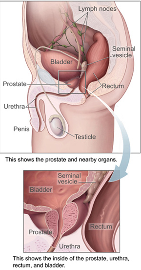
The prostate is both an accessory gland of the male reproductive system and a muscle-driven mechanical switch between urination and ejaculation. It is found in all male mammals. It differs between species anatomically, chemically, and physiologically. Anatomically, the prostate is found below the bladder, with the urethra passing through it. It is described in gross anatomy as consisting of lobes and in microanatomy by zone. It is surrounded by an elastic, fibromuscular capsule and contains glandular tissue, as well as connective tissue.

In female human anatomy, Skene's glands or the Skene glands are glands located around the lower end of the urethral meatus. The glands are surrounded by tissue that swells with blood during sexual arousal, and secrete a fluid from openings near the urethra, particularly during orgasm.

Pre-ejaculate is a clear, colorless, viscous fluid that is emitted from the urethra of the penis during sexual arousal. It is similar in composition to semen but has distinct chemical differences. The presence of sperm in the fluid is variable from low to absent. Pre-ejaculate functions as a lubricant and an acid neutralizer.

Retrograde ejaculation occurs when semen which would be ejaculated via the urethra is redirected to the urinary bladder. Normally, the sphincter of the bladder contracts before ejaculation, sealing the bladder which besides inhibiting the release of urine also prevents a reflux of seminal fluids into the male bladder during ejaculation. The semen is forced to exit via the urethra, the path of least resistance. When the bladder sphincter does not function properly, retrograde ejaculation may occur. It can also be induced deliberately by a male as a primitive form of male birth control or as part of certain alternative medicine practices. The retrograde-ejaculated semen, which goes into the bladder, is excreted with the next urination.

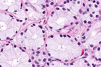
The seminal vesicles are a pair of convoluted tubular accessory glands that lie behind the urinary bladder of male mammals. They secrete fluid that partly composes the semen.
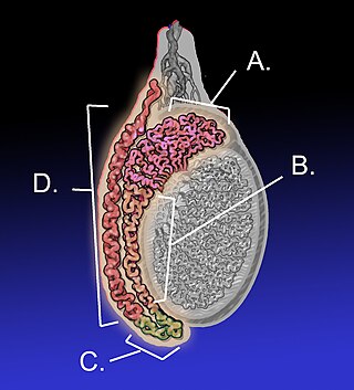
The epididymis is an elongated tubular structure attached to the posterior side of each one of the two male reproductive glands, the testicles. It is a single, narrow, tightly coiled tube in adult humans, 6 to 7 centimetres in length; uncoiled the tube would be approximately 6 m long. It connects the testicle to the vas deferens in the male reproductive system. The epididymis serves as an interconnection between the multiple efferent ducts at the rear of a testicle (proximally), and the vas deferens (distally). Its primary function is the storage, maturation and transport of sperm cells.

The vas deferens, with the more modern name ductus deferens, is part of the male reproductive system of many vertebrates. The ducts transport sperm from the epididymides to the ejaculatory ducts in anticipation of ejaculation. The vas deferens is a partially coiled tube which exits the abdominal cavity through the inguinal canal.
The ejaculatory ducts are paired structures in the male reproductive system. Each ejaculatory duct is formed by the union of the vas deferens with the duct of the seminal vesicle. They pass through the prostate, and open into the urethra above the seminal colliculus. During ejaculation, semen passes through the prostate gland, enters the urethra and exits the body via the urinary meatus.
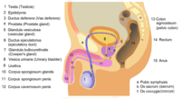
The male reproductive system consists of a number of sex organs that play a role in the process of human reproduction. These organs are located on the outside of the body, and within the pelvis.

The human reproductive system includes the male reproductive system which functions to produce and deposit sperm; and the female reproductive system which functions to produce egg cells, and to protect and nourish the fetus until birth. Humans have a high level of sexual differentiation. In addition to differences in nearly every reproductive organ, there are numerous differences in typical secondary sex characteristics.
Aspermia is the complete lack of semen with ejaculation. It is associated with infertility.

In human anatomy, the penis is an external male sex organ that additionally serves as the urinary duct. The main parts are the root, body, the epithelium of the penis including the shaft skin, and the foreskin covering the glans. The body of the penis is made up of three columns of tissue: two corpora cavernosa on the dorsal side and corpus spongiosum between them on the ventral side. The human male urethra passes through the prostate gland, where it is joined by the ejaculatory duct, and then through the penis. The urethra traverses the corpus spongiosum, and its opening, the meatus, lies on the tip of the glans. It is a passage both for urination and ejaculation of semen.
The development of the reproductive system is the part of embryonic growth that results in the sex organs and contributes to sexual differentiation. Due to its large overlap with development of the urinary system, the two systems are typically described together as the genitourinary system.

Semen, also known as seminal fluid, is an organic bodily fluid that contains spermatozoa. Spermatozoa are secreted by the male gonads and other sexual organs of male or hermaphroditic animals and can fertilize the female ovum. Semen is produced and originates from the seminal vesicle, which is located in the pelvis. The process that results in the discharge of semen from the urethral orifice is called ejaculation. In humans, seminal fluid contains several components besides spermatozoa: proteolytic and other enzymes as well as fructose are elements of seminal fluid which promote the survival of spermatozoa and provide a medium through which they can move or "swim". The fluid is adapted to be discharged deep into the vagina, so the spermatozoa can pass into the uterus and form a zygote with an egg.
The reproductive system of an organism, also known as the genital system, is the biological system made up of all the anatomical organs involved in sexual reproduction. Many non-living substances such as fluids, hormones, and pheromones are also important accessories to the reproductive system. Unlike most organ systems, the sexes of differentiated species often have significant differences. These differences allow for a combination of genetic material between two individuals, which allows for the possibility of greater genetic fitness of the offspring.

Ejaculation is the discharge of semen from the male reproductive tract. It is normally linked with orgasm, which involves involuntary contractions of the pelvic floor. It is the final stage and natural objective of male sexual stimulation, and an essential component of natural conception. Ejaculation can occur spontaneously during sleep, and is a normal part of human sexual development. In rare cases, ejaculation occurs because of prostatic disease. Anejaculation is the condition of being unable to ejaculate. Ejaculation is normally intensely pleasurable for men; dysejaculation is an ejaculation that is painful or uncomfortable. Retrograde ejaculation is the condition where semen travels backwards into the bladder rather than out of the urethra.

Male accessory glands (MAG) in humans are the seminal vesicles, prostate gland, and the bulbourethral glands. In insects, male accessory glands produce products that mix with the sperm to protect and preserve them, including seminal fluid proteins. Some insecticides can induce an increase in the protein content of the male accessory glands of certain types of insects. This has the unintended effect of increasing the number of offspring they produce.

The reproductive system of planarians is broadly similar among different families, although the associated structures can vary in complexity.
The monotremes represent the order of extant mammals most distantly related to humans. The platypus is indigenous to eastern Australia; the short-beaked echidna is indigenous to Australia and Papua New Guinea; whereas the long-beaked echidna is restricted to Papua New Guinea and Irian Jaya. Since monotremes exhibit characteristics common with both reptiles and therian mammals, they are of great interest for the study of mammalian evolution.