Cell-based therapies for Parkinson's disease

Last updated
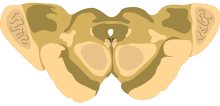
A diagram of the rostral midbrain at the level of the superior colliculi. An area of the brain which degenerates in Parkinson's disease, called the substantia nigra pars compacta, is visualized as two dark brown bands in the lower half of the image. Midbrain - superior colliculus.svg
A diagram of the rostral midbrain at the level of the superior colliculi. An area of the brain which degenerates in Parkinson's disease, called the substantia nigra pars compacta, is visualized as two dark brown bands in the lower half of the image.

Cell-based therapies for Parkinson's disease include various investigational procedures which transplant specific populations of cells into the brains of people with Parkinson's disease. The investigation of cell transplantation therapies followed the discovery that the death of dopaminergic neurons in the substantia nigra pars compacta resulted in the motor symptoms of the disease. Thus, cell transplantation has focused on various dopamine producing cells throughout the body. [1] [2]

Contents

List of cell-based sources

Fetal ventral mesencephalic tissue

Human

Porcine

Histological section through the adrenal gland. The black pointer indicates the innermost layer known as the adrenal medulla. Adrenal gland (medulla).JPG
Histological section through the adrenal gland. The black pointer indicates the innermost layer known as the adrenal medulla.

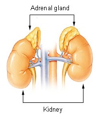
Adrenal medulla

The first cell-based therapy investigated for Parkinson's disease utilized the adrenal medulla. The adrenal medulla is the innermost part of the adrenal gland and contains neural crest derived chromaffin cells which secrete norepinephrine, epinephrine and to a far lesser extent dopamine into the blood. Autotransplantation of adrenal medullary tissue into the brains of animal models of Parkinson's disease showed minimal benefits. [3] [4] Despite this, open-label trials were undergone in humans which showed only modest benefits. [5] [6] Following these initial disappointing results however, a trial in Mexico demonstrated significant motor benefits in two patients with Parkinson's disease who had undergone the procedure. [7] This publication incited widespread interest in the field and over the next few years hundreds of patients received adrenal medulla transplants. [1] It was only when a registry was set up to consolidate all the data was it revealed that most patients did not benefit from the procedure to any significant extent. [8] [9] Furthermore, postoperative complications such as psychiatric disturbances were realized. These combined findings eventually led to the abandonment of this transplant procedure, which was largely flawed from the start. [2]

Sympathetic ganglia

A diagram of the arterial supply to the head and neck. The common carotid artery can be seen bifurcating into the internal carotid artery and external carotid artery. The carotid body is found at the bifurcation of the common carotid artery, in the same location as the carotid sinus. 2122 Common Carotid Artery.jpg
A diagram of the arterial supply to the head and neck. The common carotid artery can be seen bifurcating into the internal carotid artery and external carotid artery. The carotid body is found at the bifurcation of the common carotid artery, in the same location as the carotid sinus.

Carotid body

The carotid body is a group of chemoreceptor cells located at the bifurcation of the common carotid artery. It includes two populations of cells; glomus (type I) cells and sustentacular (type II) cells. Glomus cells are derived from the neural crest and secrete dopamine in response to hypoxemia (low level of oxygen in the blood). [10] Based on their ability to secrete dopamine and also glial cell-derived neurotrophic factor (GDNF), [11] these cells have been investigated as an intrastriatal autograft therapy for patients with Parkinson's disease. [12] [13] A clinical trial exploring this initially demonstrated motor benefits, unfortunately these benefits disappeared after 6–12 months, in correlation with poor survival of the grafted cells. [14]

Retinal pigment epithelium

The retinal pigment epithelium (RPE) is a single layer of melanin containing cells located between the neural retina and the choroid. Retinal pigment epithelial cells synthesize dopamine and secrete the neurotrophic factors glial-cell derived neurotrophic factor (GDNF) and brain-derived neurotrophic factor (BDNF). [15] Initial trials of intrastriatal allografts of cultured human retinal pigment epithelial cells attached to microcarriers (Spheramine, Bayer Schering Pharma AG) demonstrated

Stem cells

Researchers have differentiated ESCs into dopamine-producing cells with the hope that these neurons could be used in the treatment of Parkinson's disease.

Related Research Articles

<span class="mw-page-title-main">Adrenal gland</span> Endocrine gland

The adrenal glands are endocrine glands that produce a variety of hormones including adrenaline and the steroids aldosterone and cortisol. They are found above the kidneys. Each gland has an outer cortex which produces steroid hormones and an inner medulla. The adrenal cortex itself is divided into three main zones: the zona glomerulosa, the zona fasciculata and the zona reticularis.

<span class="mw-page-title-main">Vagus nerve</span> Main nerve of the parasympathetic nervous system

The vagus nerve, also known as the tenth cranial nerve, cranial nerve X, or simply CN X, is a cranial nerve that carries sensory fibers that create a pathway that interfaces with the parasympathetic control of the heart, lungs, and digestive tract. It comprises two nerves—the left and right vagus nerves—but they are typically referred to collectively as a single subsystem.

<span class="mw-page-title-main">Catecholamine</span> Class of chemical compounds

A catecholamine is a monoamine neurotransmitter, an organic compound that has a catechol and a side-chain amine.

<span class="mw-page-title-main">Adrenal medulla</span> Central part of the adrenal gland

The adrenal medulla is the inner part of the adrenal gland. It is located at the center of the gland, being surrounded by the adrenal cortex. It is the innermost part of the adrenal gland, consisting of chromaffin cells that secrete catecholamines, including epinephrine (adrenaline), norepinephrine (noradrenaline), and a small amount of dopamine, in response to stimulation by sympathetic preganglionic neurons.

<span class="mw-page-title-main">Chromaffin cell</span> Neuroendocrine cells found in adrenal medulla in mammals

Chromaffin cells, also called pheochromocytes, are neuroendocrine cells found mostly in the medulla of the adrenal glands in mammals. These cells serve a variety of functions such as serving as a response to stress, monitoring carbon dioxide and oxygen concentrations in the body, maintenance of respiration and the regulation of blood pressure. They are in close proximity to pre-synaptic sympathetic ganglia of the sympathetic nervous system, with which they communicate, and structurally they are similar to post-synaptic sympathetic neurons. In order to activate chromaffin cells, the splanchnic nerve of the sympathetic nervous system releases acetylcholine, which then binds to nicotinic acetylcholine receptors on the adrenal medulla. This causes the release of catecholamines. The chromaffin cells release catecholamines: ~80% of adrenaline (epinephrine) and ~20% of noradrenaline (norepinephrine) into systemic circulation for systemic effects on multiple organs, and can also send paracrine signals. Hence they are called neuroendocrine cells.

<span class="mw-page-title-main">Glomus cell</span>

Glomus cells are the cell type mainly located in the carotid bodies and aortic bodies. Glomus type I cells are peripheral chemoreceptors which sense the oxygen, carbon dioxide and pH levels of the blood. When there is a decrease in the blood's pH, a decrease in oxygen (pO2), or an increase in carbon dioxide (pCO2), the carotid bodies and the aortic bodies signal the dorsal respiratory group in the medulla oblongata to increase the volume and rate of breathing. The glomus cells have a high metabolic rate and good blood perfusion and thus are sensitive to changes in arterial blood gas tension. Glomus type II cells are sustentacular cells having a similar supportive function to glial cells.

Autotransplantation is the transplantation of organs, tissues, or even particular proteins from one part of the body to another in the same person.

An amniotic epithelial cell is a form of stem cell extracted from the lining of the inner membrane of the placenta. Amniotic epithelial cells start to develop around 8 days post fertilization. These cells are known to have some of the same markers as embryonic stem cells, more specifically, Oct-4 and nanog. These transcription factors are the basis of the pluripotency of stem cells. Amniotic epithelial cells have the ability to develop into any of the three germ layers: endoderm, mesoderm, and ectoderm. They can develop into several organ tissues specific to these germ layers including heart, brain, and liver. The pluripotency of the human amniotic epithelial cells makes them useful in treating and fighting diseases and disorders of the nervous system as well as other tissues of the human body. Artificial heart valves and working tracheas, as well as muscle, fat, bone, heart, neural and liver cells have all been engineered using amniotic stem cells. Tissues obtained from amniotic cell lines show promise for patients with congenital diseases or malformations of the heart, liver, lungs, kidneys, and cerebral tissue.

In the management of Parkinson's disease, due to the chronic nature of Parkinson's disease (PD), a broad-based program is needed that includes patient and family education, support-group services, general wellness maintenance, exercise, and nutrition. At present, no cure for the disease is known, but medications or surgery can provide relief from the symptoms.
<span class="mw-page-title-main">APUD cell</span> Type of endocrine cells

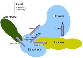
APUD cells (DNES cells) constitute a group of apparently unrelated endocrine cells, which were named by the scientist A.G.E. Pearse, who developed the APUD concept in the 1960s based on calcitonin-secreting parafollicular C cells of dog thyroid. These cells share the common function of secreting a low molecular weight polypeptide hormone. There are several different types which secrete the hormones secretin, cholecystokinin and several others. The name is derived from an acronym, referring to the following:

<span class="mw-page-title-main">Paraganglion</span>

A paraganglion is a group of non-neuronal cells derived of the neural crest. They are named for being generally in close proximity to sympathetic ganglia. They are essentially of two types: (1) chromaffin or sympathetic paraganglia made of chromaffin cells and (2) nonchromaffin or parasympathetic paraganglia made of glomus cells. They are neuroendocrine cells, the former with primary endocrine functions and the latter with primary chemoreceptor functions.
<span class="mw-page-title-main">PC12 cell line</span>

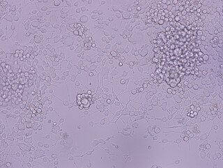
PC12 is a cell line derived from a pheochromocytoma of the rat adrenal medulla, that have an embryonic origin from the neural crest that has a mixture of neuroblastic cells and eosinophilic cells.

<span class="mw-page-title-main">PEDF</span> Protein-coding gene in the species Homo sapiens

Pigment epithelium-derived factor (PEDF) also known as serpin F1 (SERPINF1), is a multifunctional secreted protein that has anti-angiogenic, anti-tumorigenic, and neurotrophic functions. Found in vertebrates, this 50 kDa protein is being researched as a therapeutic candidate for treatment of such conditions as choroidal neovascularization, heart disease, and cancer. In humans, pigment epithelium-derived factor is encoded by the SERPINF1 gene.

<span class="mw-page-title-main">Parkinson's disease</span> Long-term degenerative neurological disorder

Parkinson's disease (PD), or simply Parkinson's, is a long-term neurodegenerative disease of the central nervous system that affects both the motor system and non-motor systems. The symptoms usually emerge slowly, and as the disease progresses, non-motor symptoms become more common. Usual symptoms are tremor, rigidity, slowness of movement, and difficulty with walking, collectively known as parkinsonism. Parkinson's disease dementia, falls and neuropsychiatric problems such as sleep abnormalities, psychosis, mood swings or behavioral changes may arise in advanced stages.

<span class="mw-page-title-main">Ivar Mendez</span> Canadian neurosurgeon and neuroscientist

Ivar Mendez is a neurosurgeon, neuroscientist and Professor of Surgery at the University of Saskatchewan. He is internationally known for his work in cell transplantation for Parkinson's disease and the use of remote presence robotics in neurosurgery and primary health care. In December 2022, Mendez was appointed an officer of the Order of Canada for his pioneering work in the use of remote telemedicine and robotics to revolutionize the delivery of health and patient care.

<span class="mw-page-title-main">Sympathoadrenal system</span>

The sympathoadrenal system is a physiological connection between the sympathetic nervous system and the adrenal medulla and is crucial in an organism's physiological response to outside stimuli. When the body receives sensory information, the sympathetic nervous system sends a signal to preganglionic nerve fibers, which activate the adrenal medulla through acetylcholine. Once activated, norepinephrine and epinephrine are released directly into the blood by adrenomedullary cells where they act as the bodily mechanism for "fight-or-flight" responses. Because of this, the sympathoadrenal system plays a large role in maintaining glucose levels, sodium levels, blood pressure, and various other metabolic pathways that couple with bodily responses to the environment. During numerous diseased states, such as hypoglycemia or even stress, the body's metabolic processes are skewed. The sympathoadrenal system works to return the body to homeostasis through the activation or inactivation of the adrenal gland. However, more severe disorders of the sympathoadrenal system such as pheochromocytoma can affect the body's ability to maintain a homeostatic state. In these cases, curative agents such as adrenergic agonists and antagonists are used to modify epinephrine and norepinephrine levels released by the adrenal medulla.

Gene therapy in Parkinson's disease consists of the creation of new cells that produce a specific neurotransmitter (dopamine), protect the neural system, or the modification of genes that are related to the disease. Then these cells are transplanted to a patient with the disease. There are different kinds of treatments that focus on reducing the symptoms of the disease but currently there is no cure.

Translational neuroscience is the field of study which applies neuroscience research to translate or develop into clinical applications and novel therapies for nervous system disorders. The field encompasses areas such as deep brain stimulation, brain machine interfaces, neurorehabilitation and the development of devices for the sensory nervous system such as the use of auditory implants, retinal implants, and electronic skins.

<span class="mw-page-title-main">Anders Björklund</span> Swedish histologist (born 1945)

Anders Björklund' is a Swedish neuroscientist and pioneer in the study of cell- and gene-based reparative and neuroprotective mechanisms in the brain. He has spent his academic career at Lund University in Sweden, as professor since 1983 and as senior professor at the Wallenberg Neuroscience Center since his formal retirement in 2012.

Foetal brain cell graft is a surgical procedure that can be used as a regenerative treatment for various neurological conditions, but was mainly explored and used specifically for treating Parkinson's disease (PD). A standardised procedure is followed: the cells are usually obtained from a 7–8 weeks old foetus and the collected cells undergo testing to examine whether they are free from infectious agents and safe for transplantation. It is found that this procedure results in an overall improvement in motor functions and a reduction in reliance on medication for PD patients.
References

  1. 1 2 Jankovic, Joseph; Tolosa, Eduardo (2015). "49". Parkinson's Disease and Movement Disorders (6th ed.). Philadelphia: Wolters Kluwer. pp. 518–525. ISBN   9781496317636. OCLC   953863020.
  2. 1 2 Barker, Roger A.; Drouin-Ouellet, Janelle; Parmar, Malin (September 2015). "Cell-based therapies for Parkinson disease—past insights and future potential". Nature Reviews Neurology. 11 (9): 492–503. doi:10.1038/nrneurol.2015.123. ISSN   1759-4758. PMID   26240036. S2CID   9256654.
  3. Freed, W. J.; Morihisa, J. M.; Spoor, E.; Hoffer, B. J.; Olson, L.; Seiger, A.; Wyatt, R. J. (1981-07-23). "Transplanted adrenal chromaffin cells in rat brain reduce lesion-induced rotational behaviour". Nature. 292 (5821): 351–352. Bibcode:1981Natur.292..351F. doi:10.1038/292351a0. ISSN   0028-0836. PMID   7254334. S2CID   37266235.
  4. Morihisa, John M.; Nakamura, Richard K.; Freed, William J.; Mishkin, Mortimer; Wyatt, Richard J. (1987). "Transplantation Techniques and the Survival of Adrenal Medulla Autografts in the Primate Brain". Annals of the New York Academy of Sciences. 495 (1): 599–604. Bibcode:1987NYASA.495..599M. doi:10.1111/j.1749-6632.1987.tb23703.x. ISSN   1749-6632. PMID   3111329. S2CID   45849626.
  5. Lindvall, O.; Backlund, E. O.; Farde, L.; Sedvall, G.; Freedman, R.; Hoffer, B.; Nobin, A.; Seiger, A.; Olson, L. (Oct 1987). "Transplantation in Parkinson's disease: two cases of adrenal medullary grafts to the putamen". Annals of Neurology. 22 (4): 457–468. doi:10.1002/ana.410220403. ISSN   0364-5134. PMID   3435067. S2CID   26259544.
  6. Backlund, E. O.; Granberg, P. O.; Hamberger, B.; Knutsson, E.; Mårtensson, A.; Sedvall, G.; Seiger, A.; Olson, L. (Feb 1985). "Transplantation of adrenal medullary tissue to striatum in parkinsonism. First clinical trials". Journal of Neurosurgery. 62 (2): 169–173. doi:10.3171/jns.1985.62.2.0169. ISSN   0022-3085. PMID   2578558.
  7. Madrazo, Ignacio; Drucker-Colín, René; Díaz, Víctor; Martínez-Mata, Juan; Torres, César; Becerril, Juan José (1987-04-02). "Open Microsurgical Autograft of Adrenal Medulla to the Right Caudate Nucleus in Two Patients with Intractable Parkinson's Disease". New England Journal of Medicine. 316 (14): 831–834. doi:10.1056/NEJM198704023161402. ISSN   0028-4793. PMID   3821826.
  8. Freed, W. J.; Poltorak, M.; Becker, J. B. (Nov 1990). "Intracerebral adrenal medulla grafts: a review" (PDF). Experimental Neurology. 110 (2): 139–166. doi:10.1016/0014-4886(90)90026-O. hdl: 2027.42/28322 . ISSN   0014-4886. PMID   1977606. S2CID   17512394.
  9. Goetz, C. G.; Stebbins, G. T.; Klawans, H. L.; Koller, W. C.; Grossman, R. G.; Bakay, R. A.; Penn, R. D. (Nov 1991). "United Parkinson Foundation Neurotransplantation Registry on adrenal medullary transplants: presurgical, and 1- and 2-year follow-up". Neurology. 41 (11): 1719–1722. doi:10.1212/WNL.41.11.1719. ISSN   0028-3878. PMID   1944898. S2CID   9262224.
  10. Iturriaga, R.; Alcayaga, J.; Gonzalez, C. (2009), Gonzalez, Constancio; Nurse, Colin A.; Peers, Chris (eds.), "Neurotransmitters in Carotid Body Function: The Case of Dopamine – Invited Article", Arterial Chemoreceptors, Advances in Experimental Medicine and Biology, vol. 648, Springer Netherlands, pp. 137–143, doi:10.1007/978-90-481-2259-2_16, ISBN   9789048122585, PMID   19536475
  11. Villadiego, Javier; Méndez-Ferrer, Simón; Valdés-Sánchez, Teresa; Silos-Santiago, Inmaculada; Fariñas, Isabel; López-Barneo, José; Toledo-Aral, Juan J. (2005-04-20). "Selective glial cell line-derived neurotrophic factor production in adult dopaminergic carotid body cells in situ and after intrastriatal transplantation" (PDF). The Journal of Neuroscience. 25 (16): 4091–4098. doi:10.1523/JNEUROSCI.4312-04.2005. ISSN   1529-2401. PMC   6724965 . PMID   15843611.
  12. Arjona, Ventura; Mínguez-Castellanos, Adolfo; Montoro, Rafael J.; Ortega, Angel; Escamilla, Francisco; Toledo-Aral, Juan José; Pardal, Ricardo; Méndez-Ferrer, Simón; Martín, José M. (August 2003). "Autotransplantation of human carotid body cell aggregates for treatment of Parkinson's disease". Neurosurgery. 53 (2): 321–328, discussion 328–330. doi:10.1227/01.NEU.0000073315.88827.72. ISSN   0148-396X. PMID   12925247. S2CID   14003906.
  13. Espejo, Emilio F; Montoro, Rafael J; Armengol, José A; López-Barneo, José (February 1998). "Cellular and Functional Recovery of Parkinsonian Rats after Intrastriatal Transplantation of Carotid Body Cell Aggregates". Neuron. 20 (2): 197–206. doi: 10.1016/S0896-6273(00)80449-3 . PMID   9491982.
  14. Mínguez-Castellanos, Adolfo; Escamilla-Sevilla, Francisco; Hotton, Gary R; Toledo-Aral, Juan J; Ortega-Moreno, Ángel; Méndez-Ferrer, Simón; Martín-Linares, José M; Katati, Majed J; Mir, Pablo (August 2007). "Carotid body autotransplantation in Parkinson disease: a clinical and positron emission tomography study". Journal of Neurology, Neurosurgery, and Psychiatry. 78 (8): 825–831. doi:10.1136/jnnp.2006.106021. ISSN   0022-3050. PMC   2117739 . PMID   17220289.
  15. Ming, Ming; Li, Xuping; Fan, Xiaolan; Yang, Dehua; Li, Liang; Chen, Sheng; Gu, Qing; Le, Weidong (2009-06-28). "Retinal pigment epithelial cells secrete neurotrophic factors and synthesize dopamine: possible contribution to therapeutic effects of RPE cell transplantation in Parkinson's disease". Journal of Translational Medicine. 7: 53. doi: 10.1186/1479-5876-7-53 . ISSN   1479-5876. PMC   2709608 . PMID   19558709.