Coronary CT calcium scan

Last updated
Coronary CT calcium scan
Purposeassessment of severity of coronary artery disease

A coronary CT calcium scan is a computed tomography (CT) scan of the heart for the assessment of severity of coronary artery disease. Specifically, it looks for calcium deposits in atherosclerotic plaques in the coronary arteries that can narrow arteries and increase the risk of heart attack. [1] These plaques are the cause of most heart attacks, and become calcified as they develop.

Contents

These calcifications can be detected by CT imaging because of their opacity to x-rays. This severity can be presented as an Agatston score or coronary artery calcium (CAC) score. The CAC score is an independent marker of risk for cardiac events, cardiac mortality, and all-cause mortality. [2] In addition, it provides additional prognostic information to other cardiovascular risk markers. [2]

Obstructions may be present even with an Agatston score of zero, especially in younger patients. [3] A typical coronary CT calcium scan is done without the use of radiocontrast agent but it can also be performed using contrast-enhanced images as well, such as in coronary CT angiography. [4] The exam is best performed with cardiac gating to eliminate motion but can also be estimated in the presence of motion.

Indications

The well-established indications for the use of the CAC score include stratification of global cardiovascular risk for asymptomatic patients: intermediate risk based on the Framingham risk score (class I); low risk based on a family history of early coronary artery disease (CAD) (class IIa); and low-risk patients with diabetes (class IIa). [2]

In symptomatic patients, the pre-test probability should always be given weight in the interpretation of the CAC score as a filter or tool to indicate the best method to facilitate the diagnosis. Therefore, the use of the CAC score alone is limited in symptomatic patients. [2]

In patients with diabetes, the CAC score helps identify the individuals most at risk, who could benefit from screening for silent ischemia and from more aggressive clinical treatment. [2]

However, coronary CT angiography (CTA) is superior to coronary CT calcium scanning in determining the risk of Major Adverse Cardiac Events (MACE). [5]

There is potential to measure CAC on chest radiographs taken for other indications, possibly allowing some primary screening for coronary artery disease without adding to radiation exposure and with minimal marginal cost. [6]

Agatston score

Lesion specific calcium score Lesionspecificcalciumscore.png
Lesion specific calcium score

The Agatston score, named after its developer Arthur Agatston, is a measure of calcium on a coronary CT calcium scan. [7] The original work, published in 1990, [8] was based on electron beam computed tomography (also known as ultrafast CT or EBCT). The score is calculated using a weighted value assigned to the highest density of calcification in a given coronary artery.

The density is measured in Hounsfield units, and score of 1 for 130–199 HU, 2 for 200–299 HU, 3 for 300–399 HU, and 4 for 400 HU and greater. This weighted score is then multiplied by the area (in square millimeters) of the coronary calcification. For example, a "speck" of coronary calcification in the left anterior descending artery measures 4 square millimeters and has a peak density of 270 HU. The score is therefore 8 (4 square millimeters × weighted score of 2).

The tomographic slices of the heart are 3 millimeters thick and average about 50–60 slices from the coronary artery ostia to the inferior wall of the heart. The calcium score of every calcification in each coronary artery for all of the tomographic slices is then summed up to give the total coronary artery calcium score (CAC score). [9]

The Agatston score is frequently used today because of its long history of clinical validation. Several variations of the Agatston score have been described, including mass-based calcium scoring, volume-based calcium scoring, or lesion-specific calcium-scoring have been developed. [10]

Lesion-specific calcium score

A lesion-specific calcium score has been developed. [11] Each individual calcified lesion is characterized and measured using parameters including the width, length, density, and distance from the entrance of the major coronary arteries. [12] Research has shown that the lesion-specific calcium scoring method is superior to the traditional Agatston score for the prediction of significant blockages in the heart. [10]

Radiation dosage

On average, a single scan will expose a patient to about 2.3 millisieverts of radiation, equivalent to 23 chest x-rays (front and side views). [13] [14] That average covers a wide range of doses depending on equipment type and scanning protocol. Using modern equipment and protocols, a 1 millisievert exposure is possible. [15] Because the exact radiation exposure for a specific patient depends on the equipment type in use, the patients build and a variety of scanning options (such as retrospective vs prospective gating) it is difficult for a patient to know what their radiation exposure will be.

A 2009 study indicated that for every 100,000 people screened with CAC testing every 5 years between ages 45 to 75 years (men) or 55 to 75 years (women), there would be 42 (men) or 62 (women) additional radiation induced cancer cases. [13]

Related Research Articles

<span class="mw-page-title-main">CT scan</span> Medical imaging procedure using X-rays to produce cross-sectional images

A computed tomography scan is a medical imaging technique used to obtain detailed internal images of the body. The personnel that perform CT scans are called radiographers or radiology technologists.

<span class="mw-page-title-main">Coronary artery bypass surgery</span> Surgical procedure to restore normal blood flow to an obstructed coronary artery

Coronary artery bypass surgery, also known as coronary artery bypass graft, is a surgical procedure to treat coronary artery disease (CAD), the buildup of plaques in the arteries of the heart. It can relieve chest pain caused by CAD, slow the progression of CAD, and increase life expectancy. It aims to bypass narrowings in heart arteries by using arteries or veins harvested from other parts of the body, thus restoring adequate blood supply to the previously ischemic heart.

<span class="mw-page-title-main">Angiography</span> Medical imaging technique

Angiography or arteriography is a medical imaging technique used to visualize the inside, or lumen, of blood vessels and organs of the body, with particular interest in the arteries, veins, and the heart chambers. Modern angiography is performed by injecting a radio-opaque contrast agent into the blood vessel and imaging using X-ray based techniques such as fluoroscopy.

<span class="mw-page-title-main">Coronary catheterization</span> Radiography of heart and blood vessels

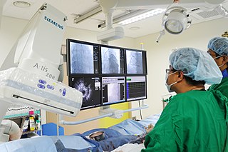
A coronary catheterization is a minimally invasive procedure to access the coronary circulation and blood filled chambers of the heart using a catheter. It is performed for both diagnostic and interventional (treatment) purposes.

<span class="mw-page-title-main">Cardiac stress test</span> Measures the hearts ability to respond to external stress in a controlled clinical environment

A cardiac stress test is a cardiological test that measures the heart's ability to respond to external stress in a controlled clinical environment. The stress response is induced by exercise, intravenous pharmacological (drug) stimulation, or in some cases, a combination of both.

<span class="mw-page-title-main">Atheroma</span> Accumulation of degenerative material in the inner layer of artery walls

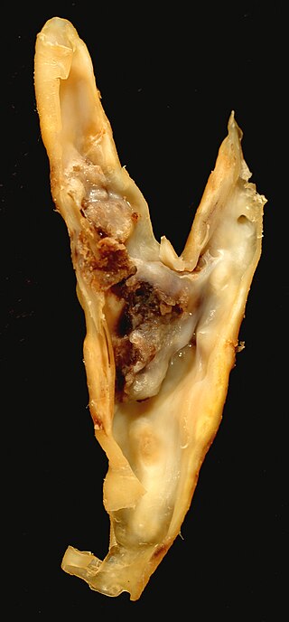
An atheroma, or atheromatous plaque, is an abnormal accumulation of material in the inner layer of an arterial wall.

Intravascular ultrasound (IVUS) or intravascular echocardiography is a medical imaging methodology using a specially designed catheter with a miniaturized ultrasound probe attached to the distal end of the catheter. The proximal end of the catheter is attached to computerized ultrasound equipment. It allows the application of ultrasound technology, such as piezoelectric transducer or CMUT, to see from inside blood vessels out through the surrounding blood column, visualizing the endothelium of blood vessels.

<span class="mw-page-title-main">Electron beam computed tomography</span> Medical diagnostic technology

Electron beam computed tomography (EBCT) is a specific form of computed tomography (CT) in which the X-ray tube is not mechanically spun in order to rotate the source of X-ray photons. This different design was explicitly developed to better image heart structures that never stop moving, performing a complete cycle of movement with each heartbeat.

<span class="mw-page-title-main">Myocardial perfusion imaging</span> Nuclear medicine imaging method

Myocardial perfusion imaging or scanning is a nuclear medicine procedure that illustrates the function of the heart muscle (myocardium).

<span class="mw-page-title-main">Computed tomography angiography</span>

Computed tomography angiography is a computed tomography technique used for angiography—the visualization of arteries and veins—throughout the human body. Using contrast injected into the blood vessels, images are created to look for blockages, aneurysms, dissections, and stenosis. CTA can be used to visualize the vessels of the heart, the aorta and other large blood vessels, the lungs, the kidneys, the head and neck, and the arms and legs. CTA can also be used to localise arterial or venous bleed of the gastrointestinal system.

<span class="mw-page-title-main">CT pulmonary angiogram</span>

A CT pulmonary angiogram (CTPA) is a medical diagnostic test that employs computed tomography (CT) angiography to obtain an image of the pulmonary arteries. Its main use is to diagnose pulmonary embolism (PE). It is a preferred choice of imaging in the diagnosis of PE due to its minimally invasive nature for the patient, whose only requirement for the scan is an intravenous line.

<span class="mw-page-title-main">Spontaneous coronary artery dissection</span> Uncommon cause of heart attacks mostly affecting younger, healthy women

Spontaneous coronary artery dissection (SCAD) is an uncommon but potentially lethal condition in which one of the coronary arteries that supply the heart, spontaneously develops a blood collection, or hematoma, within the artery wall due to a tear in the wall. SCAD is one of the arterial dissections that can occur.

<span class="mw-page-title-main">Cardiac magnetic resonance imaging perfusion</span>

Cardiac magnetic resonance imaging perfusion, also known as stress CMR perfusion, is a clinical magnetic resonance imaging test performed on patients with known or suspected coronary artery disease to determine if there are perfusion defects in the myocardium of the left ventricle that are caused by narrowing of one or more of the coronary arteries.

<span class="mw-page-title-main">Contrast CT</span>

Contrast CT, or contrast enhanced computed tomography (CECT), is X-ray computed tomography (CT) using radiocontrast. Radiocontrasts for X-ray CT are generally iodine-based types. This is useful to highlight structures such as blood vessels that otherwise would be difficult to delineate from their surroundings. Using contrast material can also help to obtain functional information about tissues. Often, images are taken both with and without radiocontrast. CT images are called precontrast or native-phase images before any radiocontrast has been administered, and postcontrast after radiocontrast administration.

<span class="mw-page-title-main">Coronary CT angiography</span> Use of computed tomography angiography to assess the coronary arteries of the heart

Coronary CT angiography is the use of computed tomography (CT) angiography to assess the coronary arteries of the heart. The patient receives an intravenous injection of radiocontrast and then the heart is scanned using a high speed CT scanner, allowing physicians to assess the extent of occlusion in the coronary arteries, usually in order to diagnose coronary artery disease.

<span class="mw-page-title-main">Cardiac imaging</span>

Cardiac imaging refers to minimally invasive imaging of the heart using ultrasound, magnetic resonance imaging (MRI), computed tomography (CT), or nuclear medicine (NM) imaging with PET or SPECT. These cardiac techniques are otherwise referred to as echocardiography, Cardiac MRI, Cardiac CT, Cardiac PET and Cardiac SPECT including myocardial perfusion imaging.

<span class="mw-page-title-main">Hybrid cardiac surgery</span>

A hybrid cardiac surgical procedure in a narrow sense is defined as a procedure that combines a conventional, more invasive surgical part with an interventional part, using some sort of catheter-based procedure guided by fluoroscopy imaging in a hybrid operating room (OR) without interruption. The hybrid technique has a reduced risk of surgical complications and has shown decreased recovery time. It can be used to treat numerous heart diseases and conditions and with the increasing complexity of each case, the hybrid surgical technique is becoming more common.

Overscreening, also called unnecessary screening, is the performance of medical screening without a medical indication to do so. Screening is a medical test in a healthy person who is showing no symptoms of a disease and is intended to detect a disease so that a person may prepare to respond to it. Screening is indicated in people who have some threshold risk for getting a disease, but is not indicated in people who are unlikely to develop a disease. Overscreening is a type of unnecessary health care.

The BaleDoneen Method is a risk assessment and treatment protocol aimed at preventing heart attack and stroke. The method also seeks to prevent or reduce the effects of type 2 diabetes. The method was developed by Bradley Field Bale and Amy Doneen.

<span class="mw-page-title-main">Cardiac allograft vasculopathy</span> Medical condition

Cardiac allograft vasculopathy (CAV) is a progressive type of coronary artery disease in people who have had a heart transplant. As the donor heart has lost its nerve supply there is typically no chest pain, and CAV is usually detected on routine testing. It may present with symptoms such as tiredness and breathlessness.

References

  1. "Heart scan (coronary calcium scan)". Mayo Clinic. Retrieved 9 August 2015.
  2. 1 2 3 4 5 Neves PO, Andrade J, Monção H (2017). "Coronary artery calcium score: current status". Radiologia Brasileira. 50 (3): 182–189. doi:10.1590/0100-3984.2015.0235. PMC   5487233 . PMID   28670030. CC BY 4.0
  3. Bødtker Mortensen M, et al, "Association of age with the diagnostic value of coronary artery calcium score for ruling out coronary stenosis in symptomatic patients", JAMA Cardiol, October 27, 2021 DOI:10.1001/jamacardio.2021.4406.
  4. van der Bijl N, Joemai RM, Geleijns J, Bax JJ, Schuijf JD, de Roos A, Kroft LJ (December 2010). "Assessment of Agatston coronary artery calcium score using contrast-enhanced CT coronary angiography". AJR. American Journal of Roentgenology. 195 (6): 1299–305. doi:10.2214/AJR.09.3734. PMID   21098187.
  5. Zhi-hui Hou; Bin Lu; Yang Gao; Shi-liang Jiang; Yang Wang; Wei Li; Matthew J. Budoff (2012). "Prognostic Value of Coronary CT Angiography and Calcium Score for Major Adverse Cardiac Events in Outpatients". JACC: Cardiovascular Imaging. 5 (10): 990–999. doi:10.1016/j.jcmg.2012.06.006. PMID   23058065 . Retrieved April 9, 2015.
  6. Hughes-Austin JM, Dominguez A, Allison MA, Wassel CL, Rifkin DE, Morgan CG, Daniels MR, Ikram U, Knox JB, Wright CM, Criqui MH, Ix JH (February 2016). "Relationship of Coronary Calcium on Standard Chest CT Scans With Mortality". JACC: Cardiovascular Imaging. 9 (2): 152–9. doi:10.1016/j.jcmg.2015.06.030. PMC   4744104 . PMID   26777213.
  7. Hoffmann U, Brady TJ, Muller J (August 2003). "Cardiology patient page. Use of new imaging techniques to screen for coronary artery disease". Circulation. 108 (8): e50-3. doi: 10.1161/01.CIR.0000085363.88377.F2 . PMID   12939244.
  8. Agatston, Arthur S.; Janowitz, Warren R.; Hildner, Frank J.; Zusmer, Noel R.; Viamonte, Manuel; Detrano, Robert (March 1990). "Quantification of coronary artery calcium using ultrafast computed tomography". Journal of the American College of Cardiology. 15 (4): 827–832. doi: 10.1016/0735-1097(90)90282-t . PMID   2407762.
  9. Divakaran, S.; Cheezum, M. K.; Hulten, E. A.; Bittencourt, M. S.; Silverman, M. G.; Nasir, K.; Blankstein, R. (February 2015). "Use of cardiac CT and calcium scoring for detecting coronary plaque: implications on prognosis and patient management". The British Journal of Radiology. 88 (1046): 20140594. doi:10.1259/bjr.20140594. ISSN   1748-880X. PMC   4614250 . PMID   25494818.
  10. 1 2 Qian Z, Anderson H, Marvasty I, Akram K, Vazquez G, Rinehart S, Voros S (2010). "Lesion- and vessel-specific coronary artery calcium scores are superior to whole-heart Agatston and volume scores in the diagnosis of obstructive coronary artery disease". Journal of Cardiovascular Computed Tomography. 4 (6): 391–9. doi:10.1016/j.jcct.2010.09.001. PMID   21035423.
  11. Akram K, Voros S (October 2008). "Absolute coronary artery calcium scores are superior to MESA percentile rank in predicting obstructive coronary artery disease". The International Journal of Cardiovascular Imaging. 24 (7): 743–9. doi:10.1007/s10554-008-9305-5. PMID   18351440. S2CID   565168.
  12. Qian Z, Marvasty I, Anderson H, Rinehart S, Voros S (June 2009). "Lesion-specific coronary artery calcium quantification better predicts cardiac events". 2009 IEEE International Symposium on Biomedical Imaging: From Nano to Macro. pp. 237–240. doi:10.1109/ISBI.2009.5193027. ISBN   978-1-4244-3931-7. S2CID   14169702.{{cite book}}: |journal= ignored (help)
  13. 1 2 Kim, Kwang Pyo; Einstein, Andrew J.; Berrington de González, Amy (2009-07-13). "Coronary Artery Calcification Screening". Archives of Internal Medicine. American Medical Association (AMA). 169 (13): 1188–94. doi:10.1001/archinternmed.2009.162. ISSN   0003-9926. PMC   2765044 . PMID   19597067.
  14. Radiation Dose in X-Ray and CT Exams, Radiologyinfo.org
  15. Patel, Amish; Budoff, Matthew; Fine, Jeffrey J. (2016). "Coronary Calcium Scans And Radiation Exposure In The Society For Heart Attack Prevention And Eradication Cohort". Journal of the American College of Cardiology. Elsevier BV. 67 (13): 1733. doi: 10.1016/s0735-1097(16)31734-x . ISSN   0735-1097.