Cortical thymic epithelial cells

Last updated
A figure depicting the process of T cell / thymocyte positive and negative selection in the thymus. cTEC shown in yellow. Thymic selection.pdf
A figure depicting the process of T cell / thymocyte positive and negative selection in the thymus. cTEC shown in yellow.

Cortical thymic epithelial cells (cTECs) form unique parenchyma cell population of the thymus which critically contribute to the development of T cells.

Contents

Thymus tissue is compartmentalized into cortex and medulla and each of these two compartments comprises its specific thymic epithelial cell subset. cTECs reside in the outer part- cortex, which mostly serves as a developmental site for T cells. Precursors of T cells originate in the bone marrow from which they migrate via bloodstream into thymic cortex, where they encounter stromal cells including cTECs, which form the microenvironment crucial for proliferation and development of T cells by expression of DLL4 (delta-like notch ligand 4), cytokines IL-7, TGFβ or stem cell factor and chemokines CCL25, CXCL12 or CCRL1 etc. [1] Essential part of T cell development forms process called VDJ recombination, mediated by RAG recombinases, that stochastically changes DNA sequences of T cell receptors (TCR) and endows them with diverse recognition specificity. Thanks to this process, T cells can recognize vast repertoire of pathogens, but also self-peptides or even their TCRs don't respond to any surrounding signals. Major role of thymic epithelial cells is to test, whether TCRs are "functional" and on the other hand "harmless" to our body. While cTECs control the functionality of TCRs during the process called positive selection, Medullary thymic epithelial cells (mTECs) that home in the inner part of the thymus- medulla, present on their MHC molecules self-peptides, generated mostly by protein Autoimmune regulator, to eliminate T cells with self-reactive TCRs via processes of central tolerance e.g. negative selection and protect the body against development of autoimmunity. [2]

Positive selection of T cells

Major function of cTECs is to positively select those T cells that are capable to recognize and interact with MHC molecules on their surface [3] . Once T cell precursors enter the thymic cortex, they start their transformation from double negative stages (T cell without surface expression of CD4 and CD8 co-receptors) to a double positive stage (T cell with surface expression of both co-receptors) that expresses fully recombined TCR. [4] This stage undergoes above mentioned selection process. [5]

Double positive–single positive transition

Interaction between TCR of double positive T cell and MHC I molecule leads to loss of CD4 expression and double positive T cell becomes CD8 single positive T cell, conversely, engagement of MHC II molecule leads to the development into CD4 single positive T cell. [6] It was also described that CD8/CD4 restriction is influenced by transcription factors Runx3, in the case of CD8 restriction, [7] and Th-POK [8] which directs the development into CD4 T cell lineage and represses the expression of Runx3. [9] More than 90% of double positive T cells are unable to reach this interaction and they die by neglect. [10]

Cortex–medulla migration

Besides double positive-single positive transition, TCR-MHC interaction also triggers the expression of CCR7, chemokine receptor which recognizes chemokines CCL19 and CCL21, that are largely produced by mTECs in the medulla, and positively selected T cells start to migrate to medulla via their gradient. [11] [12]

Unique proteolytic pathways

It is incompletely understood whether presence of peptide ligands on MHC molecules of cTECs plays some role in positive selection. But it is likely that these peptide-MHC complexes are unique and different from self-peptides presented by mTECs, since cTECs developed unique proteolytic pathways. Indeed, there is slight evidence focused on unique cTEC peptide ligands, [13] [14] [15] nevertheless, its more systematic characterization is still required.

Thymoproteasome (β5t)

Enzymatic machinery for MHC I antigen processing and presentation in cTECs involves thymoproteasome, which is defined by the presence of β5t subunit encoded by Psmb11 gene. [16] Knockout of this gene revealed only slight reduction in positive selection of CD8 T cells, but TCR repertoire of these cells was shown to be limited [17] and they revealed impaired immunological properties e.g. bad antigen responsiveness and failure to maintain naive population in the periphery. [18] β5t subunit was shown to reduce chymotrypsin-like activity of thymoproteasomes, resulting in generation of low affinity peptides. [16] Such finding was confirmed by study that was focused on properties of thymoproteasome- chopped peptides. [15] Importantly, low affinity interactions are considered to result in positive selection, whereas high affinity interactions are typical for negative selection and interaction with mTECs. [3]

Cathepsin L

MHC II processing and presentation in cTECs took advantage of several proteolytic pathways including cathepsin L, encoded by Ctsl gene. Cathepsin S which is produced by most of the antigen- presenting cells along with mTECs is absent in cTECs. [19] Cathepsin L not only cleaves invariant chain as other cathepsins, nevertheless was shown to cleave peptides for MHC II presentation and enlarge the pool of cTEC unique peptide ligands. [20] Ctsl knockout mouse revealed severe reduction in frequency and repertoire of CD4 T cells and impairment of invariant chain degradation. [19] Another study revealed that reduction of T cell repertoire wasn't caused by absence of invariant chain degradation, rather due to alterations in repertoire of cathepsin L cleaved peptides. [20]

Thymus specific serine protease

Thymus specific serine protease is another cTEC specific enzyme, encoded by Prss16 gene, which is also involved in MHC II peptide processing. [21] Prss16 knockout mice revealed reduced repertoire of positively selected CD4 T cells. [22]

Macroautophagy

Common feature of cTECs and mTECs is constitutive macroautophagy. [23] This process involves engulfment of portion of cytoplasm that contains organelles and vesicles into autophagosome that fuses with late endosomes or lysosomes and its content is chopped to small peptides. [24] cTECs and mTECs utilize this endogenous pathway for MHC II presentation during selection processes, instead of common loading of exogenous peptides. Mouse with deficient macroautophagy, specifically in the thymus, revealed reduced numbers and repertoire of CD4 T cells. [25]

Development

cTECs and mTECs originate from endoderm, more specifically from the third pharyngeal pouch [26] and it has been shown that they share common progenitor cell. [27] [28] Importantly, mTECs during their development possess classical markers of cTECs including CD205 [29] and β5t [30] which are completely absent in mature mTECs, [31] suggesting another possible cTEC function, namely they might serve as a progenitor cell reservoir for mTECs. Indeed, several lineage tracing studies confirmed that cTEC progenitors [32] or even mature cTECs [33] [34] are capable to give rise to mTECs.

Nevertheless, there is available series of publications which suggests different mTEC progenitor pools [35] [36] or even argue that cTECs and mTECs reveal distinct unipotent progenitor cells. [37] [38]

Related Research Articles

<span class="mw-page-title-main">Thymus</span> Endocrine gland

The thymus is a specialized primary lymphoid organ of the immune system. Within the thymus, thymus cell lymphocytes or T cells mature. T cells are critical to the adaptive immune system, where the body adapts to specific foreign invaders. The thymus is located in the upper front part of the chest, in the anterior superior mediastinum, behind the sternum, and in front of the heart. It is made up of two lobes, each consisting of a central medulla and an outer cortex, surrounded by a capsule.

<span class="mw-page-title-main">T cell</span> White blood cells of the immune system

T cells are one of the important types of white blood cells of the immune system and play a central role in the adaptive immune response. T cells can be distinguished from other lymphocytes by the presence of a T-cell receptor (TCR) on their cell surface.

<span class="mw-page-title-main">Cytotoxic T cell</span> T cell that kills infected, damaged or cancerous cells

A cytotoxic T cell (also known as TC, cytotoxic T lymphocyte, CTL, T-killer cell, cytolytic T cell, CD8+ T-cell or killer T cell) is a T lymphocyte (a type of white blood cell) that kills cancer cells, cells that are infected by intracellular pathogens (such as viruses or bacteria), or cells that are damaged in other ways.

The regulatory T cells (Tregs or Treg cells), formerly known as suppressor T cells, are a subpopulation of T cells that modulate the immune system, maintain tolerance to self-antigens, and prevent autoimmune disease. Treg cells are immunosuppressive and generally suppress or downregulate induction and proliferation of effector T cells. Treg cells express the biomarkers CD4, FOXP3, and CD25 and are thought to be derived from the same lineage as naïve CD4+ cells. Because effector T cells also express CD4 and CD25, Treg cells are very difficult to effectively discern from effector CD4+, making them difficult to study. Research has found that the cytokine transforming growth factor beta (TGF-β) is essential for Treg cells to differentiate from naïve CD4+ cells and is important in maintaining Treg cell homeostasis.

Cross-presentation is the ability of certain professional antigen-presenting cells (mostly dendritic cells) to take up, process and present extracellular antigens with MHC class I molecules to CD8 T cells (cytotoxic T cells). Cross-priming, the result of this process, describes the stimulation of naive cytotoxic CD8+ T cells into activated cytotoxic CD8+ T cells. This process is necessary for immunity against most tumors and against viruses that infect dendritic cells and sabotage their presentation of virus antigens. Cross presentation is also required for the induction of cytotoxic immunity by vaccination with protein antigens, for example, tumour vaccination.

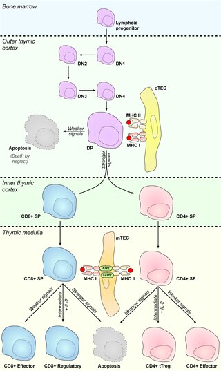
In immunology, central tolerance is the process of eliminating any developing T or B lymphocytes that are autoreactive, i.e. reactive to the body itself. Through elimination of autoreactive lymphocytes, tolerance ensures that the immune system does not attack self peptides. Lymphocyte maturation occurs in primary lymphoid organs such as the bone marrow and the thymus. In mammals, B cells mature in the bone marrow and T cells mature in the thymus.

A thymocyte is an immune cell present in the thymus, before it undergoes transformation into a T cell. Thymocytes are produced as stem cells in the bone marrow and reach the thymus via the blood.

<span class="mw-page-title-main">Antigen presentation</span> Vital immune process that is essential for T cell immune response triggering

Antigen presentation is a vital immune process that is essential for T cell immune response triggering. Because T cells recognize only fragmented antigens displayed on cell surfaces, antigen processing must occur before the antigen fragment can be recognized by a T-cell receptor. Specifically, the fragment, bound to the major histocompatibility complex (MHC), is transported to the surface of the cell, a process known as presentation. If there has been an infection with viruses or bacteria, the cell will present an endogenous or exogenous peptide fragment derived from the antigen by MHC molecules. There are two types of MHC molecules which differ in the behaviour of the antigens: MHC class I molecules (MHC-I) bind peptides from the cell cytosol, while peptides generated in the endocytic vesicles after internalisation are bound to MHC class II (MHC-II). Cellular membranes separate these two cellular environments - intracellular and extracellular. Each T cell can only recognize tens to hundreds of copies of a unique sequence of a single peptide among thousands of other peptides presented on the same cell, because an MHC molecule in one cell can bind to quite a large range of peptides. Predicting which antigens will be presented to the immune system by a certain MHC/HLA type is difficult, but the technology involved is improving.

MHC-restricted antigen recognition, or MHC restriction, refers to the fact that a T cell can interact with a self-major histocompatibility complex molecule and a foreign peptide bound to it, but will only respond to the antigen when it is bound to a particular MHC molecule.

Self-protein refers to all proteins endogenously produced by DNA-level transcription and translation within an organism of interest. This does not include proteins synthesized due to viral infection, but may include those synthesized by commensal bacteria within the intestines. Proteins that are not created within the body of the organism of interest, but nevertheless enter through the bloodstream, a breach in the skin, or a mucous membrane, may be designated as “non-self” and subsequently targeted and attacked by the immune system. Tolerance to self-protein is crucial for overall wellbeing; when the body erroneously identifies self-proteins as “non-self”, the subsequent immune response against endogenous proteins may lead to the development of an autoimmune disease.

<span class="mw-page-title-main">Minor histocompatibility antigen</span>

Minor histocompatibility antigen are peptides presented on the cellular surface of donated organs that are known to give an immunological response in some organ transplants. They cause problems of rejection less frequently than those of the major histocompatibility complex (MHC). Minor histocompatibility antigens (MiHAs) are diverse, short segments of proteins and are referred to as peptides. These peptides are normally around 9-12 amino acids in length and are bound to both the major histocompatibility complex (MHC) class I and class II proteins. Peptide sequences can differ among individuals and these differences arise from SNPs in the coding region of genes, gene deletions, frameshift mutations, or insertions. About a third of the characterized MiHAs come from the Y chromosome. Prior to becoming a short peptide sequence, the proteins expressed by these polymorphic or diverse genes need to be digested in the proteasome into shorter peptides. These endogenous or self peptides are then transported into the endoplasmic reticulum with a peptide transporter pump called TAP where they encounter and bind to the MHC class I molecule. This contrasts with MHC class II molecules's antigens which are peptides derived from phagocytosis/endocytosis and molecular degradation of non-self entities' proteins, usually by antigen-presenting cells. MiHA antigens are either ubiquitously expressed in most tissue like skin and intestines or restrictively expressed in the immune cells.

In immunology, peripheral tolerance is the second branch of immunological tolerance, after central tolerance. It takes place in the immune periphery. Its main purpose is to ensure that self-reactive T and B cells which escaped central tolerance do not cause autoimmune disease. Peripheral tolerance prevents immune response to harmless food antigens and allergens, too.

In immunology, clonal deletion is the removal through apoptosis of B cells and T cells that have expressed receptors for self before developing into fully immunocompetent lymphocytes. This prevents recognition and destruction of self host cells, making it a type of negative selection or central tolerance. Central tolerance prevents B and T lymphocytes from reacting to self. Thus, clonal deletion can help protect individuals against autoimmunity. Clonal deletion is thought to be the most common type of negative selection. It is one method of immune tolerance.

Thymic nurse cells (TNCs) are large epithelial cells found in the cortex of the thymus and also in cortico-medullary junction. They have their own nucleus and are known to internalize thymocytes through extensions of plasma membrane. The cell surfaces of TNCs and their cytoplasmic vacuoles express MHC Class I and MHC Class II antigens. The interaction of these antigens with the developing thymocytes determines whether the thymocytes undergo positive or negative selection.

<span class="mw-page-title-main">Medullary thymic epithelial cells</span>

Medullary thymic epithelial cells (mTECs) represent a unique stromal cell population of the thymus which plays an essential role in the establishment of central tolerance. Therefore, mTECs rank among cells relevant for the development of functional mammal immune system.

Antigen transfer in the thymus is the transmission of self-antigens between thymic antigen-presenting cells which contributes to the establishment of T cell central tolerance.

Thymic epithelial cells (TECs) are specialized cells with high degree of anatomic, phenotypic and functional heterogeneity that are located in the outer layer (epithelium) of the thymic stroma. The thymus, as a primary lymphoid organ, mediates T cell development and maturation. The thymic microenvironment is established by TEC network filled with thymocytes in different developing stages. TECs and thymocytes are the most important components in the thymus, that are necessary for production of functionally competent T lymphocytes and self tolerance. Dysfunction of TECs causes several immunodeficiencies and autoimmune diseases.

Promiscuous gene expression (PGE), formerly referred to as ectopic expression, is a process specific to the thymus that plays a pivotal role in the establishment of central tolerance. This phenomenon enables generation of self-antigens, so called tissue-restricted antigens (TRAs), which are in the body expressed only by one or few specific tissues. These antigens are represented for example by insulin from the pancreas or defensins from the gastrointestinal tract. Antigen-presenting cells (APCs) of the thymus, namely medullary thymic epithelial cells (mTECs), dendritic cells (DCs) and B cells are capable to present peptides derived from TRAs to developing T cells and hereby test, whether their T cell receptors (TCRs) engage self entities and therefore their occurrence in the body can potentially lead to the development of autoimmune disease. In that case, thymic APCs either induce apoptosis in these autoreactive T cells or they deviate them to become T regulatory cells, which suppress self-reactive T cells in the body that escaped negative selection in the thymus. Thus, PGE is crucial for tissues protection against autoimmunity.

Thymoproteasome is a special kind of proteasome, which is present in vertebrates. In the body it is located in thymus, exclusively in cortical thymic epithelial cells (cTECs). But in thymus we can also find another type of specific proteasome, immunoproteasome, which is present in thymocytes, dendritic cells and medular thymic epithelial cells. It was first described in 2007 during a search for non-intronic sequence proximal to PSMB5 locus in mouse genome. The PSMB5 locus encodes the standard β5 proteasome subunit, while this sequence encodes a variant subunit β5t (PSMB11) specific to thymoproteasome. The importance of this protein complex is its involvement in positive selection of T cells.

Thymus stromal cells are subsets of specialized cells located in different areas of the thymus. They include all non-T-lineage cells, such as thymic epithelial cells (TECs), endothelial cells, mesenchymal cells, dendritic cells, and B lymphocytes, and provide signals essential for thymocyte development and the homeostasis of the thymic stroma.

References

  1. Ohigashi, Izumi; Kozai, Mina; Takahama, Yousuke (2016-04-18). "Development and developmental potential of cortical thymic epithelial cells". Immunological Reviews. 271 (1): 10–22. doi:10.1111/imr.12404. ISSN   0105-2896. PMID   27088904. S2CID   205213554.
  2. Klein, Ludger; Hinterberger, Maria; Wirnsberger, Gerald; Kyewski, Bruno (December 2009). "Antigen presentation in the thymus for positive selection and central tolerance induction". Nature Reviews Immunology. 9 (12): 833–844. doi:10.1038/nri2669. ISSN   1474-1733. PMID   19935803. S2CID   205490965.
  3. 1 2 Klein, Ludger; Kyewski, Bruno; Allen, Paul M.; Hogquist, Kristin A. (2014-05-16). "Positive and negative selection of the T cell repertoire: what thymocytes see (and don't see)". Nature Reviews Immunology. 14 (6): 377–391. doi:10.1038/nri3667. ISSN   1474-1733. PMC   4757912 . PMID   24830344.
  4. Petrie, Howard T. (November 2002). "Role of thymic organ structure and stromal composition in steady-state postnatal T-cell production". Immunological Reviews. 189: 8–19. doi:10.1034/j.1600-065X.2002.18902.x. ISSN   0105-2896. PMID   12445261. S2CID   34924894.
  5. Starr, Timothy K.; Jameson, Stephen C.; Hogquist, Kristin A. (April 2003). "Positive Andnegativeselection Oft Cells". Annual Review of Immunology. 21 (1): 139–176. doi:10.1146/annurev.immunol.21.120601.141107. ISSN   0732-0582. PMID   12414722.
  6. Germain, Ronald N. (May 2002). "T-cell development and the CD4–CD8 lineage decision". Nature Reviews Immunology. 2 (5): 309–322. doi:10.1038/nri798. ISSN   1474-1733. PMID   12033737. S2CID   21479833.
  7. Setoguchi, Ruka; Tachibana, Masashi; Naoe, Yoshinori; Muroi, Sawako; Akiyama, Kaori; Tezuka, Chieko; Okuda, Tsukasa; Taniuchi, Ichiro (2008-02-08). "Repression of the Transcription Factor Th-POK by Runx Complexes in Cytotoxic T Cell Development". Science. 319 (5864): 822–825. Bibcode:2008Sci...319..822S. doi:10.1126/science.1151844. ISSN   0036-8075. PMID   18258917. S2CID   6695125.
  8. He, Xiao; He, Xi; Dave, Vibhuti P.; Zhang, Yi; Hua, Xiang; Nicolas, Emmanuelle; Xu, Weihong; Roe, Bruce A.; Kappes, Dietmar J. (February 2005). "The zinc finger transcription factor Th-POK regulates CD4 versus CD8 T-cell lineage commitment". Nature. 433 (7028): 826–833. Bibcode:2005Natur.433..826H. doi: 10.1038/nature03338 . ISSN   0028-0836. PMID   15729333.
  9. Luckey, Megan A; Kimura, Motoko Y; Waickman, Adam T; Feigenbaum, Lionel; Singer, Alfred; Park, Jung-Hyun (2014-06-01). "The transcription factor ThPOK suppresses Runx3 and imposes CD4+ lineage fate by inducing the SOCS suppressors of cytokine signaling". Nature Immunology. 15 (7): 638–645. doi:10.1038/ni.2917. ISSN   1529-2908. PMC   6693509 . PMID   24880459.
  10. Palmer, Ed (May 2003). "Negative selection — clearing out the bad apples from the T-cell repertoire". Nature Reviews Immunology. 3 (5): 383–391. doi:10.1038/nri1085. ISSN   1474-1733. PMID   12766760. S2CID   28321309.
  11. Ueno, Tomoo; Saito, Fumi; Gray, Daniel H. D.; Kuse, Sachiyo; Hieshima, Kunio; Nakano, Hideki; Kakiuchi, Terutaka; Lipp, Martin; Boyd, Richard L. (2004-08-16). "CCR7 Signals Are Essential for Cortex–Medulla Migration of Developing Thymocytes". Journal of Experimental Medicine. 200 (4): 493–505. doi:10.1084/jem.20040643. ISSN   0022-1007. PMC   2211934 . PMID   15302902.
  12. Kurobe, Hirotsugu; Liu, Cunlan; Ueno, Tomoo; Saito, Fumi; Ohigashi, Izumi; Seach, Natalie; Arakaki, Rieko; Hayashi, Yoshio; Kitagawa, Tetsuya (February 2006). "CCR7-Dependent Cortex-to-Medulla Migration of Positively Selected Thymocytes Is Essential for Establishing Central Tolerance". Immunity. 24 (2): 165–177. doi: 10.1016/j.immuni.2005.12.011 . ISSN   1074-7613. PMID   16473829.
  13. Lo, Wan-Lin; Felix, Nathan J; Walters, James J; Rohrs, Henry; Gross, Michael L; Allen, Paul M (2009-10-04). "An endogenous peptide positively selects and augments the activation and survival of peripheral CD4+ T cells". Nature Immunology. 10 (11): 1155–1161. doi:10.1038/ni.1796. ISSN   1529-2908. PMC   2764840 . PMID   19801984.
  14. Santori, Fabio R.; Kieper, William C.; Brown, Stuart M.; Lu, Yun; Neubert, Thomas A.; Johnson, Kenneth L.; Naylor, Stephen; Vukmanović, Stanislav; Hogquist, Kristin A. (August 2002). "Rare, structurally homologous self-peptides promote thymocyte positive selection". Immunity. 17 (2): 131–142. doi: 10.1016/S1074-7613(02)00361-8 . ISSN   1074-7613. PMID   12196285.
  15. 1 2 Sasaki, Katsuhiro; Takada, Kensuke; Ohte, Yuki; Kondo, Hiroyuki; Sorimachi, Hiroyuki; Tanaka, Keiji; Takahama, Yousuke; Murata, Shigeo (2015-06-23). "Thymoproteasomes produce unique peptide motifs for positive selection of CD8+ T cells". Nature Communications. 6 (1): 7484. Bibcode:2015NatCo...6.7484S. doi:10.1038/ncomms8484. ISSN   2041-1723. PMC   4557289 . PMID   26099460.
  16. 1 2 Murata, Shigeo; Sasaki, Katsuhiro; Kishimoto, Toshihiko; Niwa, Shin-ichiro; Hayashi, Hidemi; Takahama, Yousuke; Tanaka, Keiji (2007-06-01). "Regulation of CD8+ T Cell Development by Thymus-Specific Proteasomes". Science. 316 (5829): 1349–1353. Bibcode:2007Sci...316.1349M. doi:10.1126/science.1141915. ISSN   0036-8075. PMID   17540904. S2CID   37185716.
  17. Nitta, Takeshi; Murata, Shigeo; Sasaki, Katsuhiro; Fujii, Hideki; Ripen, Adiratna Mat; Ishimaru, Naozumi; Koyasu, Shigeo; Tanaka, Keiji; Takahama, Yousuke (January 2010). "Thymoproteasome Shapes Immunocompetent Repertoire of CD8+ T Cells". Immunity. 32 (1): 29–40. doi: 10.1016/j.immuni.2009.10.009 . ISSN   1074-7613. PMID   20045355.
  18. Takada, Kensuke; Van Laethem, Francois; Xing, Yan; Akane, Kazuyuki; Suzuki, Haruhiko; Murata, Shigeo; Tanaka, Keiji; Jameson, Stephen C; Singer, Alfred (2015-08-24). "TCR affinity for thymoproteasome-dependent positively selecting peptides conditions antigen responsiveness in CD8+ T cells". Nature Immunology. 16 (10): 1069–1076. doi:10.1038/ni.3237. ISSN   1529-2908. PMC   4810782 . PMID   26301566.
  19. 1 2 Nakagawa, T.; Roth, W.; Wong, P.; Nelson, A.; Farr, A.; Deussing, J.; Villadangos, J. A.; Ploegh, H.; Peters, C. (1998-04-17). "Cathepsin L: critical role in Ii degradation and CD4 T cell selection in the thymus". Science. 280 (5362): 450–453. Bibcode:1998Sci...280..450N. doi:10.1126/science.280.5362.450. ISSN   0036-8075. PMID   9545226.
  20. 1 2 Honey, Karen; Nakagawa, Terry; Peters, Christoph; Rudensky, Alexander (2002-05-20). "Cathepsin L Regulates CD4+ T Cell Selection Independently of Its Effect on Invariant Chain". The Journal of Experimental Medicine. 195 (10): 1349–1358. doi:10.1084/jem.20011904. ISSN   0022-1007. PMC   2193748 . PMID   12021314.
  21. Bowlus, Christopher L.; Ahn, Jung; Chu, Tom; Gruen, Jeffrey R. (September 1999). "Cloning of a Novel MHC-Encoded Serine Peptidase Highly Expressed by Cortical Epithelial Cells of the Thymus". Cellular Immunology. 196 (2): 80–86. doi:10.1006/cimm.1999.1543. ISSN   0008-8749. PMID   10527559.
  22. Gommeaux, Julien; Grégoire, Claude; Nguessan, Prudence; Richelme, Mireille; Malissen, Marie; Guerder, Sylvie; Malissen, Bernard; Carrier, Alice (April 2009). "Thymus-specific serine protease regulates positive selection of a subset of CD4+thymocytes". European Journal of Immunology. 39 (4): 956–964. doi: 10.1002/eji.200839175 . ISSN   0014-2980. PMID   19283781.
  23. Mizushima, Noboru; Yamamoto, Akitsugu; Matsui, Makoto; Yoshimori, Tamotsu; Ohsumi, Yoshinori (March 2004). "In vivo analysis of autophagy in response to nutrient starvation using transgenic mice expressing a fluorescent autophagosome marker". Molecular Biology of the Cell. 15 (3): 1101–1111. doi:10.1091/mbc.e03-09-0704. ISSN   1059-1524. PMC   363084 . PMID   14699058.
  24. Feng, Yuchen; He, Ding; Yao, Zhiyuan; Klionsky, Daniel J (2013-12-24). "The machinery of macroautophagy". Cell Research. 24 (1): 24–41. doi:10.1038/cr.2013.168. ISSN   1001-0602. PMC   3879710 . PMID   24366339.
  25. Nedjic, Jelena; Aichinger, Martin; Emmerich, Jan; Mizushima, Noboru; Klein, Ludger (2008-08-13). "Autophagy in thymic epithelium shapes the T-cell repertoire and is essential for tolerance". Nature. 455 (7211): 396–400. Bibcode:2008Natur.455..396N. CiteSeerX   10.1.1.655.8545 . doi:10.1038/nature07208. ISSN   0028-0836. PMID   18701890. S2CID   4390542.
  26. Gordon, Julie; Wilson, Valerie A; Blair, Natalie F; Sheridan, Julie; Farley, Alison; Wilson, Linda; Manley, Nancy R; Blackburn, C Clare (2004-04-18). "Functional evidence for a single endodermal origin for the thymic epithelium". Nature Immunology. 5 (5): 546–553. doi:10.1038/ni1064. ISSN   1529-2908. PMID   15098031. S2CID   10282524.
  27. Rossi, Simona W.; Jenkinson, William E.; Anderson, Graham; Jenkinson, Eric J. (June 2006). "Clonal analysis reveals a common progenitor for thymic cortical and medullary epithelium". Nature. 441 (7096): 988–991. Bibcode:2006Natur.441..988R. doi:10.1038/nature04813. ISSN   0028-0836. PMID   16791197. S2CID   2358330.
  28. Bleul, Conrad C.; Corbeaux, Tatiana; Reuter, Alexander; Fisch, Paul; Mönting, Jürgen Schulte; Boehm, Thomas (June 2006). "Formation of a functional thymus initiated by a postnatal epithelial progenitor cell". Nature. 441 (7096): 992–996. Bibcode:2006Natur.441..992B. doi:10.1038/nature04850. ISSN   0028-0836. PMID   16791198. S2CID   4396922.
  29. Baik, Song; Jenkinson, Eric J.; Lane, Peter J. L.; Anderson, Graham; Jenkinson, William E. (2013-02-11). "Generation of both cortical and Aire+medullary thymic epithelial compartments from CD205+progenitors". European Journal of Immunology. 43 (3): 589–594. doi:10.1002/eji.201243209. ISSN   0014-2980. PMC   3960635 . PMID   23299414.
  30. Ohigashi, Izumi; Zuklys, Saulius; Sakata, Mie; Mayer, Carlos E.; Zhanybekova, Saule; Murata, Shigeo; Tanaka, Keiji; Holländer, Georg A.; Takahama, Yousuke (2013-06-11). "Aire-expressing thymic medullary epithelial cells originate from β5t-expressing progenitor cells". Proceedings of the National Academy of Sciences. 110 (24): 9885–9890. Bibcode:2013PNAS..110.9885O. doi: 10.1073/pnas.1301799110 . PMC   3683726 . PMID   23720310.
  31. Ohigashi, Izumi; Zuklys, Saulius; Sakata, Mie; Mayer, Carlos E.; Hamazaki, Yoko; Minato, Nagahiro; Hollander, Georg A.; Takahama, Yousuke (November 2015). "Adult Thymic Medullary Epithelium Is Maintained and Regenerated by Lineage-Restricted Cells Rather Than Bipotent Progenitors". Cell Reports. 13 (7): 1432–1443. doi: 10.1016/j.celrep.2015.10.012 . hdl: 2433/216325 . ISSN   2211-1247. PMID   26549457.
  32. Mayer, Carlos E.; Žuklys, Saulius; Zhanybekova, Saule; Ohigashi, Izumi; Teh, Hong-Ying; Sansom, Stephen N.; Shikama-Dorn, Noriko; Hafen, Katrin; Macaulay, Iain C. (2016-01-18). "Dynamic spatio-temporal contribution of single β5t+ cortical epithelial precursors to the thymus medulla". European Journal of Immunology. 46 (4): 846–856. doi:10.1002/eji.201545995. ISSN   0014-2980. PMC   4832341 . PMID   26694097.
  33. Meireles, Catarina; Ribeiro, Ana R.; Pinto, Rute D.; Leitão, Catarina; Rodrigues, Pedro M.; Alves, Nuno L. (2017-04-13). "Thymic crosstalk restrains the pool of cortical thymic epithelial cells with progenitor properties". European Journal of Immunology. 47 (6): 958–969. doi: 10.1002/eji.201746922 . hdl: 10216/111812 . ISSN   0014-2980. PMID   28318017.
  34. Brunk, Fabian; Michel, Chloé; Holland-Letz, Tim; Slynko, Alla; Kopp-Schneider, Annette; Kyewski, Bruno; Pinto, Sheena (2017-05-22). "Dissecting and modeling the emergent murine TEC compartment during ontogeny". European Journal of Immunology. 47 (7): 1153–1159. doi: 10.1002/eji.201747006 . ISSN   0014-2980. PMID   28439878.
  35. Ucar, Ahmet; Ucar, Olga; Klug, Paula; Matt, Sonja; Brunk, Fabian; Hofmann, Thomas G.; Kyewski, Bruno (August 2014). "Adult Thymus Contains FoxN1− Epithelial Stem Cells that Are Bipotent for Medullary and Cortical Thymic Epithelial Lineages". Immunity. 41 (2): 257–269. doi:10.1016/j.immuni.2014.07.005. ISSN   1074-7613. PMC   4148705 . PMID   25148026.
  36. Ulyanchenko, Svetlana; O’Neill, Kathy E.; Medley, Tanya; Farley, Alison M.; Vaidya, Harsh J.; Cook, Alistair M.; Blair, Natalie F.; Blackburn, C. Clare (March 2016). "Identification of a Bipotent Epithelial Progenitor Population in the Adult Thymus". Cell Reports. 14 (12): 2819–2832. doi:10.1016/j.celrep.2016.02.080. ISSN   2211-1247. PMC   4819909 . PMID   26997270.
  37. Hamazaki, Yoko; Fujita, Harumi; Kobayashi, Takashi; Choi, Yongwon; Scott, Hamish S; Matsumoto, Mitsuru; Minato, Nagahiro (2007-02-04). "Medullary thymic epithelial cells expressing Aire represent a unique lineage derived from cells expressing claudin". Nature Immunology. 8 (3): 304–311. doi:10.1038/ni1438. ISSN   1529-2908. PMID   17277780. S2CID   7775843.
  38. Sekai, Miho; Hamazaki, Yoko; Minato, Nagahiro (November 2014). "Medullary Thymic Epithelial Stem Cells Maintain a Functional Thymus to Ensure Lifelong Central T Cell Tolerance". Immunity. 41 (5): 753–761. doi: 10.1016/j.immuni.2014.10.011 . ISSN   1074-7613. PMID   25464854.