Medullary thymic epithelial cells

Last updated
A figure depicting the process of T cell / thymocyte positive and negative selection in the thymus. mTEC shown in orange. Thymic selection.pdf
A figure depicting the process of T cell / thymocyte positive and negative selection in the thymus. mTEC shown in orange.

Medullary thymic epithelial cells (mTECs) represent a unique stromal cell population of the thymus which plays an essential role in the establishment of central tolerance. Therefore, mTECs rank among cells relevant for the development of functional mammal immune system.

Contents

T cell precursors rise in bone marrow and migrate through the bloodstream to the thymus for further development. During their maturation in the thymus, they undergo a process called V(D)J recombination which conducts the development of T cell receptors (TCRs). The mechanism of this stochastic process enables on one hand the generation of vast repertoire of TCRs, however, on the other hand causes also origin of so called "autoreactive T cells" which recognize self antigens via their TCRs. Autoreactive T cells must be eliminated from the body or skewed into the T Regulatory cells (TRegs) lineage to prevent manifestations of autoimmunity. mTECs possess the ability to deal with these autoreactive clones via mediation of the processes of central tolerance, namely clonal deletion or T regulatory cells selection, respectively.

N.B.: All the below cited references utilized mouse as a model organism.

Self-antigens generation and presentation

In 1989, two scientific groups came up with the hypothesis that the thymus expresses genes which are in the periphery, strictly expressed by specific tissues (e.g.: Insulin produced by β cells of the pancreas) to subsequently present these so-called "tissue restricted antigens" (TRAs) from almost all parts of the body to developing T cells in order to test which TCRs recognize self-tissues and can be therefore harmful to the body. [1] It was found, after more than a decade, that this phenomenon is managed specifically by mTECs in the thymus and was named Promiscuous gene expression (PGE). [2]

Autoimmune regulator

Aire is a protein called autoimmune regulator (Aire) which is also specifically expressed by mTECs. [3] and its expression is completely dependent on NF- kappa B signaling pathway. [4] Aire recognizes target genes of TRAs via specific methylation marks [5] [6] and requires about 50 partner molecules for activation of their expression. [7] Moreover, Aire-dependent activation of TRA genes expression is accompanied by formation of DNA double-strand breaks. [8] which probably results in very short lifespan of mTECs between 2–3 days [9]

Mutations of Aire gene in human cause a rare autoimmune disorder called Autoimmune Polyendocrinopathy Candidiasis Ectodermal Distrophy (APECED)., [10] [11] which usually manifests in combination with other autoimmune diseases e.g.: diabetes mellitus type 1. Dysfunction of murine Aire gene results in comparable scenario and therefore mouse is used as the model organism for investigation of APECED.

mTECs in numbers

mTECs as a population are capable to express more than 19000 genes (about 80% of mouse genome) among which approximately 4000 belong to Aire-dependent TRAs. It is important to emphasize that single mTEC expresses about 150 Aire-dependent TRAs and approximately 600 Aire-independent TRAs, [12] indicating that other still unknown PGE regulators exist. Indeed, another protein called Fezf2 was suggested to be the second regulator of PGE. [13]

It was shown that each mTEC expresses stochastically 1-3% of TRA pool. [14] However, more recent studies discovered stable co-expression patterns between TRA genes which are localized in close proximity, suggesting "order in this stochastic process". [15] [16]

Tissues protection against autoreactive T cells

T cell precursors extravasate from the bloodstream in cortico-medullary junction and they first migrate to the thymic cortex, where they undergo construction of TCRs and subsequently a process called T cell positive selection which is mediated by mTEC-related cells: cortical thymic epithelial cells (cTECs). This process verifies, whether newly generated TCRs are functional. [17] About 90% of T cells displays badly rearranged TCRs, they cannot reach the positive selection and they die by neglect in the cortex. [18] The rest starts to express CCR7, which is a receptor for mTEC-generated chemokine CCL21, and migrate after concentration gradient to the thymic medulla to encounter mTECs. [19]

Two modes of central tolerance

mTECs are not only mediators of PGE and "factories of TRAs". They also express high levels of MHC II and costimulatory molecules CD80/CD86 and rank among efficient antigen-presenting cells (APCs). [2] Moreover, they utilize macroautophagy to load self antigens on MHCII molecules. [20] Thus, mTECs are capable to present self-generated TRAs on their MHC molecules to select potential autoreactive T cells. It was published that mTECs mediate clonal deletion (recessive tolerance), via presentation of TRAs, which leads to the apoptosis of autoreactive T cells, [21] [22] [23] as well as they are competent to skew autoreactive T cells into TRegs, also through the presentation of TRAs, which then migrate to the periphery to protect tissues against autoreactive T cells that occasionally avoid selection processes in the thymus (dominant tolerance). [24] [25]

How mTECs discriminate between these two modes of tolerance? It was shown that prospective TRegs interact with presented TRAs with lower affinity than those which are clonally deleted. [17] Furthermore, it was also revealed that specific TRAs skew autoreactive T cells into TRegs with much higher efficiency than they do in the case of clonal deletion. [26]

Antigen transfer in the thymus

mTECs form rare population which is composed of approximately 100000 cells per thymus of 2-week-old mice. [27] Thus, there is low probability of encounter between autoreactive T cell and mTEC. Unidirectional antigen transfer from mTECs to thymic dendritic cells (DCs), which itself can't express TRAs, extends the network of TRA presentation, enables TRA processing by different microenvironments and increases the probability of encounter between autoreactive T cell and its appropriate self-antigen. [28] [29] [30] Moreover, DCs competently induce both recessive and dominant tolerance as well as mTECs. [29]

In contrast, another seminal study reveals that mTECs itself suffice to establish both recessive and dominant tolerance without help of additional APCs. [31]

Development

Subsets

mTEC population is not homogenous and basically could be subdivided into more numerous population of mTECs which express low number of MHCII and CD80/CD86, namely mTECsLo and smaller population of mTECsHi which express higher amounts of these molecules. [32] PGE regulator Aire is expressed only by part of mTECsHi. [9] However, this claim does not mean that mTECsLo don't contribute to PGE, mTECsHi, especially that expressing Aire, are just much more efficient in this process. [32]

There is evidence that mTECsLo serve as precursors of mTECsHi in the embryonic thymus [33] [34] Nevertheless, situation changes after birth, where only part of mTECsLo pool represents immature mTECsHi reservoir [33] and another part is constituted by mature mTECs which are specialized for expression of chemokine CCL21, [35] discussed above. Further subset of mTECsLo pool is formed by terminally differentiated cells called Post- Aire mTECs which already downregulated the expression of Aire, MHCII and CD80/CD86. [36]

mTECs can develop into Thymic mimetic cells, which combine the mTEC identity with lineage specific transcription factors. These cells exhibit the phenotype of differentiated peripheral cells and produce their corresponding TRAs. The most famous example is Hassall's corpuscles. [37]

Progenitor cells

TECs (mTECs and cTECs) originate from the third pharyngeal pouch which is a product of endoderm. [38] Their common origin points to the fact that both mTECs and cTECs rise from one bipotent progenitor. This notion was confirmed by several studies of embryonic thymus. [39] [40] and was further developed by finding that these bipotent progenitors express cTEC markers. [41] [42] Nevertheless, another sources document existence of mTEC unipotent progenitors that express claudin 3 and 4 (Cld3/4). [43] [44] These two opposite findings were interfaced by observation of unipotent mTEC progenitors in the postnatal thymus that previously expressed cTEC markers and concurrently express Cld3/4. [45] On the other hand, several other studies describe appearance of bipotent progenitors in postnatal thymus. [46] [47] [48] [49] Thus, embryonic as well as postnatal thymus might shelter both bipotent TEC or unipotent mTEC progenitors.

Similarly to Aire expression, mTECs development is highly dependent on NF- kappa B signaling pathway. [50]

Related Research Articles

<span class="mw-page-title-main">Thymus</span> Endocrine gland

The thymus is a specialized primary lymphoid organ of the immune system. Within the thymus, thymus cell lymphocytes or T cells mature. T cells are critical to the adaptive immune system, where the body adapts to specific foreign invaders. The thymus is located in the upper front part of the chest, in the anterior superior mediastinum, behind the sternum, and in front of the heart. It is made up of two lobes, each consisting of a central medulla and an outer cortex, surrounded by a capsule.

<span class="mw-page-title-main">T cell</span> White blood cells of the immune system

T cells are one of the important types of white blood cells of the immune system and play a central role in the adaptive immune response. T cells can be distinguished from other lymphocytes by the presence of a T-cell receptor (TCR) on their cell surface.

The regulatory T cells (Tregs or Treg cells), formerly known as suppressor T cells, are a subpopulation of T cells that modulate the immune system, maintain tolerance to self-antigens, and prevent autoimmune disease. Treg cells are immunosuppressive and generally suppress or downregulate induction and proliferation of effector T cells. Treg cells express the biomarkers CD4, FOXP3, and CD25 and are thought to be derived from the same lineage as naïve CD4+ cells. Because effector T cells also express CD4 and CD25, Treg cells are very difficult to effectively discern from effector CD4+, making them difficult to study. Research has found that the cytokine transforming growth factor beta (TGF-β) is essential for Treg cells to differentiate from naïve CD4+ cells and is important in maintaining Treg cell homeostasis.

Cross-presentation is the ability of certain professional antigen-presenting cells (mostly dendritic cells) to take up, process and present extracellular antigens with MHC class I molecules to CD8 T cells (cytotoxic T cells). Cross-priming, the result of this process, describes the stimulation of naive cytotoxic CD8+ T cells into activated cytotoxic CD8+ T cells. This process is necessary for immunity against most tumors and against viruses that infect dendritic cells and sabotage their presentation of virus antigens. Cross presentation is also required for the induction of cytotoxic immunity by vaccination with protein antigens, for example, tumour vaccination.

In immunology, central tolerance is the process of eliminating any developing T or B lymphocytes that are autoreactive, i.e. reactive to the body itself. Through elimination of autoreactive lymphocytes, tolerance ensures that the immune system does not attack self peptides. Lymphocyte maturation occurs in primary lymphoid organs such as the bone marrow and the thymus. In mammals, B cells mature in the bone marrow and T cells mature in the thymus.

Immune tolerance, or immunological tolerance, or immunotolerance, is a state of unresponsiveness of the immune system to substances or tissues that would otherwise have the capacity to elicit an immune response in a given organism. It is induced by prior exposure to that specific antigen and contrasts with conventional immune-mediated elimination of foreign antigens. Tolerance is classified into central tolerance or peripheral tolerance depending on where the state is originally induced—in the thymus and bone marrow (central) or in other tissues and lymph nodes (peripheral). The mechanisms by which these forms of tolerance are established are distinct, but the resulting effect is similar.

A thymocyte is an immune cell present in the thymus, before it undergoes transformation into a T cell. Thymocytes are produced as stem cells in the bone marrow and reach the thymus via the blood.

<span class="mw-page-title-main">Hassall's corpuscles</span>

Hassall's corpuscles (or thymic corpuscles (bodies)) are structures found in the medulla of the human thymus, formed from eosinophilic type VI epithelial reticular cells arranged concentrically. These concentric corpuscles are composed of a central mass, consisting of one or more granular cells, and of a capsule formed of epithelioid cells. They vary in size with diameters from 20 to more than 100μm, and tend to grow larger with age. They can be spherical or ovoid and their epithelial cells contain keratohyalin and bundles of cytoplasmic fibres. Later studies indicate that Hassall's corpuscles differentiate from medullary thymic epithelial cells after they lose autoimmune regulator (AIRE) expression. This makes them an example of Thymic mimetic cells. They are named for Arthur Hill Hassall, who discovered them in 1846.

<span class="mw-page-title-main">Autoimmune regulator</span> Immune system protein

The autoimmune regulator (AIRE) is a protein that in humans is encoded by the AIRE gene. It is a 13kb gene on chromosome 21q22.3 that has 545 amino acids. AIRE is a transcription factor expressed in the medulla of the thymus. It is part of the mechanism which eliminates self-reactive T cells that would cause autoimmune disease. It exposes T cells to normal, healthy proteins from all parts of the body, and T cells that react to those proteins are destroyed.

Self-protein refers to all proteins endogenously produced by DNA-level transcription and translation within an organism of interest. This does not include proteins synthesized due to viral infection, but may include those synthesized by commensal bacteria within the intestines. Proteins that are not created within the body of the organism of interest, but nevertheless enter through the bloodstream, a breach in the skin, or a mucous membrane, may be designated as “non-self” and subsequently targeted and attacked by the immune system. Tolerance to self-protein is crucial for overall wellbeing; when the body erroneously identifies self-proteins as “non-self”, the subsequent immune response against endogenous proteins may lead to the development of an autoimmune disease.

In immunology, peripheral tolerance is the second branch of immunological tolerance, after central tolerance. It takes place in the immune periphery. Its main purpose is to ensure that self-reactive T and B cells which escaped central tolerance do not cause autoimmune disease. Peripheral tolerance prevents immune response to harmless food antigens and allergens, too.

<span class="mw-page-title-main">IKZF1</span> Protein-coding gene in the species Homo sapiens

DNA-binding protein Ikaros also known as Ikaros family zinc finger protein 1 is a protein that in humans is encoded by the IKZF1 gene.

In immunology, clonal deletion is the removal through apoptosis of B cells and T cells that have expressed receptors for self before developing into fully immunocompetent lymphocytes. This prevents recognition and destruction of self host cells, making it a type of negative selection or central tolerance. Central tolerance prevents B and T lymphocytes from reacting to self. Thus, clonal deletion can help protect individuals against autoimmunity. Clonal deletion is thought to be the most common type of negative selection. It is one method of immune tolerance.

Thymic nurse cells (TNCs) are large epithelial cells found in the cortex of the thymus and also in cortico-medullary junction. They have their own nucleus and are known to internalize thymocytes through extensions of plasma membrane. The cell surfaces of TNCs and their cytoplasmic vacuoles express MHC Class I and MHC Class II antigens. The interaction of these antigens with the developing thymocytes determines whether the thymocytes undergo positive or negative selection.

Antigen transfer in the thymus is the transmission of self-antigens between thymic antigen-presenting cells which contributes to the establishment of T cell central tolerance.

<span class="mw-page-title-main">Cortical thymic epithelial cells</span>

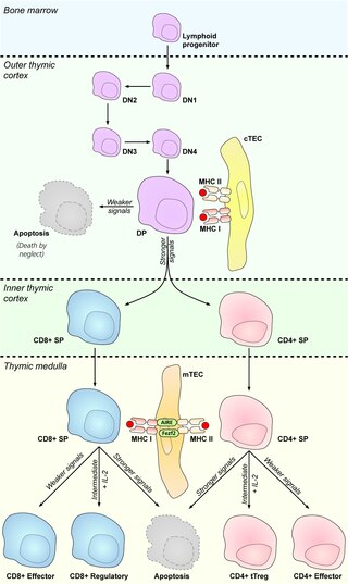
Cortical thymic epithelial cells (cTECs) form unique parenchyma cell population of the thymus which critically contribute to the development of T cells.

Thymic epithelial cells (TECs) are specialized cells with high degree of anatomic, phenotypic and functional heterogeneity that are located in the outer layer (epithelium) of the thymic stroma. The thymus, as a primary lymphoid organ, mediates T cell development and maturation. The thymic microenvironment is established by TEC network filled with thymocytes in different developing stages. TECs and thymocytes are the most important components in the thymus, that are necessary for production of functionally competent T lymphocytes and self tolerance. Dysfunction of TECs causes several immunodeficiencies and autoimmune diseases.

Promiscuous gene expression (PGE), formerly referred to as ectopic expression, is a process specific to the thymus that plays a pivotal role in the establishment of central tolerance. This phenomenon enables generation of self-antigens, so called tissue-restricted antigens (TRAs), which are in the body expressed only by one or few specific tissues. These antigens are represented for example by insulin from the pancreas or defensins from the gastrointestinal tract. Antigen-presenting cells (APCs) of the thymus, namely medullary thymic epithelial cells (mTECs), dendritic cells (DCs) and B cells are capable to present peptides derived from TRAs to developing T cells and hereby test, whether their T cell receptors (TCRs) engage self entities and therefore their occurrence in the body can potentially lead to the development of autoimmune disease. In that case, thymic APCs either induce apoptosis in these autoreactive T cells or they deviate them to become T regulatory cells, which suppress self-reactive T cells in the body that escaped negative selection in the thymus. Thus, PGE is crucial for tissues protection against autoimmunity.

Thymus stromal cells are subsets of specialized cells located in different areas of the thymus. They include all non-T-lineage cells, such as thymic epithelial cells (TECs), endothelial cells, mesenchymal cells, dendritic cells, and B lymphocytes, and provide signals essential for thymocyte development and the homeostasis of the thymic stroma.

Thymic mimetic cells are a heterogeneous population of cells located in the thymus that exhibit phenotypes of a wide variety of differentiated peripheral cells. They arise from medullary thymic epithelial cells (mTECs) and also function in negative selection of self-reactive T cells.

References

  1. Linsk R, Gottesman M, Pernis B (October 1989). "Are tissues a patch quilt of ectopic gene expression?". Science. 246 (4927): 261. Bibcode:1989Sci...246..261L. doi:10.1126/science.2799388. PMID   2799388.
  2. 1 2 Derbinski J, Schulte A, Kyewski B, Klein L (November 2001). "Promiscuous gene expression in medullary thymic epithelial cells mirrors the peripheral self". Nature Immunology. 2 (11): 1032–9. doi:10.1038/ni723. PMID   11600886. S2CID   20155713.
  3. Anderson MS, Venanzi ES, Klein L, Chen Z, Berzins SP, Turley SJ, von Boehmer H, Bronson R, Dierich A, Benoist C, Mathis D (November 2002). "Projection of an immunological self shadow within the thymus by the aire protein". Science. 298 (5597): 1395–401. Bibcode:2002Sci...298.1395A. doi:10.1126/science.1075958. PMID   12376594. S2CID   13989491.
  4. Haljasorg U, Bichele R, Saare M, Guha M, Maslovskaja J, Kõnd K, Remm A, Pihlap M, Tomson L, Kisand K, Laan M, Peterson P (December 2015). "A highly conserved NF-κB-responsive enhancer is critical for thymic expression of Aire in mice". European Journal of Immunology. 45 (12): 3246–56. doi: 10.1002/eji.201545928 . PMID   26364592.
  5. Org T, Chignola F, Hetényi C, Gaetani M, Rebane A, Liiv I, Maran U, Mollica L, Bottomley MJ, Musco G, Peterson P (April 2008). "The autoimmune regulator PHD finger binds to non-methylated histone H3K4 to activate gene expression". EMBO Reports. 9 (4): 370–6. doi:10.1038/embor.2008.11. PMC   2261226 . PMID   18292755.
  6. Koh AS, Kuo AJ, Park SY, Cheung P, Abramson J, Bua D, Carney D, Shoelson SE, Gozani O, Kingston RE, Benoist C, Mathis D (October 2008). "Aire employs a histone-binding module to mediate immunological tolerance, linking chromatin regulation with organ-specific autoimmunity". Proceedings of the National Academy of Sciences of the United States of America. 105 (41): 15878–83. Bibcode:2008PNAS..10515878K. doi: 10.1073/pnas.0808470105 . PMC   2572939 . PMID   18840680.
  7. Abramson J, Giraud M, Benoist C, Mathis D (January 2010). "Aire's partners in the molecular control of immunological tolerance". Cell. 140 (1): 123–35. doi: 10.1016/j.cell.2009.12.030 . PMID   20085707.
  8. Guha M, Saare M, Maslovskaja J, Kisand K, Liiv I, Haljasorg U, Tasa T, Metspalu A, Milani L, Peterson P (April 2017). "DNA breaks and chromatin structural changes enhance the transcription of autoimmune regulator target genes". The Journal of Biological Chemistry. 292 (16): 6542–6554. doi: 10.1074/jbc.m116.764704 . PMC   5399106 . PMID   28242760.
  9. 1 2 Gray D, Abramson J, Benoist C, Mathis D (October 2007). "Proliferative arrest and rapid turnover of thymic epithelial cells expressing Aire". The Journal of Experimental Medicine. 204 (11): 2521–8. doi:10.1084/jem.20070795. PMC   2118482 . PMID   17908938.
  10. Nagamine K, Peterson P, Scott HS, Kudoh J, Minoshima S, Heino M, Krohn KJ, Lalioti MD, Mullis PE, Antonarakis SE, Kawasaki K, Asakawa S, Ito F, Shimizu N (December 1997). "Positional cloning of the APECED gene". Nature Genetics. 17 (4): 393–8. doi:10.1038/ng1297-393. PMID   9398839. S2CID   1583134.
  11. Kisand K, Peterson P (July 2015). "Autoimmune polyendocrinopathy candidiasis ectodermal dystrophy". Journal of Clinical Immunology. 35 (5): 463–78. doi:10.1007/s10875-015-0176-y. PMID   26141571. S2CID   8080647.
  12. Sansom SN, Shikama-Dorn N, Zhanybekova S, Nusspaumer G, Macaulay IC, Deadman ME, Heger A, Ponting CP, Holländer GA (December 2014). "Population and single-cell genomics reveal the Aire dependency, relief from Polycomb silencing, and distribution of self-antigen expression in thymic epithelia". Genome Research. 24 (12): 1918–31. doi:10.1101/gr.171645.113. PMC   4248310 . PMID   25224068.
  13. Takaba H, Morishita Y, Tomofuji Y, Danks L, Nitta T, Komatsu N, Kodama T, Takayanagi H (November 2015). "Fezf2 Orchestrates a Thymic Program of Self-Antigen Expression for Immune Tolerance". Cell. 163 (4): 975–87. doi: 10.1016/j.cell.2015.10.013 . PMID   26544942.
  14. Derbinski J, Pinto S, Rösch S, Hexel K, Kyewski B (January 2008). "Promiscuous gene expression patterns in single medullary thymic epithelial cells argue for a stochastic mechanism". Proceedings of the National Academy of Sciences of the United States of America. 105 (2): 657–62. Bibcode:2008PNAS..105..657D. doi: 10.1073/pnas.0707486105 . PMC   2206592 . PMID   18180458.
  15. Brennecke P, Reyes A, Pinto S, Rattay K, Nguyen M, Küchler R, Huber W, Kyewski B, Steinmetz LM (September 2015). "Single-cell transcriptome analysis reveals coordinated ectopic gene-expression patterns in medullary thymic epithelial cells". Nature Immunology. 16 (9): 933–41. doi:10.1038/ni.3246. PMC   4675844 . PMID   26237553.
  16. Rattay K, Meyer HV, Herrmann C, Brors B, Kyewski B (February 2016). "Evolutionary conserved gene co-expression drives generation of self-antigen diversity in medullary thymic epithelial cells". Journal of Autoimmunity. 67: 65–75. doi: 10.1016/j.jaut.2015.10.001 . PMID   26481130.
  17. 1 2 Klein L, Kyewski B, Allen PM, Hogquist KA (June 2014). "Positive and negative selection of the T cell repertoire: what thymocytes see (and don't see)". Nature Reviews. Immunology. 14 (6): 377–91. doi:10.1038/nri3667. PMC   4757912 . PMID   24830344.
  18. Palmer E (May 2003). "Negative selection--clearing out the bad apples from the T-cell repertoire". Nature Reviews. Immunology. 3 (5): 383–91. doi:10.1038/nri1085. PMID   12766760. S2CID   28321309.
  19. Kurobe H, Liu C, Ueno T, Saito F, Ohigashi I, Seach N, Arakaki R, Hayashi Y, Kitagawa T, Lipp M, Boyd RL, Takahama Y (February 2006). "CCR7-dependent cortex-to-medulla migration of positively selected thymocytes is essential for establishing central tolerance". Immunity. 24 (2): 165–77. doi: 10.1016/j.immuni.2005.12.011 . PMID   16473829.
  20. Aichinger M, Wu C, Nedjic J, Klein L (February 2013). "Macroautophagy substrates are loaded onto MHC class II of medullary thymic epithelial cells for central tolerance". The Journal of Experimental Medicine. 210 (2): 287–300. doi:10.1084/jem.20122149. PMC   3570095 . PMID   23382543.
  21. Liston A, Lesage S, Wilson J, Peltonen L, Goodnow CC (April 2003). "Aire regulates negative selection of organ-specific T cells". Nature Immunology. 4 (4): 350–4. doi:10.1038/ni906. PMID   12612579. S2CID   4561402.
  22. Anderson MS, Venanzi ES, Chen Z, Berzins SP, Benoist C, Mathis D (August 2005). "The cellular mechanism of Aire control of T cell tolerance". Immunity. 23 (2): 227–39. doi: 10.1016/j.immuni.2005.07.005 . PMID   16111640.
  23. Taniguchi RT, DeVoss JJ, Moon JJ, Sidney J, Sette A, Jenkins MK, Anderson MS (May 2012). "Detection of an autoreactive T-cell population within the polyclonal repertoire that undergoes distinct autoimmune regulator (Aire)-mediated selection". Proceedings of the National Academy of Sciences of the United States of America. 109 (20): 7847–52. Bibcode:2012PNAS..109.7847T. doi: 10.1073/pnas.1120607109 . PMC   3356674 . PMID   22552229.
  24. Aschenbrenner K, D'Cruz LM, Vollmann EH, Hinterberger M, Emmerich J, Swee LK, Rolink A, Klein L (April 2007). "Selection of Foxp3+ regulatory T cells specific for self antigen expressed and presented by Aire+ medullary thymic epithelial cells". Nature Immunology. 8 (4): 351–8. doi:10.1038/ni1444. PMID   17322887. S2CID   21052344.
  25. Malchow S, Leventhal DS, Nishi S, Fischer BI, Shen L, Paner GP, Amit AS, Kang C, Geddes JE, Allison JP, Socci ND, Savage PA (March 2013). "Aire-dependent thymic development of tumor-associated regulatory T cells". Science. 339 (6124): 1219–24. Bibcode:2013Sci...339.1219M. doi:10.1126/science.1233913. PMC   3622085 . PMID   23471412.
  26. Malchow S, Leventhal DS, Lee V, Nishi S, Socci ND, Savage PA (May 2016). "Aire Enforces Immune Tolerance by Directing Autoreactive T Cells into the Regulatory T Cell Lineage". Immunity. 44 (5): 1102–13. doi:10.1016/j.immuni.2016.02.009. PMC   4871732 . PMID   27130899.
  27. Klein L (August 2009). "Dead man walking: how thymocytes scan the medulla". Nature Immunology. 10 (8): 809–11. doi:10.1038/ni0809-809. PMID   19621041. S2CID   28668641.
  28. Koble C, Kyewski B (July 2009). "The thymic medulla: a unique microenvironment for intercellular self-antigen transfer". The Journal of Experimental Medicine. 206 (7): 1505–13. doi:10.1084/jem.20082449. PMC   2715082 . PMID   19564355.
  29. 1 2 Perry JS, Lio CJ, Kau AL, Nutsch K, Yang Z, Gordon JI, Murphy KM, Hsieh CS (September 2014). "Distinct contributions of Aire and antigen-presenting-cell subsets to the generation of self-tolerance in the thymus". Immunity. 41 (3): 414–426. doi:10.1016/j.immuni.2014.08.007. PMC   4175925 . PMID   25220213.
  30. Kroger CJ, Spidale NA, Wang B, Tisch R (January 2017). "Thymic Dendritic Cell Subsets Display Distinct Efficiencies and Mechanisms of Intercellular MHC Transfer". Journal of Immunology. 198 (1): 249–256. doi:10.4049/jimmunol.1601516. PMC   5173434 . PMID   27895179.
  31. Hinterberger M, Aichinger M, Prazeres da Costa O, Voehringer D, Hoffmann R, Klein L (June 2010). "Autonomous role of medullary thymic epithelial cells in central CD4(+) T cell tolerance". Nature Immunology. 11 (6): 512–9. doi:10.1038/ni.1874. PMID   20431619. S2CID   33154019.
  32. 1 2 Derbinski J, Gäbler J, Brors B, Tierling S, Jonnakuty S, Hergenhahn M, Peltonen L, Walter J, Kyewski B (July 2005). "Promiscuous gene expression in thymic epithelial cells is regulated at multiple levels". The Journal of Experimental Medicine. 202 (1): 33–45. doi:10.1084/jem.20050471. PMC   2212909 . PMID   15983066.
  33. 1 2 Gäbler J, Arnold J, Kyewski B (December 2007). "Promiscuous gene expression and the developmental dynamics of medullary thymic epithelial cells". European Journal of Immunology. 37 (12): 3363–72. doi: 10.1002/eji.200737131 . PMID   18000951.
  34. Rossi SW, Kim MY, Leibbrandt A, Parnell SM, Jenkinson WE, Glanville SH, McConnell FM, Scott HS, Penninger JM, Jenkinson EJ, Lane PJ, Anderson G (June 2007). "RANK signals from CD4(+)3(-) inducer cells regulate development of Aire-expressing epithelial cells in the thymic medulla". The Journal of Experimental Medicine. 204 (6): 1267–72. doi:10.1084/jem.20062497. PMC   2118623 . PMID   17502664.
  35. Lkhagvasuren E, Sakata M, Ohigashi I, Takahama Y (May 2013). "Lymphotoxin β receptor regulates the development of CCL21-expressing subset of postnatal medullary thymic epithelial cells". Journal of Immunology. 190 (10): 5110–7. doi: 10.4049/jimmunol.1203203 . PMID   23585674.
  36. Metzger TC, Khan IS, Gardner JM, Mouchess ML, Johannes KP, Krawisz AK, Skrzypczynska KM, Anderson MS (October 2013). "Lineage tracing and cell ablation identify a post-Aire-expressing thymic epithelial cell population". Cell Reports. 5 (1): 166–79. doi:10.1016/j.celrep.2013.08.038. PMC   3820422 . PMID   24095736.
  37. Michelson, Daniel A.; Mathis, Diane (October 2022). "Thymic mimetic cells: tolerogenic masqueraders". Trends in Immunology. 43 (10): 782–791. doi:10.1016/j.it.2022.07.010. ISSN   1471-4906. PMC   9509455 . PMID   36008259.
  38. Gordon J, Wilson VA, Blair NF, Sheridan J, Farley A, Wilson L, Manley NR, Blackburn CC (May 2004). "Functional evidence for a single endodermal origin for the thymic epithelium". Nature Immunology. 5 (5): 546–53. doi:10.1038/ni1064. PMID   15098031. S2CID   10282524.
  39. Bleul CC, Corbeaux T, Reuter A, Fisch P, Mönting JS, Boehm T (June 2006). "Formation of a functional thymus initiated by a postnatal epithelial progenitor cell". Nature. 441 (7096): 992–6. Bibcode:2006Natur.441..992B. doi:10.1038/nature04850. PMID   16791198. S2CID   4396922.
  40. Rossi SW, Jenkinson WE, Anderson G, Jenkinson EJ (June 2006). "Clonal analysis reveals a common progenitor for thymic cortical and medullary epithelium". Nature. 441 (7096): 988–91. Bibcode:2006Natur.441..988R. doi:10.1038/nature04813. PMID   16791197. S2CID   2358330.
  41. Baik S, Jenkinson EJ, Lane PJ, Anderson G, Jenkinson WE (March 2013). "Generation of both cortical and Aire(+) medullary thymic epithelial compartments from CD205(+) progenitors". European Journal of Immunology. 43 (3): 589–94. doi:10.1002/eji.201243209. PMC   3960635 . PMID   23299414.
  42. Ohigashi I, Zuklys S, Sakata M, Mayer CE, Zhanybekova S, Murata S, Tanaka K, Holländer GA, Takahama Y (June 2013). "Aire-expressing thymic medullary epithelial cells originate from β5t-expressing progenitor cells". Proceedings of the National Academy of Sciences of the United States of America. 110 (24): 9885–90. Bibcode:2013PNAS..110.9885O. doi: 10.1073/pnas.1301799110 . PMC   3683726 . PMID   23720310.
  43. Hamazaki Y, Fujita H, Kobayashi T, Choi Y, Scott HS, Matsumoto M, Minato N (March 2007). "Medullary thymic epithelial cells expressing Aire represent a unique lineage derived from cells expressing claudin". Nature Immunology. 8 (3): 304–11. doi:10.1038/ni1438. PMID   17277780. S2CID   7775843.
  44. Sekai M, Hamazaki Y, Minato N (November 2014). "Medullary thymic epithelial stem cells maintain a functional thymus to ensure lifelong central T cell tolerance". Immunity. 41 (5): 753–61. doi: 10.1016/j.immuni.2014.10.011 . PMID   25464854.
  45. Ohigashi I, Zuklys S, Sakata M, Mayer CE, Hamazaki Y, Minato N, Hollander GA, Takahama Y (November 2015). "Adult Thymic Medullary Epithelium Is Maintained and Regenerated by Lineage-Restricted Cells Rather Than Bipotent Progenitors". Cell Reports. 13 (7): 1432–1443. doi: 10.1016/j.celrep.2015.10.012 . hdl: 2433/216325 . PMID   26549457.
  46. Ucar A, Ucar O, Klug P, Matt S, Brunk F, Hofmann TG, Kyewski B (August 2014). "Adult thymus contains FoxN1(-) epithelial stem cells that are bipotent for medullary and cortical thymic epithelial lineages". Immunity. 41 (2): 257–69. doi:10.1016/j.immuni.2014.07.005. PMC   4148705 . PMID   25148026.
  47. Wong K, Lister NL, Barsanti M, Lim JM, Hammett MV, Khong DM, Siatskas C, Gray DH, Boyd RL, Chidgey AP (August 2014). "Multilineage potential and self-renewal define an epithelial progenitor cell population in the adult thymus". Cell Reports. 8 (4): 1198–209. doi: 10.1016/j.celrep.2014.07.029 . PMID   25131206.
  48. Ulyanchenko S, O'Neill KE, Medley T, Farley AM, Vaidya HJ, Cook AM, Blair NF, Blackburn CC (March 2016). "Identification of a Bipotent Epithelial Progenitor Population in the Adult Thymus". Cell Reports. 14 (12): 2819–32. doi:10.1016/j.celrep.2016.02.080. PMC   4819909 . PMID   26997270.
  49. Meireles C, Ribeiro AR, Pinto RD, Leitão C, Rodrigues PM, Alves NL (June 2017). "Thymic crosstalk restrains the pool of cortical thymic epithelial cells with progenitor properties". European Journal of Immunology. 47 (6): 958–969. doi: 10.1002/eji.201746922 . hdl: 10216/111812 . PMID   28318017.
  50. van Delft MA, Huitema LF, Tas SW (May 2015). "The contribution of NF-κB signalling to immune regulation and tolerance". European Journal of Clinical Investigation. 45 (5): 529–39. doi:10.1111/eci.12430. PMID   25735405.