Diagnosis of myocardial infarction

Last updated
Myocardial infarction diagnosis
PurposeDiagnose myocardial infarct via physical exam and EKG (plus blood test)

A diagnosis of myocardial infarction is created by integrating the history of the presenting illness and physical examination with electrocardiogram findings and cardiac markers (blood tests for heart muscle cell damage). [1] [2] A coronary angiogram allows visualization of narrowings or obstructions on the heart vessels, and therapeutic measures can follow immediately. At autopsy, a pathologist can diagnose a myocardial infarction based on anatomopathological findings.

Contents

A chest radiograph and routine blood tests may indicate complications or precipitating causes and are often performed upon arrival to an emergency department. New regional wall motion abnormalities on an echocardiogram are also suggestive of a myocardial infarction. Echo may be performed in equivocal cases by the on-call cardiologist. [3] In stable patients whose symptoms have resolved by the time of evaluation, Technetium (99mTc) sestamibi (i.e. a "MIBI scan"), thallium-201 chloride or Rubidium-82 Chloride can be used in nuclear medicine to visualize areas of reduced blood flow in conjunction with physiologic or pharmacologic stress. [3] [4] Thallium may also be used to determine viability of tissue, distinguishing whether non-functional myocardium is actually dead or merely in a state of hibernation or of being stunned. [5]

Diagnostic criteria

According to the WHO criteria as revised in 2000, [6] a cardiac troponin rise accompanied by either typical symptoms, pathological Q waves, ST elevation or depression or coronary intervention are diagnostic of MI.

Previous WHO criteria [7] formulated in 1979 put less emphasis on cardiac biomarkers; according to these, a patient is diagnosed with myocardial infarction if two (probable) or three (definite) of the following criteria are satisfied:

  1. Clinical history of ischaemic type chest pain lasting for more than 20 minutes
  2. Changes in serial ECG tracings
  3. Rise and fall of serum cardiac biomarkers such as creatine kinase-MB fraction and troponin

Physical examination

The general appearance of patients may vary according to the experienced symptoms; the patient may be comfortable, or restless and in severe distress with an increased respiratory rate. A cool and pale skin is common and points to vasoconstriction. Some patients have low-grade fever (38–39 °C). Blood pressure may be elevated or decreased, and the pulse can become irregular. [8] [9] :1444

If heart failure ensues, elevated jugular venous pressure and hepatojugular reflux, or swelling of the legs due to peripheral edema may be found on inspection. Rarely, a cardiac bulge with a pace different from the pulse rhythm can be felt on precordial examination. Various abnormalities can be found on auscultation, such as a third and fourth heart sound, systolic murmurs, paradoxical splitting of the second heart sound, a pericardial friction rub and rales over the lung. [8] [9] :1450

Electrocardiogram

12-lead electrocardiogram showing ST-segment elevation (orange) in I, aVL and V1-V5 with reciprocal changes (blue) in the inferior leads, indicative of an anterior wall myocardial infarction. 12 Lead EKG ST Elevation tracing color coded.jpg
12-lead electrocardiogram showing ST-segment elevation (orange) in I, aVL and V1-V5 with reciprocal changes (blue) in the inferior leads, indicative of an anterior wall myocardial infarction.

The primary purpose of the electrocardiogram is to detect ischemia or acute coronary injury in broad, symptomatic emergency department populations. A serial ECG may be used to follow rapid changes in time. The standard 12 lead ECG does not directly examine the right ventricle, and is relatively poor at examining the posterior basal and lateral walls of the left ventricle. In particular, acute myocardial infarction in the distribution of the circumflex artery is likely to produce a nondiagnostic ECG. [10] The use of additional ECG leads like right-sided leads V3R and V4R and posterior leads V7, V8, and V9 may improve sensitivity for right ventricular and posterior myocardial infarction.[ citation needed ]

The 12 lead ECG is used to classify patients into one of three groups: [11]

  1. those with ST segment elevation or new bundle branch block (suspicious for acute injury and a possible candidate for acute reperfusion therapy with thrombolytics or primary PCI),
  2. those with ST segment depression or T wave inversion (suspicious for ischemia), and
  3. those with a so-called non-diagnostic or normal ECG.

A normal ECG does not rule out acute myocardial infarction. Mistakes in interpretation are relatively common, and the failure to identify high risk features has a negative effect on the quality of patient care. [12]

It should be determined if a person is at high risk for myocardial infarction before conducting imaging tests to make a diagnosis. [13] People who have a normal ECG and who are able to exercise, for example, do not merit routine imaging. [13] Imaging tests such as stress radionuclide myocardial perfusion imaging or stress echocardiography can confirm a diagnosis when a person's history, physical exam, ECG and cardiac biomarkers suggest the likelihood of a problem. [13]

Cardiac markers

Cardiac markers or cardiac enzymes are proteins that leak out of injured myocardial cells through their damaged cell membranes into the bloodstream. Until the 1980s, the enzymes SGOT and LDH were used to assess cardiac injury. Now, the markers most widely used in detection of MI are MB subtype of the enzyme creatine kinase and cardiac troponins T and I as they are more specific for myocardial injury. The cardiac troponins T and I which are released within 4–6 hours of an attack of MI and remain elevated for up to 2 weeks, have nearly complete tissue specificity and are now the preferred markers for assessing myocardial damage. [14] Heart-type fatty acid binding protein is another marker, used in some home test kits. Elevated troponins in the setting of chest pain may accurately predict a high likelihood of a myocardial infarction in the near future. [15] New markers such as glycogen phosphorylase isoenzyme BB are under investigation. [16] Note that only the cardiac troponins are used clinically for myocardial infarction as creatine kinase adds little value in diagnosing MI while adding to system cost. [17] [18] [19]

The diagnosis of myocardial infarction requires two out of three components (history, ECG, and enzymes). When damage to the heart occurs, levels of cardiac markers rise over time, which is why blood tests for them are taken over a 24-hour period. Because these enzyme levels are not elevated immediately following a heart attack, patients presenting with chest pain are generally treated with the assumption that a myocardial infarction has occurred and then evaluated for a more precise diagnosis. [20]

Angiography

Angiogram of the coronary arteries. Ha1.jpg
Angiogram of the coronary arteries.

In difficult cases or in situations where intervention to restore blood flow is appropriate, coronary angiography can be performed. A catheter is inserted into an artery (typically the radial or femoral artery [21] ) and pushed to the vessels supplying the heart. A radio-opaque dye is administered through the catheter and a sequence of x-rays (fluoroscopy) is performed. Obstructed or narrowed arteries can be identified, and angioplasty applied as a therapeutic measure (see below). Angioplasty requires extensive skill, especially in emergency settings. It is performed by a physician trained in interventional cardiology.[ citation needed ]

Histopathology

Micrograph of a myocardial infarction (ca. 400x H&E stain ) with prominent contraction band necrosis. MI with contraction bands very high mag.jpg
Micrograph of a myocardial infarction (ca. 400x H&E stain ) with prominent contraction band necrosis.

Histopathological examination of the heart may reveal infarction at autopsy. Gross examination may reveal signs of myocardial infarction.[ citation needed ]

Under the microscope, myocardial infarction presents as a circumscribed area of ischemic, coagulative necrosis (cell death). On gross examination, the infarct is not identifiable within the first 12 hours. [22]

Although earlier changes can be discerned using electron microscopy, one of the earliest changes under a normal microscope are so-called wavy fibers. [23] Subsequently, the myocyte cytoplasm becomes more eosinophilic (pink) and the cells lose their transversal striations, with typical changes and eventually loss of the cell nucleus. [24] The interstitium at the margin of the infarcted area is initially infiltrated with neutrophils, then with lymphocytes and macrophages, who phagocytose ("eat") the myocyte debris. The necrotic area is surrounded and progressively invaded by granulation tissue, which will replace the infarct with a fibrous (collagenous) scar (which are typical steps in wound healing). The interstitial space (the space between cells outside of blood vessels) may be infiltrated with red blood cells. [22]

These features can be recognized in cases where the perfusion was not restored; reperfused infarcts can have other hallmarks, such as contraction band necrosis. [25]

These tables gives an overview of the histopathology seen in myocardial infarction by time after obstruction.[ citation needed ]

By individual parameters

Myocardial histologic parameters (HE staining) [26] Earliest manifestation [26] Full development [26] Decrease/disappearance [26] Image
Stretched/wavy fibres1–2 h Histopathology of myofiber waviness in myocardial infarction.jpg
Coagulative necrosis: cytoplasmic hypereosinophilia 1–3 h1–3 days; cytoplasmic hypereosinophilia and loss of striations> 3 days: disintegration Histopathology of coagulative necrosis of cardiomyocytes.jpg
Interstitial edema4–12 h Histopathology of interstitial edema in myocardial infarction.jpg
Coagulative necrosis: 'nuclear changes'12–24 (pyknosis, karyorrhexis)1–3 days (loss of nuclei)Depends on size of infarction Histopathology of myocardial infarction with loss of nuclei.jpg
Neutrophil infiltration12–24 h1–3 days5–7 days Histopathology of neutrophil infiltration in myocardial infarction.jpg
Karyorrhexis of neutrophils1.5–2 days3–5 days Histopathology of myocardial infarction with karyorrhexis and few lymphocytes.jpg
Macrophages and lymphocytes 3–5 days5–10 days (including 'siderophages')10 days to 2 months Histopathology of macrophages and lymphocyte infiltration with early removal of necrotic debris in myocardial infarction.jpg
Vessel/endothelial sprouts*5–10 days10 days–4 weeks4 weeks: disappearance of capillaries; some large dilated vessels persist Histopathology of granulation tissue with formation of microvessels in myocardial infarction.jpg
Fibroblast and young collagen*5–10 days2–4 weeksAfter 4 weeks; depends on size of infarction; Histopathology of fibroblast proliferation and early collagen deposition in myocardial infarction.jpg
Dense fibrosis4 weeks2–3 monthsNo Histopathology of dense fibrous scar replacing myocyte loss in myocardial infarction.jpg

Differential diagnoses for myocardial fibrosis:

Chronological

Time Gross examination Histopathology
by light microscopy
0 - 0.5 hoursNone [note 1] None [note 1]
0.5 – 4 hoursNone [note 2]
  • Glycogen Depletion, as seen with a PAS Stain
  • Possibly waviness of fibers at border
4 – 12 hours
12 – 24 hours
  • Dark mottling
1 – 3 days
  • Infarct center becomes yellow-tan
  • Continued coagulation necrosis
  • Loss of nuclei and striations
  • Increased infiltration of neutrophils to interstitium
3 – 7 days
  • Hyperemia at border
  • Softening yellow-tan center
  • Beginning of disintegration of dead muscle fibers
  • Apoptosis of neutrophils
  • Beginning of macrophage removal of dead cells at border
7 – 10 days
  • Maximally soft and yellow-tan
  • Red-tan margins
  • Increased phagocytosis of dead cells at border
  • Beginning of granulation tissue formation at margins
10 – 14 days
  • Red-gray and depressed borders
2 – 8 weeks
  • Gray-white granulation tissue
  • Increased collagen deposition
  • Decreased cellularity
More than 2 monthsCompleted scarring [note 3] Dense collagenous scar formed [note 3]
If not else specified in boxes, then reference is nr [31]

See also

Notes

  1. 1 2 For the first ~30 minutes no change at all can be seen by gross examination or by light microscopy in histopathology. However, in electron microscopy relaxed myofibrils, as well as glycogen loss and mitochondrial swelling can be observered.
  2. It is often possible, however, to highlight the area of necrosis that first becomes apparent after 2 to 3 hours by immersion of tissue slices in a solution of triphenyltetrazolium chloride. This dye imparts a brick-red color to intact, noninfarcted myocardium where the dehydrogenase activity is preserved. Because dehydrogenases are depleted in the area of ischemic necrosis (i.e., they leak out through the damaged cell membranes), an infarcted area is revealed as an unstained pale zone. Instead of a triphenyltetrazolium chloride dye, a LDH (lactate dehydrogenase) dye can also be used to visualize an area of necrosis.
  3. 1 2 Once scarring is completed, there is yet no common method of discerning the actual age of the infarct, since e.g. a scar that is four months old looks identical to a scar that is ten years old.

Related Research Articles

<span class="mw-page-title-main">Coronary artery disease</span> Reduction of blood flow to the heart

Coronary artery disease (CAD), also called coronary heart disease (CHD), ischemic heart disease (IHD), myocardial ischemia, or simply heart disease, involves the reduction of blood flow to the cardiac muscle due to build-up of atherosclerotic plaque in the arteries of the heart. It is the most common of the cardiovascular diseases. Types include stable angina, unstable angina, and myocardial infarction.

<span class="mw-page-title-main">Angina</span> Chest discomfort that is generally brought on by inadequate blood flow to the cardiac muscle

Angina, also known as angina pectoris, is chest pain or pressure, usually caused by insufficient blood flow to the heart muscle (myocardium). It is most commonly a symptom of coronary artery disease.

<span class="mw-page-title-main">Chest pain</span> Discomfort or pain in the chest as a medical symptom

Chest pain is pain or discomfort in the chest, typically the front of the chest. It may be described as sharp, dull, pressure, heaviness or squeezing. Associated symptoms may include pain in the shoulder, arm, upper abdomen, or jaw, along with nausea, sweating, or shortness of breath. It can be divided into heart-related and non-heart-related pain. Pain due to insufficient blood flow to the heart is also called angina pectoris. Those with diabetes or the elderly may have less clear symptoms.

<span class="mw-page-title-main">Troponin</span> Protein complex

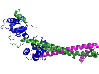
Troponin, or the troponin complex, is a complex of three regulatory proteins that are integral to muscle contraction in skeletal muscle and cardiac muscle, but not smooth muscle. Measurements of cardiac-specific troponins I and T are extensively used as diagnostic and prognostic indicators in the management of myocardial infarction and acute coronary syndrome. Blood troponin levels may be used as a diagnostic marker for stroke or other myocardial injury that is ongoing, although the sensitivity of this measurement is low.

<span class="mw-page-title-main">Cardiac stress test</span> Measures the hearts ability to respond to external stress in a controlled clinical environment

A cardiac stress test is a cardiological examination that evaluates the cardiovascular system's response to external stress within a controlled clinical setting. This stress response can be induced through physical exercise or intravenous pharmacological stimulation of heart rate.

<span class="mw-page-title-main">Cardiac marker</span>

Cardiac markers are biomarkers measured to evaluate heart function. They can be useful in the early prediction or diagnosis of disease. Although they are often discussed in the context of myocardial infarction, other conditions can lead to an elevation in cardiac marker level.

<span class="mw-page-title-main">Acute coronary syndrome</span> Medical condition

Acute coronary syndrome (ACS) is a syndrome due to decreased blood flow in the coronary arteries such that part of the heart muscle is unable to function properly or dies. The most common symptom is centrally located pressure-like chest pain, often radiating to the left shoulder or angle of the jaw, and associated with nausea and sweating. Many people with acute coronary syndromes present with symptoms other than chest pain, particularly women, older people, and people with diabetes mellitus.

<span class="mw-page-title-main">Unstable angina</span> Medical condition

Unstable angina is a type of angina pectoris that is irregular or more easily provoked. It is classified as a type of acute coronary syndrome.

<span class="mw-page-title-main">Acute pericarditis</span> Medical condition

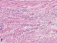
Acute pericarditis is a type of pericarditis usually lasting less than 6 weeks. It is the most common condition affecting the pericardium.

<span class="mw-page-title-main">Takotsubo cardiomyopathy</span> Sudden temporary weakening of the heart muscle

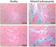
Takotsubo cardiomyopathy or takotsubo syndrome (TTS), also known as stress cardiomyopathy, is a type of non-ischemic cardiomyopathy in which there is a sudden temporary weakening of the muscular portion of the heart. It usually appears after a significant stressor, either physical or emotional; when caused by the latter, the condition is sometimes called broken heart syndrome.

Door-to-balloon is a time measurement in emergency cardiac care (ECC), specifically in the treatment of ST segment elevation myocardial infarction. The interval starts with the patient's arrival in the emergency department, and ends when a catheter guidewire crosses the culprit lesion in the cardiac cath lab. Because of the adage that "time is muscle", meaning that delays in treating a myocardial infarction increase the likelihood and amount of cardiac muscle damage due to localised hypoxia, ACC/AHA guidelines recommend a door-to-balloon interval of no more than 90 minutes. As of 2006 in the United States, fewer than half of STEMI patients received reperfusion with primary percutaneous coronary intervention (PCI) within the guideline-recommended timeframe. It has become a core quality measure for the Joint Commission on Accreditation of Healthcare Organizations (TJC).

<span class="mw-page-title-main">Troponin I</span> Muscle protein

Troponin I is a cardiac and skeletal muscle protein family. It is a part of the troponin protein complex, where it binds to actin in thin myofilaments to hold the actin-tropomyosin complex in place. Troponin I prevents myosin from binding to actin in relaxed muscle. When calcium binds to the troponin C, it causes conformational changes which lead to dislocation of troponin I. Afterwards, tropomyosin leaves the binding site for myosin on actin leading to contraction of muscle. The letter I is given due to its inhibitory character. It is a useful marker in the laboratory diagnosis of heart attack. It occurs in different plasma concentration but the same circumstances as troponin T - either test can be performed for confirmation of cardiac muscle damage and laboratories usually offer one test or the other.

<span class="mw-page-title-main">Myocardial perfusion imaging</span> Nuclear medicine imaging method

Myocardial perfusion imaging or scanning is a nuclear medicine procedure that illustrates the function of the heart muscle (myocardium).

<span class="mw-page-title-main">Myocardial infarction</span> Interruption of blood supply to a part of the heart

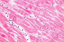
A myocardial infarction (MI), commonly known as a heart attack, occurs when blood flow decreases or stops in one of the coronary arteries of the heart, causing infarction to the heart muscle. The most common symptom is chest pain or discomfort which may travel into the shoulder, arm, back, neck or jaw. Often such pain occurs in the center or left side of the chest and lasts for more than a few minutes. The discomfort may occasionally feel like heartburn.

<span class="mw-page-title-main">Heart-type fatty acid binding protein</span> Protein-coding gene in the species Homo sapiens

Heart-type fatty acid binding protein (hFABP) also known as mammary-derived growth inhibitor is a protein that in humans is encoded by the FABP3 gene.

<span class="mw-page-title-main">Electrocardiography in myocardial infarction</span>

Electrocardiography in suspected myocardial infarction has the main purpose of detecting ischemia or acute coronary injury in emergency department populations coming for symptoms of myocardial infarction (MI). Also, it can distinguish clinically different types of myocardial infarction.

<span class="mw-page-title-main">Francis M. Fesmire</span> American emergency physician (1969–2014)

Francis Miller Fesmire was an American emergency physician and a nationally recognized expert in myocardial infarction. He authored numerous academic articles and assisted in the development of clinical guidelines on the standard of care in treating patients with suspected myocardial infarction by the American College of Emergency Physicians and the American Heart Association/American College of Cardiology. He performed numerous research investigations in chest pain patients, reporting the usefulness of continuous 12-lead ECG monitoring, two-hour delta cardiac marker testing, and nuclear cardiac stress testing in the emergency department. The culmination of his studies was The Erlanger Chest Pain Evaluation Protocol published in the Annals of Emergency Medicine in 2002. In 2011 he published a novel Nashville Skyline that received a 5 star review by ForeWord Reviews. His most recent research involved the risk stratification of chest pain patients in the emergency department.

<span class="mw-page-title-main">Management of acute coronary syndrome</span>

Management of acute coronary syndrome is targeted against the effects of reduced blood flow to the affected area of the heart muscle, usually because of a blood clot in one of the coronary arteries, the vessels that supply oxygenated blood to the myocardium. This is achieved with urgent hospitalization and medical therapy, including drugs that relieve chest pain and reduce the size of the infarct, and drugs that inhibit clot formation; for a subset of patients invasive measures are also employed. Basic principles of management are the same for all types of acute coronary syndrome. However, some important aspects of treatment depend on the presence or absence of elevation of the ST segment on the electrocardiogram, which classifies cases upon presentation to either ST segment elevation myocardial infarction (STEMI) or non-ST elevation acute coronary syndrome (NST-ACS); the latter includes unstable angina and non-ST elevation myocardial infarction (NSTEMI). Treatment is generally more aggressive for STEMI patients, and reperfusion therapy is more often reserved for them. Long-term therapy is necessary for prevention of recurrent events and complications.

Kounis syndrome is defined as acute coronary syndrome caused by an allergic reaction or a strong immune reaction to a drug or other substance. It is a rare syndrome with authentic cases reported in 130 males and 45 females, as reviewed in 2017; however, the disorder is suspected of being commonly overlooked and therefore much more prevalent. Mast cell activation and release of inflammatory cytokines as well as other inflammatory agents from the reaction leads to spasm of the arteries leading to the heart muscle or a plaque breaking free and blocking one or more of those arteries.

Remote ischemic conditioning (RIC) is an experimental medical procedure that aims to reduce the severity of ischaemic injury to an organ such as the heart or the brain, most commonly in the situation of a heart attack or a stroke, or during procedures such as heart surgery when the heart may temporary suffer ischaemia during the operation, by triggering the body's natural protection against tissue injury. Although noted to have some benefits in experimental models in animals, this is still an experimental procedure in humans and initial evidence from small studies have not been replicated in larger clinical trials. Successive clinical trials have failed to identify evidence supporting a protective role in humans.

References

  1. Mallinson T (2010). "Myocardial Infarction". Focus on First Aid (15): 15. Archived from the original on 2010-05-21. Retrieved 2010-06-08.
  2. "Myocardial infarction: diagnosis and investigations". GPnotebook . Retrieved 27 November 2006.
  3. 1 2 Fenton DE, Stahmer S, Baumann DM. McNamara RM, Talavera F, Halamka J, Feied C (eds.). "Myocardial infarction]". eMedicine . Archived from the original on 25 December 2006. Retrieved 27 November 2006.
  4. "HEART SCAN". University College London . Archived from the original on 16 February 2009. Retrieved 27 November 2006.
  5. Skoufis E, McGhie AI (1998). "Radionuclide techniques for the assessment of myocardial viability". Texas Heart Institute Journal. 25 (4): 272–279. PMC   325572 . PMID   9885104.
  6. Alpert JS, Thygesen K, Antman E, Bassand JP (September 2000). "Myocardial infarction redefined--a consensus document of The Joint European Society of Cardiology/American College of Cardiology Committee for the redefinition of myocardial infarction". Journal of the American College of Cardiology. 36 (3): 959–969. doi: 10.1016/S0735-1097(00)00804-4 . PMID   10987628.
  7. Anonymous (March 1979). "Nomenclature and criteria for diagnosis of ischemic heart disease. Report of the Joint International Society and Federation of Cardiology/World Health Organization task force on standardization of clinical nomenclature". Circulation. 59 (3): 607–609. doi: 10.1161/01.CIR.59.3.607 . PMID   761341.
  8. 1 2 Garas S, Zafari AM, Garas S. Vanderbush E, Talavera F, Runge MS, Mylonakis E, Zevitz ME (eds.). "Myocardial Infarction". eMedicine . Archived from the original on 11 December 2006. Retrieved 22 November 2006.
  9. 1 2 Kasper DL, Braunwald E, Fauci AS, Hauser SL, Longo DL, Jameson JL (2005). Harrison's Principles of Internal Medicine. New York: McGraw-Hill. ISBN   0-07-139140-1.
  10. Cannon CP at al. Management of Acute Coronary Syndromes. p. 175. New Jersey: Humana Press, 1999. ISBN   0-89603-552-2.
  11. ECC Committee, Subcommittees and Task Forces of the American Heart Association (December 2005). "2005 American Heart Association Guidelines for Cardiopulmonary Resuscitation and Emergency Cardiovascular Care". Circulation. 112 (24 Suppl): IV1-203. doi: 10.1161/CIRCULATIONAHA.105.166561 . PMID   16314375.
  12. Masoudi FA, Magid DJ, Vinson DR, Tricomi AJ, Lyons EE, Crounse L, et al. (October 2006). "Implications of the failure to identify high-risk electrocardiogram findings for the quality of care of patients with acute myocardial infarction: results of the Emergency Department Quality in Myocardial Infarction (EDQMI) study". Circulation. 114 (15): 1565–1571. doi: 10.1161/CIRCULATIONAHA.106.623652 . PMID   17015790.
  13. 1 2 3 American Society of Nuclear Cardiology, "Five Things Physicians and Patients Should Question" (PDF), Choosing Wisely: an initiative of the ABIM Foundation , American Society of Nuclear Cardiology, archived from the original (PDF) on April 16, 2012, retrieved August 17, 2012, which cites
  14. Eisenman A (July 2006). "Troponin assays for the diagnosis of myocardial infarction and acute coronary syndrome: where do we stand?". Expert Review of Cardiovascular Therapy. 4 (4): 509–514. doi:10.1586/14779072.4.4.509. PMID   16918269. S2CID   38481075.
  15. Aviles RJ, Askari AT, Lindahl B, Wallentin L, Jia G, Ohman EM, et al. (June 2002). "Troponin T levels in patients with acute coronary syndromes, with or without renal dysfunction". The New England Journal of Medicine. 346 (26): 2047–2052. doi: 10.1056/NEJMoa013456 . PMID   12087140.. Summary for laymen
  16. Apple FS, Wu AH, Mair J, Ravkilde J, Panteghini M, Tate J, et al. (May 2005). "Future biomarkers for detection of ischemia and risk stratification in acute coronary syndrome". Clinical Chemistry. 51 (5): 810–824. doi: 10.1373/clinchem.2004.046292 . PMID   15774573.
  17. Mechanic OJ, Gavin M, Grossman SA (2024). "Acute Myocardial Infarction". StatPearls. Treasure Island (FL): StatPearls Publishing. PMID   29083808 . Retrieved 2024-02-05.
  18. Wiens EJ, Arbour J, Thompson K, Seifer CM (July 2019). "Routine creatine kinase testing does not provide clinical utility in the emergency department for diagnosis of acute coronary syndromes". BMC Emergency Medicine. 19 (1): 37. doi: 10.1186/s12873-019-0251-4 . PMC   6617848 . PMID   31288735.
  19. Taggart C, Wereski R, Mills NL, Chapman AR (May 2021). "Diagnosis, Investigation and Management of Patients with Acute and Chronic Myocardial Injury". Journal of Clinical Medicine. 10 (11): 2331. doi: 10.3390/jcm10112331 . PMC   8199345 . PMID   34073539.
  20. Braunwald E, Antman EM, Beasley JW, Califf RM, Cheitlin MD, Hochman JS, et al. (October 2002). "ACC/AHA 2002 guideline update for the management of patients with unstable angina and non-ST-segment elevation myocardial infarction--summary article: a report of the American College of Cardiology/American Heart Association task force on practice guidelines (Committee on the Management of Patients With Unstable Angina)". Journal of the American College of Cardiology. 40 (7): 1366–1374. doi:10.1016/s0735-1097(02)02336-7. PMID   12383588.
  21. Kolkailah AA, Alreshq RS, Muhammed AM, Zahran ME, Anas El-Wegoud M, Nabhan AF (April 2018). "Transradial versus transfemoral approach for diagnostic coronary angiography and percutaneous coronary intervention in people with coronary artery disease". The Cochrane Database of Systematic Reviews. 2018 (4): CD012318. doi:10.1002/14651858.CD012318.pub2. PMC   6494633 . PMID   29665617.
  22. 1 2 Rubin E, Gorstein F, Rubin R, Schwarting R, Strayer D (2001). Rubin's Pathology — Clinicopathological Foundations of Medicine. Maryland: Lippincott Williams & Wilkins. p. 546. ISBN   978-0-7817-4733-2.
  23. Eichbaum FW (1975). "'Wavy' myocardial fibers in spontaneous and experimental adrenergic cardiopathies". Cardiology. 60 (6): 358–365. doi:10.1159/000169735. PMID   782705.
  24. Roy S. "Myocardial infarction" . Retrieved 28 November 2006.
  25. Fishbein MC (March 1990). "Reperfusion injury". Clinical Cardiology. 13 (3): 213–217. doi:10.1002/clc.4960130312. PMID   2182247. S2CID   23499085.
  26. 1 2 3 4 Unless otherwise specified in boxes, reference is: Michaud K, Basso C, d'Amati G, Giordano C, Kholová I, Preston SD, et al. (February 2020). "Diagnosis of myocardial infarction at autopsy: AECVP reappraisal in the light of the current clinical classification". Virchows Archiv. 476 (2): 179–194. doi: 10.1007/s00428-019-02662-1 . PMC   7028821 . PMID   31522288.
  27. Chute M, Aujla P, Jana S, Kassiri Z (September 2019). "The Non-Fibrillar Side of Fibrosis: Contribution of the Basement Membrane, Proteoglycans, and Glycoproteins to Myocardial Fibrosis". Journal of Cardiovascular Development and Disease. 6 (4): 35. doi: 10.3390/jcdd6040035 . PMC   6956278 . PMID   31547598.
  28. Gräni C, Eichhorn C, Bière L, Kaneko K, Murthy VL, Agarwal V, et al. (February 2019). "Comparison of myocardial fibrosis quantification methods by cardiovascular magnetic resonance imaging for risk stratification of patients with suspected myocarditis". Journal of Cardiovascular Magnetic Resonance. 21 (1): 14. doi: 10.1186/s12968-019-0520-0 . PMC   6393997 . PMID   30813942.
  29. Bhaskaran A, Tung R, Stevenson WG, Kumar S (January 2019). "Catheter Ablation of VT in Non-Ischaemic Cardiomyopathies: Endocardial, Epicardial and Intramural Approaches". Heart, Lung & Circulation. 28 (1): 84–101. doi:10.1016/j.hlc.2018.10.007. PMID   30385114. S2CID   54349750.
  30. Bishop JE, Greenbaum R, Gibson DG, Yacoub M, Laurent GJ. Enhanced deposition of predominantly type I collagen in myocardial disease. J Mol Cell Cardiol. 1990;22:1157–1165
  31. Table 11-2 in: Mitchell, Richard Sheppard, Kumar, Vinay, Abbas, Abul K., Fausto, Nelson (1997). Robbins Basic Pathology . Philadelphia: Saunders. ISBN   1-4160-2973-7. 8th edition.