
Magnetic resonance imaging (MRI) is a medical imaging technique used in radiology to form pictures of the anatomy and the physiological processes inside the body. MRI scanners use strong magnetic fields, magnetic field gradients, and radio waves to generate images of the organs in the body. MRI does not involve X-rays or the use of ionizing radiation, which distinguishes it from computed tomography (CT) and positron emission tomography (PET) scans. MRI is a medical application of nuclear magnetic resonance (NMR) which can also be used for imaging in other NMR applications, such as NMR spectroscopy.

The Scripps Institution of Oceanography (SIO) is the center for oceanography and Earth science based at the University of California, San Diego. Its main campus is located in La Jolla, with additional facilities in Point Loma.

Medical imaging is the technique and process of imaging the interior of a body for clinical analysis and medical intervention, as well as visual representation of the function of some organs or tissues (physiology). Medical imaging seeks to reveal internal structures hidden by the skin and bones, as well as to diagnose and treat disease. Medical imaging also establishes a database of normal anatomy and physiology to make it possible to identify abnormalities. Although imaging of removed organs and tissues can be performed for medical reasons, such procedures are usually considered part of pathology instead of medical imaging.
Paul Christian Lauterbur was an American chemist who shared the Nobel Prize in Physiology or Medicine in 2003 with Peter Mansfield for his work which made the development of magnetic resonance imaging (MRI) possible.
The Visible Human Project is an effort to create a detailed data set of cross-sectional photographs of the human body, in order to facilitate anatomy visualization applications. It is used as a tool for the progression of medical findings, in which these findings link anatomy to its audiences. A male and a female cadaver were cut into thin slices, which were then photographed and digitized. The project is run by the U.S. National Library of Medicine (NLM) under the direction of Michael J. Ackerman. Planning began in 1986; the data set of the male was completed in November 1994 and the one of the female in November 1995. The project can be viewed today at the NLM in Bethesda, Maryland. There are currently efforts to repeat this project with higher resolution images but only with parts of the body instead of a cadaver.
Raymond Vahan Damadian was an American physician, medical practitioner, and inventor of the first nuclear magnetic resonance (NMR) scanning machine.
Image-guided surgery (IGS) is any surgical procedure where the surgeon uses tracked surgical instruments in conjunction with preoperative or intraoperative images in order to directly or indirectly guide the procedure. Image guided surgery systems use cameras, ultrasonic, electromagnetic or a combination of fields to capture and relay the patient's anatomy and the surgeon's precise movements in relation to the patient, to computer monitors in the operating room or to augmented reality headsets. This is generally performed in real-time though there may be delays of seconds or minutes depending on the modality and application.

Opahs, also commonly known as moonfish, sunfish, kingfish, and redfin ocean pan are large, colorful, deep-bodied pelagic lampriform fishes comprising the small family Lampridae.

Analysis of Functional NeuroImages (AFNI) is an open-source environment for processing and displaying functional MRI data—a technique for mapping human brain activity.

The National High Magnetic Field Laboratory (MagLab) is a facility at Florida State University, the University of Florida, and Los Alamos National Laboratory in New Mexico, that performs magnetic field research in physics, biology, bioengineering, chemistry, geochemistry, biochemistry. It is the only such facility in the US, and is among twelve high magnetic facilities worldwide. The lab is supported by the National Science Foundation and the state of Florida, and works in collaboration with private industry.

Magnetic resonance angiography (MRA) is a group of techniques based on magnetic resonance imaging (MRI) to image blood vessels. Magnetic resonance angiography is used to generate images of arteries in order to evaluate them for stenosis, occlusions, aneurysms or other abnormalities. MRA is often used to evaluate the arteries of the neck and brain, the thoracic and abdominal aorta, the renal arteries, and the legs.

Cardiac magnetic resonance imaging, also known as cardiovascular MRI, is a magnetic resonance imaging (MRI) technology used for non-invasive assessment of the function and structure of the cardiovascular system. Conditions in which it is performed include congenital heart disease, cardiomyopathies and valvular heart disease, diseases of the aorta such as dissection, aneurysm and coarctation, coronary heart disease. It can also be used to look at pulmonary veins.
3D Slicer (Slicer) is a free and open source software package for image analysis and scientific visualization. Slicer is used in a variety of medical applications, including autism, multiple sclerosis, systemic lupus erythematosus, prostate cancer, lung cancer, breast cancer, schizophrenia, orthopedic biomechanics, COPD, cardiovascular disease and neurosurgery.

Henry William Menard was an American geologist.

RV Horizon, ex Auxiliary Fleet Tug ATA-180, was a Scripps Institution of Oceanography research vessel from 1949 through 1968. During that time she made 267 cruises and logging 610,522 miles (982,540 km) spending 4,207 days at sea.

Positron emission tomography–magnetic resonance imaging (PET–MRI) is a hybrid imaging technology that incorporates magnetic resonance imaging (MRI) soft tissue morphological imaging and positron emission tomography (PET) functional imaging.

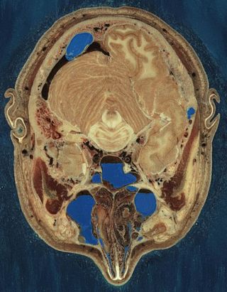
Magnetic resonance imaging of the brain uses magnetic resonance imaging (MRI) to produce high quality two-dimensional or three-dimensional images of the brain and brainstem as well as the cerebellum without the use of ionizing radiation (X-rays) or radioactive tracers.
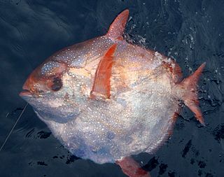
Lampris guttatus, commonly known as the opah, cravo, moonfish, kingfish, and Jerusalem haddock, is a large, colorful, deep-bodied pelagic lampriform fish belonging to the family Lampridae, which comprises the genus Lampris.
The following outline is provided as an overview of and topical guide to brain mapping:

An MRI pulse sequence in magnetic resonance imaging (MRI) is a particular setting of pulse sequences and pulsed field gradients, resulting in a particular image appearance.
