Related Research Articles

The retina is the innermost, light-sensitive layer of tissue of the eye of most vertebrates and some molluscs. The optics of the eye create a focused two-dimensional image of the visual world on the retina, which translates that image into electrical neural impulses to the brain to create visual perception. The retina serves a function analogous to that of the film or image sensor in a camera.

Eyes are organs of the visual system. They provide animals with vision, the ability to receive and process visual detail, as well as enabling several photo response functions that are independent of vision. Eyes detect light and convert it into electro-chemical impulses in neurons. In higher organisms, the eye is a complex optical system which collects light from the surrounding environment, regulates its intensity through a diaphragm, focuses it through an adjustable assembly of lenses to form an image, converts this image into a set of electrical signals, and transmits these signals to the brain through complex neural pathways that connect the eye via the optic nerve to the visual cortex and other areas of the brain. Eyes with resolving power have come in ten fundamentally different forms, and 96% of animal species possess a complex optical system. Image-resolving eyes are present in molluscs, chordates and arthropods.

Mantis shrimp, or stomatopods, are carnivorous marine crustaceans of the order Stomatopoda, branching from other members of the class Malacostraca around 340 million years ago. Mantis shrimps typically grow to around 10 cm (3.9 in) in length, while a few can reach up to 38 cm (15 in). A mantis shrimp's carapace covers only the rear part of the head and the first four segments of the thorax. Varieties range in color from shades of brown to vivid colors, with more than 450 species of mantis shrimps being known. They are among the most important predators in many shallow, tropical and subtropical marine habitats. However, despite being common, they are poorly understood, as many species spend most of their lives tucked away in burrows and holes.

Color vision is an ability of animals to perceive differences between light composed of different wavelengths independently of light intensity. Color perception is a part of the larger visual system and is mediated by a complex process between neurons that begins with differential stimulation of different types of photoreceptors by light entering the eye. Those photoreceptors then emit outputs that are propagated through many layers of neurons and then ultimately to the brain. Color vision is found in many animals and is mediated by similar underlying mechanisms with common types of biological molecules and a complex history of evolution in different animal taxa. In primates, color vision may have evolved under selective pressure for a variety of visual tasks including the foraging for nutritious young leaves, ripe fruit, and flowers, as well as detecting predator camouflage and emotional states in other primates.

A photoreceptor cell is a specialized type of neuroepithelial cell found in the retina that is capable of visual phototransduction. The great biological importance of photoreceptors is that they convert light into signals that can stimulate biological processes. To be more specific, photoreceptor proteins in the cell absorb photons, triggering a change in the cell's membrane potential.

The lagoon triggerfish, also known as the blackbar triggerfish, the Picasso triggerfish, or the Picassofish, is a triggerfish, up to 30 cm in length, found on reefs in the Indo-Pacific region.

Tetrachromacy is the condition of possessing four independent channels for conveying color information, or possessing four types of cone cell in the eye. Organisms with tetrachromacy are called tetrachromats.

Trichromacy or trichromatism is the possessing of three independent channels for conveying color information, derived from the three different types of cone cells in the eye. Organisms with trichromacy are called trichromats.

Rod cells are photoreceptor cells in the retina of the eye that can function in lower light better than the other type of visual photoreceptor, cone cells. Rods are usually found concentrated at the outer edges of the retina and are used in peripheral vision. On average, there are approximately 92 million rod cells in the human retina. Rod cells are more sensitive than cone cells and are almost entirely responsible for night vision. However, rods have little role in color vision, which is the main reason why colors are much less apparent in dim light.
In visual physiology, adaptation is the ability of the retina of the eye to adjust to various levels of light. Natural night vision, or scotopic vision, is the ability to see under low-light conditions. In humans, rod cells are exclusively responsible for night vision as cone cells are only able to function at higher illumination levels. Night vision is of lower quality than day vision because it is limited in resolution and colors cannot be discerned; only shades of gray are seen. In order for humans to transition from day to night vision they must undergo a dark adaptation period of up to two hours in which each eye adjusts from a high to a low luminescence "setting", increasing sensitivity hugely, by many orders of magnitude. This adaptation period is different between rod and cone cells and results from the regeneration of photopigments to increase retinal sensitivity. Light adaptation, in contrast, works very quickly, within seconds.
Dichromacy is the state of having two types of functioning color receptors, called cone cells, in the eyes. Organisms with dichromacy are called dichromats. Dichromats can match any color they see with a mixture of no more than two pure spectral lights. By comparison, trichromats can perceive colors made of up to three pure spectral lights, and tetrachromats can perceive colors made of four.

Monochromacy is the ability of organisms or machines to perceive only light intensity, without respect to spectral composition (color). Organisms with monochromacy are called monochromats.

Opsins are a group of proteins made light-sensitive via the chromophore retinal found in photoreceptor cells of the retina. Five classical groups of opsins are involved in vision, mediating the conversion of a photon of light into an electrochemical signal, the first step in the visual transduction cascade. Another opsin found in the mammalian retina, melanopsin, is involved in circadian rhythms and pupillary reflex but not in vision.

The Young–Helmholtz theory, also known as the trichromatic theory, is a theory of trichromatic color vision – the manner in which the visual system gives rise to the phenomenological experience of color. In 1802, Young postulated the existence of three types of photoreceptors in the eye, each of which was sensitive to a particular range of visible light.

Many researchers have found the evolution of the eye attractive to study because the eye distinctively exemplifies an analogous organ found in many animal forms. Simple light detection is found in bacteria, single-celled organisms, plants and animals. Complex, image-forming eyes have evolved independently several times.

The evolution of color vision in primates is unique compared to most eutherian mammals. A remote vertebrate ancestor of primates possessed tetrachromacy, but nocturnal, warm-blooded, mammalian ancestors lost two of four cones in the retina at the time of dinosaurs. Most teleost fish, reptiles and birds are therefore tetrachromatic while most mammals are strictly dichromats, the exceptions being some primates and marsupials, who are trichromats, and many marine mammals, who are monochromats.

Vision is the most important sense for birds, since good eyesight is essential for safe flight. Birds have a number of adaptations which give visual acuity superior to that of other vertebrate groups; a pigeon has been described as "two eyes with wings". Birds likely being descendents of theropod dinosaurs, the avian eye resembles that of other reptiles, with ciliary muscles that can change the shape of the lens rapidly and to a greater extent than in the mammals. Birds have the largest eyes relative to their size in the animal kingdom, and movement is consequently limited within the eye's bony socket. In addition to the two eyelids usually found in vertebrates, it is protected by a third transparent movable membrane. The eye's internal anatomy is similar to that of other vertebrates, but has a structure, the pecten oculi, unique to birds.

Vision is an important sensory system for most species of fish. Fish eyes are similar to the eyes of terrestrial vertebrates like birds and mammals, but have a more spherical lens. Birds and mammals normally adjust focus by changing the shape of their lens, but fish normally adjust focus by moving the lens closer to or further from the retina. Fish retinas generally have both rod cells and cone cells, and most species have colour vision. Some fish can see ultraviolet and some are sensitive to polarised light.
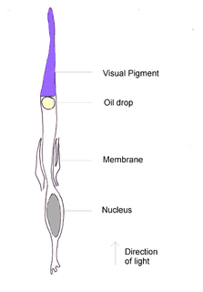
Oil droplets are found in the eyes of some animals, being located in the photoreceptor cells. They are especially common in the eyes of diurnal reptiles and birds, though are present in other taxa such as lungfish. They are found in cone cells far more often than in rods, suggesting a role in colour vision. Occurrence in rod cells may imply that they have been modified from a cone cell ancestor. They occasionally occur in double cones/double rods. Some oil droplets are coloured, while others appear colourless. They are located in the cone inner segment, where they intercept and filter light before it can pass through to the cone outer segment where the visual pigment is.
The evolution of human colour vision in Homo sapiens produced a trichromatic view of the world in comparison to a majority of other mammals that only have a dichromatic view. Early human ancestors are believed to have viewed the world using UV vision as far back as 90 million years ago. It is thought that the shift to trichromatic vision capabilities and the ability to see blue light have evolved as an adaptive trait over time.
References
- 1 2 Marchiafava, P.L. (1985). "Cell coupling in double cones of the fish retina". Proceedings of the Royal Society of London B . 226 (1243): 211–215. Bibcode:1985RSPSB.226..211M. doi:10.1098/rspb.1985.0091. S2CID 85331839.
- 1 2 3 Pignatelli, V.; Champ, C.; Marshall, J.; Vorobyev, M. (2010). "Double cones are used for colour discrimination in the reef fish, Rhinecanthus aculeatus". Biology Letters . The Royal Society. 6 (4): 537–539. doi:10.1098/rsbl.2009.1010. PMC 2936199 . PMID 20129950.
- 1 2 3 Bowmaker, J. (1990). "Visual pigments of fishes". In Douglas, R; Djamgoz, M. (eds.). The Visual System of Fish. Chapman and Hall. p. 87.
- ↑ Downing, J; Djamgoz, M; Bowmaker, J (1986). "Photoreceptors of cyprinid fish: morphological and spectral characteristics". Journal of Comparative Physiology A. 159: 859–868. doi:10.1007/bf00603739. S2CID 21456736.