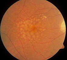
Macular hard drusen in the right eye. 65-year-old diabetic woman.
Drusen, from the German word for node or geode (singular, "Druse"), are tiny yellow or white accumulations of extracellular material that build up between Bruch's membrane and the retinal pigment epithelium of the eye. The presence of a few small ("hard") drusen is normal with advancing age, and most people over 40 have some hard drusen.[1] However, the presence of larger and more numerous drusen in the macula is a common early sign of age-related macular degeneration (AMD).
Drusen associated with aging and macular degeneration are distinct from another clinical entity, optic disc drusen, which is present on the optic nerve head.[2] Both age-related drusen and optic disc drusen can be observed by ophthalmoscopy. Optical coherence tomography scans of the orbits or head, calcification at the head of the optic nerve without change in size of globe strongly suggests drusen in a middle-age or elderly patient.
Whether drusen promote AMD or are symptomatic of an underlying process that causes both drusen and AMD is not known, but they are indicators of increased risk of the complications of AMD.[3]
'Hard drusen' may coalesce into 'soft drusen' which is a manifestation of macular degeneration.[4]
Pathophysiology
Around 1850, three authors, Carl Wedl, Franciscus Donders, and Heinrich Müller, gave drusen different labels. Drusen, the hallmark of AMD, were first described in 1854 by Wedl.[5] Wedl named them colloid bodies of the choroid and thought that they were incompletely developed cells. Franciscus Donders[6] called them "Colloidkugeln" (colloid spheres). Later, Heinrich Müller named them by the German word for geode, based on their glittering appearance.[7] He was convinced that drusen originated from the nuclei of the pigment cells, which he believed to belong to the choroid.[8] In view of their location between the retinal pigment epithelium (RPE) and its vascular supply, the choriocapillaris, it is possible that drusen deprive the RPE and photoreceptor cells of oxygen and nutrients. In some cases, drusen develop above the so-called pillars of the choriocapillaris that is the area between two micro vessels;[9] although important variations are observed between different subtypes of AMD.
The source of the proteins and lipids in drusen is also not clear, with potential contributions by both the RPE and the choroid. Several trace elements are present in drusen,[10] probably the most concentrated being zinc.[11] The protein composition of drusen includes apolipoproteins and oxidized proteins, likely originating from blood, RPE, and photoreceptors.[12] Drusen composition also includes members of the complement system. Zinc in drusen has been suggested to play a role in drusen formation by precipitating and inhibiting the elements of the complement cascade, especially complement factor H.[11]
The presence of molecules that regulate inflammation in drusen has led some investigators to conclude that these deposits are product of the immune system.[13]
Diagnosis
Usually being asymptomatic, drusen are typically found during routine eye exams where the pupils have been dilated.[14]
Treatment
Laser treatment of drusen has been studied. While it is possible to eliminate drusen with this treatment strategy, it has been shown that this fails to reduce the risk of developing the choroidal neovascularisation which causes the blindness associated with age-related macular degeneration.[15]
↑ Wedl C. Grundzüge der pathologischen Histologie Wien: Gerold, C; 1854
↑ Donders FC (March 1855). "Beitrage zur pathologischen Anatomie des Auges". Graefes Arch Clin Exp Ophthalmol. 1 (2): 106–18. doi:10.1007/BF02720791. S2CID45074362.
↑ Müller H (January 1856). "Anatomische Beiträge zur Ophthalmologie – Untersuchungen über die Glashäute des Auges, insbesondere die Glaslamelle der Chorioidea und ihre senilen Veränderungen". Graefes Arch Clin Exp Ophthalmol. 2 (2): 1–69. doi:10.1007/BF02720657. S2CID41644271.
↑ de Jong PT. A Historical Analysis of the Quest for the Origins of Aging Macula Disorder, the Tissues Involved, and Its Terminology. Ophthalmol Eye Dis. 2016;8(Suppl 1):5-14. Published 2016 Nov 1. doi:10.4137/OED.S40523
↑ van der Schaft TL, de Bruijn WC, Mooy CM, Ketelaars DA, de Jong PT (March 1992). "Element analysis of the early stages of age-related macular degeneration". Arch. Ophthalmol. 110 (3): 389–94. doi:10.1001/archopht.1992.01080150087034. PMID1543459.
1 2 Lengyel I, Flinn JM, Peto T, etal. (April 2007). "High concentration of zinc in sub-retinal pigment epithelial deposits". Exp. Eye Res. 84 (4): 772–80. doi:10.1016/j.exer.2006.12.015. PMID17313944.
↑ Hageman GS, Luthert PJ, Victor Chong NH, Johnson LV, Anderson DH, Mullins RF (November 2001). "An integrated hypothesis that considers drusen as biomarkers of immune-mediated processes at the RPE-Bruch's membrane interface in aging and age-related macular degeneration". Prog Retin Eye Res. 20 (6): 705–32. doi:10.1016/S1350-9462(01)00010-6. PMID11587915. S2CID7737763.
Mishra S, Goel S, Roy SS, Garg B, Parvin M, Saurabh K, Roy R. Multimodal imaging characteristics of refractile drusen. Indian journal of ophthalmology. 2019 Jan;67(1):128.
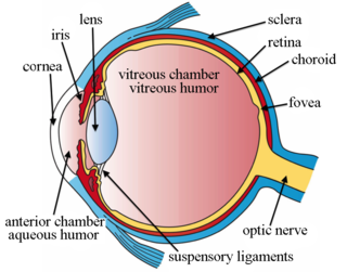
The macula (/ˈmakjʊlə/) or macula lutea is an oval-shaped pigmented area in the center of the retina of the human eye and in other animals. The macula in humans has a diameter of around 5.5 mm (0.22 in) and is subdivided into the umbo, foveola, foveal avascular zone, fovea, parafovea, and perifovea areas.
The choroid, also known as the choroidea or choroid coat, is a part of the uvea, the vascular layer of the eye. It contains connective tissues, and lies between the retina and the sclera. The human choroid is thickest at the far extreme rear of the eye, while in the outlying areas it narrows to 0.1 mm. The choroid provides oxygen and nourishment to the outer layers of the retina. Along with the ciliary body and iris, the choroid forms the uveal tract.
This is a partial list of human eye diseases and disorders.
Macular degeneration, also known as age-related macular degeneration, is a medical condition which may result in blurred or no vision in the center of the visual field. Early on there are often no symptoms. Over time, however, some people experience a gradual worsening of vision that may affect one or both eyes. While it does not result in complete blindness, loss of central vision can make it hard to recognize faces, drive, read, or perform other activities of daily life. Visual hallucinations may also occur.
Bruch's membrane or lamina vitrea is the innermost layer of the choroid of the eye. It is also called the vitreous lamina or Membrane vitriae, because of its glassy microscopic appearance. It is 2–4 μm thick.
A cone dystrophy is an inherited ocular disorder characterized by the loss of cone cells, the photoreceptors responsible for both central and color vision.
The pigmented layer of retina or retinal pigment epithelium (RPE) is the pigmented cell layer just outside the neurosensory retina that nourishes retinal visual cells, and is firmly attached to the underlying choroid and overlying retinal visual cells.
Sattler's layer, named after Hubert Sattler, an Austrian ophthalmologist, is one of five layers of medium-diameter blood vessels of the choroid, and a layer of the eye. It is situated between the Bruch's membrane, choriocapillaris below, and the Haller's layer and suprachoroidea above, respectively. The origin seems to be related to a continuous differentiation throughout the growth of the tissue and even further differentiation during adulthood.
Optic disc drusen (ODD) are globules of mucoproteins and mucopolysaccharides that progressively calcify in the optic disc. They are thought to be the remnants of the axonal transport system of degenerated retinal ganglion cells. ODD have also been referred to as congenitally elevated or anomalous discs, pseudopapilledema, pseudoneuritis, buried disc drusen, and disc hyaline bodies.
Choroidal neovascularization (CNV) is the creation of new blood vessels in the choroid layer of the eye. Choroidal neovascularization is a common cause of neovascular degenerative maculopathy commonly exacerbated by extreme myopia, malignant myopic degeneration, or age-related developments.
A maculopathy is any pathological condition of the macula, an area at the centre of the retina that is associated with highly sensitive, accurate vision.
Intraocular hemorrhage is bleeding inside the eye. Bleeding can occur from any structure of the eye where there is vasculature or blood flow, including the anterior chamber, vitreous cavity, retina, choroid, suprachoroidal space, or optic disc.
Jeffrey W. Berger was an American vitreoretinal surgeon and engineer.
Serpiginous choroiditis, also known as geographic helicoid peripapillary choroidopathy (GHPC), is a rare, chronic, progressive, and recurrent bilateral inflammatory disease involving the retinal pigment epithelium (RPE), the choriocapillaries, and the choroid. It affects adult men and women equally in the second to seventh decades of life.
Joan Whitten Miller is a Canadian-American ophthalmologist and scientist who has made notable contributions to the treatment and understanding of eye disorders. She is credited for developing photodynamic therapy (PDT) with verteporfin (Visudyne), the first pharmacologic therapy for retinal disease. She also co-discovered the role of vascular endothelial growth factor (VEGF) in eye disease and demonstrated the therapeutic potential of VEGF inhibitors, forming the scientific basis of anti-VEGF therapy for age-related macular degeneration (AMD), diabetic retinopathy, and related conditions.
Geographic atrophy (GA), also known as atrophic age-related macular degeneration (AMD) or advanced dry AMD, is an advanced form of age-related macular degeneration that can result in the progressive and irreversible loss of retinal tissue (photoreceptors, retinal pigment epithelium, choriocapillaris) which can lead to a loss of visual function over time. It is estimated that GA affects over 5 million people worldwide and approximately 1 million patients in the US, which is similar to the prevalence of neovascular (wet) AMD, the other advanced form of the disease.
Pachychoroid disorders of the macula represent a group of diseases affecting the central part of the retina of the eye, the macula. Due to thickening and congestion of the highly vascularized layer underneath the macula, the choroid, damage to the retinal pigment epithelium and the retinal photoreceptor cells ensues. This leads to impaired vision. The best known representative of the pachychoroid disease spectrum, central serous chorioretinopathy, is the fourth most common cause of irreversible damage to the macula:.
Indocyanine green angiography (ICGA) is a diagnostic procedure used to examine choroidal blood flow and associated pathology. Indocyanine green (ICG) is a water soluble cyanine dye which shows fluorescence in near-infrared (790–805 nm) range, with peak spectral absorption of 800-810 nm in blood. The near infrared light used in ICGA penetrates ocular pigments such as melanin and xanthophyll, as well as exudates and thin layers of sub-retinal vessels. Age-related macular degeneration is the third main cause of blindness worldwide, and it is the leading cause of blindness in industrialized countries. Indocyanine green angiography is widely used to study choroidal neovascularization in patients with exudative age-related macular degeneration. In nonexudative AMD, ICGA is used in classification of drusen and associated subretinal deposits.
Stem cell therapy for macular degeneration is the use of stem cells to heal, replace dead or damaged cells of the macula in the retina. Stem cell based therapies using bone marrow stem cells as well as retinal pigment epithelial transplantation are being studied. A number of trials have occurred in humans with encouraging results.
Polypoidal choroidal vasculopathy (PCV) is an eye disease primarily affecting the choroid. It may cause sudden blurring of vision or a scotoma in the central field of vision. Since Indocyanine green angiography gives better imaging of choroidal structures, it is more preferred in diagnosing PCV. Treatment options of PCV include careful observation, photodynamic therapy, thermal laser, intravitreal injection of anti-VEGF therapy, or combination therapy.
This page is based on this Wikipedia article Text is available under the CC BY-SA 4.0 license; additional terms may apply. Images, videos and audio are available under their respective licenses.