National Eye Institute

Last updated
National Eye Institute
AbbreviationNEI
Formation1968
TypeU.S. government agency
Legal statusActive
Purpose"...eliminate vision loss and improve quality of life through vision research"
Headquarters Bethesda, Maryland
Region served
United States
Official language
English
Director
Michael F. Chiang
Parent organization
National Institutes of Health
Affiliations United States Public Health Service
Website www.nei.nih.gov

The National Eye Institute (NEI) is part of the U.S. National Institutes of Health (NIH), an agency of the U.S. Department of Health and Human Services. The mission of NEI is "to eliminate vision loss and improve quality of life through vision research." [1] NEI consists of two major branches for research: an extramural branch that funds studies outside NIH and an intramural branch that funds research on the NIH campus in Bethesda, Maryland. Most of the NEI budget funds extramural research.

Contents

NEI was established in 1968 as the nation's leading supporter of eye health and vision research projects. These projects include basic science research into the fundamental biology of the eye and the visual system. NEI also funds translational and clinical research aimed at developing and testing therapies for eye diseases and disorders. This research is focused on developing therapies for leading causes of vision loss including glaucoma, diabetic retinopathy, age-related macular degeneration (AMD), cataract, myopia and amblyopia. NEI also funds research on many other causes of vision loss including retinitis pigmentosa, uveitis, retinal detachment, and rare eye diseases and disorders.

Since its founding, NEI has supported the work of several Nobel Prize recipients, including Roger Y. Tsien (2008); Peter Agre (2003); David H. Hubel (1981); and Torsten Wiesel (1981).

History

NEI Director Michael F. Chiang Michael F. Chiang, M.D., Director, National Eye Institute, NIH (50646205476).jpg
NEI Director Michael F. Chiang

National Institute of Neurological Disease and Blindness, 1950 to 1968

Before 1968, vision research at NIH was funded and overseen by the National Institute of Neurological Disease and Blindness [2] (now known as the National Institute of Neurological Disorders and Stroke), which was established in 1950, after President Harry S. Truman signed the Omnibus Medical Research Act. [2] This bill marked the beginning of vision research at the federal level.

Organizing, structuring, and separating vision and neurological research was a challenge at National Institute of Neurological Disease and Blindness. In its early years, securing funding was difficult. [3] The institute established an Ophthalmology Branch, which served primarily as an ophthalmic consultation service for NIH. [2] Ophthalmic research grew slowly throughout the 1950s and early 1960s, producing results despite small budgets. [2] One notable example was the study that identified the cause of retrolental fibroplasia (now known as retinopathy of prematurity, the leading cause of blindness among children at the time. [2]

Despite this progress, some prominent members of the vision research community asserted that too many important proposals for ophthalmic research were not granted funding. They also emphasized that ophthalmology could stand on its own as a discrete academic discipline beyond surgery and neurology. [3] [4] This prompted some leading academic ophthalmologists and vision community supporters to campaign for a separate institute focused solely on vision research. [3] These advocates included Bernard Becker, M.D.; A. Edward Maumenee, M.D.; David Glendenning Cogan, M.D.; Frank Newell, M.D.; Michael J. Hogan, M.D.; Frank C. Winter, M.D.; John M. McLean, M.D.; and Jules Stein, M.D.

The lobbying campaign for a separate ophthalmology-focused institute began in earnest in the mid-1960s and culminated in President Lyndon B. Johnson signing legislation creating NEI as part of NIH. [3] NEI was established on August 16, 1968, as the United States' first civilian governmental body focused on eye diseases, eye disorders, and vision research. [4]

NEI, 1968 to present

NEI officially began operating on December 26, 1968, and the first meeting of the National Advisory Eye Council occurred on April 3, 1969. [4] The first director of NEI, Carl Kupfer, was appointed on January 11, 1970. [4]  

From 1970 to Kupfer's retirement in 2000, NEI's budget grew from $24 million to over $500 million.[3] Kupfer expanded NEI's vision research program to focus not only on the eyes but on the entire visual system, including visual processing in the brain.  

In June 2001, Paul A. Sieving, joined NEI as its second director and served until July 2019. [5]

Under Sieving's directorship, NEI established the Audacious Goals Initiative for Regenerative Medicine. The initiative is a strategic research effort to replace cells of the retina that have been damaged by disease or injury and to restore their connections to the visual centers of the brain. Success will mean new approaches to prevent and even reverse vision loss caused by diseases such as AMD and glaucoma. [6]

Past directors

Directors dating to 1970 [7]

PortraitDirectorTook officeLeft office
Image of Dr. Carl Kupfer.jpg Carl KupferJanuary 11, 1970July 15, 2000
Noimage.svg Jack A. McLaughlin (acting)July 16, 2000June 16, 2001
Paul Sieving-4414 pp square.jpg Paul A. Sieving June 17, 2001July 19, 2019
Santa J. Tumminia.png Santa J. Tumminia (acting)July 20, 2019November 15, 2020

Organizational structure

Director

In November 2020, Michael F. Chiang, began serving as the third director of NEI. Chiang, a pediatric ophthalmologist, conducts research on the interface of biomedical informatics and clinical ophthalmology in areas such as retinopathy of prematurity, telehealth, artificial intelligence, electronic health records, data science, and genotypephenotype correlation.

Extramural Research Program

NEI supports extramural vision research through about 2,100 research grants and training awards to scientists at more than 150 medical centers, universities, and other institutions across the United States and worldwide. [1]

The NEI extramural research program is organized by anatomy and disease around core areas: retina; cornea; lens and cataract; glaucoma and optic neuropathy; strabismus, amblyopia, and visual processing; and vision rehabilitation. These core areas reflect clinical divisions of most ophthalmology and optometry departments.

In addition to these core program areas, the NEI Strategic Plan Vision for the Future 2021-2025 [8] outlines seven cross-cutting areas of emphasis: genetics, neuroscience, immunology, regenerative medicine, data science, quality of life, and public health and health disparities. These areas emphasize the methodological expertise required to address challenges across the entire visual system and facilitate translation of promising findings into clinical care and population health.

Intramural Research Program

NEI's Intramural Research Program is part of the NIH Intramural Research Program, which conducts eye and vision research on the NIH campus in Bethesda, Maryland.

Research Initiatives and Offices

In support of its extramural and intramural activities, the NEI has established several offices and research initiatives to facilitate oversight and collaboration in specific areas of emphasis.

Research achievements

NEI-supported research has contributed important knowledge about the cause, progression, and treatment of many eye diseases. Some notable examples are described below.

AMD typically happens when aging damages the macula. It is a leading cause of vision loss for older adults. It can blur the sharp central vision needed to read, see faces, and do close-up work. NEI has supported several studies investigating treatments for AMD, including:

Although AREDS2 found no overall additional benefits, it did show that two groups of participants had improved results: participants who took the AREDS formulation with no beta-carotene and participants with very low initial levels of lutein and zeaxanthin in their diets. [10]

Diabetic retinopathy

Diabetic retinopathy is an eye condition that can cause vision loss and blindness in people with diabetes. NEI has supported several studies on the treatment of diabetic retinopathy, including:

Glaucoma

Glaucoma refers to a group of eye diseases that damage the optic nerve and cause vision loss and blindness. Open-angle glaucoma is the most common form of glaucoma in the United States. Most clinical trials focus on managing open-angle glaucoma by reducing intraocular pressure (IOP), the only known modifiable risk factor for glaucoma. NEI has supported several studies, including:

Amblyopia (lazy eye)

Amblyopia is a type of visual impairment that occurs when the brain does not recognize visual signals from one eye and favors the other eye. NEI has supported research into effective treatment for amblyopia, including:

Corneal stromal keratitis

Corneal stromal keratitis or herpetic simplex keratitis is inflammation of the cornea caused by herpes infection of the eye. NEI-funded research led to a breakthrough in treatment for this condition:

Optic nerve diseases

Optic nerve diseases, like optic neuropathy and optic neuritis, can damage the connection between the eye and the visual processing centers of the brain and cause vision loss. NEI has supported studies on the treatment of optic nerve disease, including:

Retinopathy of prematurity

Retinopathy of prematurity happens when abnormal blood vessels grow in the retina and cause vision loss and blindness in babies who are premature or who weigh less than 3 pounds at birth. NEI has supported studies investigating the treatment and progression of retinopathy of prematurity, including:

Retinitis pigmentosa

Retinitis pigmentosa refers to a group of genetic eye diseases that cause cells in the retina to degenerate, leading to impaired night vision and loss of peripheral vision. NEI has supported research into therapies that slow disease progression, including:

Uveitis

Uveitis is inflammation of the uvea, the middle layer of the eye between the sclera and the retina. NEI has supported several studies to examine the causes of uveitis, including:

Leber congenital amaurosis

Leber congenital amaurosis is a rare inherited eye disease that impairs vision starting in infancy. NEI supported work leading to a gene therapy for one type of this disease:

In December 2017, Luxturna became the first directly administered gene therapy approved in the United States that targets a disease caused by mutations in a specific gene. It was approved for the treatment of patients with confirmed biallelic RPE65 mutation-associated retinal dystrophy that leads to vision loss and may cause complete blindness in certain patients. [45]

Technology development

NEI has invested in the development of technologies to support diagnosis and management. A few examples of this investment include:

Education and outreach activities

Part of NEI's mission is to educate health care providers, scientists, policymakers, and the public about advances in vision research and their impact on health and quality of life. This effort is led by NEI's National Eye Health Education Program.

National Eye Health Education Program

NEI created the National Eye Health Education Program to educate professionals and the public about the importance of eye health. The program partners with more than 60 national organizations representing health professionals, educators, and patients in accomplishing this mission. It also oversees public and professional education programs on diabetic eye disease, glaucoma, vision rehabilitation, special population outreach, and vision and aging—with a focus on individuals and populations at higher risk of eye health disorders, including older people, those with diabetes, Black/African American people, and Hispanic/Latino people.

The program also draws on research supported by NIH and NEI to identify other populations at risk (e.g., Asian American people, residents of rural communities) and to produce educational materials for professional and public audiences. It emphasizes the importance of early detection and timely treatment of eye disease and the benefits of vision rehabilitation. The program also aims to increase awareness among health professionals and the public of science-based health information that can be applied to preserving sight and preventing blindness.

Eye on the Future Teen Video Contest

In 2022, the NEI created the Eye on the Future Teen Video Contest to foster the next generation of American scientists. [47] The NEI awards American youth who create educational videos in three categories: "Science in your world," "Science in the field or lab," and "Science in your future." [48] One winner is selected in each category, each receiving a prize of $2,000 USD.

Contest Winners
Name of Person [49] YearAwardCategory
Thuy-Tien Tran 20231st PlaceScience in your world
Celia Cooley20231st PlaceScience in the field or lab
Mark Leschinsky 20231st PlaceScience in your future
Meenakshi Ambati2022WinnerN/A
Sanjana Kumar2022WinnerN/A

Related Research Articles

<span class="mw-page-title-main">Retinopathy</span> Medical condition

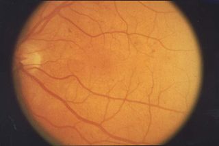
Retinopathy is any damage to the retina of the eyes, which may cause vision impairment. Retinopathy often refers to retinal vascular disease, or damage to the retina caused by abnormal blood flow. Age-related macular degeneration is technically included under the umbrella term retinopathy but is often discussed as a separate entity. Retinopathy, or retinal vascular disease, can be broadly categorized into proliferative and non-proliferative types. Frequently, retinopathy is an ocular manifestation of systemic disease as seen in diabetes or hypertension. Diabetes is the most common cause of retinopathy in the U.S. as of 2008. Diabetic retinopathy is the leading cause of blindness in working-aged people. It accounts for about 5% of blindness worldwide and is designated a priority eye disease by the World Health Organization.

<span class="mw-page-title-main">Diabetic retinopathy</span> Medical condition

Diabetic retinopathy, is a medical condition in which damage occurs to the retina due to diabetes. It is a leading cause of blindness in developed countries.

<span class="mw-page-title-main">Retinoschisis</span> Eye disease involving splitting of the retina

Retinoschisis is an eye disease characterized by the abnormal splitting of the retina's neurosensory layers, usually in the outer plexiform layer. Retinoschisis can be divided into degenerative forms which are very common and almost exclusively involve the peripheral retina and hereditary forms which are rare and involve the central retina and sometimes the peripheral retina. The degenerative forms are asymptomatic and involve the peripheral retina only and do not affect the visual acuity. Some rarer forms result in a loss of vision in the corresponding visual field.

<span class="mw-page-title-main">Macular edema</span> Medical condition

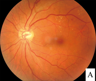
Macular edema occurs when fluid and protein deposits collect on or under the macula of the eye and causes it to thicken and swell (edema). The swelling may distort a person's central vision, because the macula holds tightly packed cones that provide sharp, clear, central vision to enable a person to see detail, form, and color that is directly in the centre of the field of view.

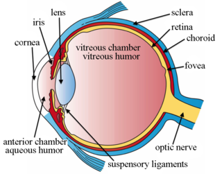
This is a partial list of human eye diseases and disorders.

<span class="mw-page-title-main">Visual impairment</span> Decreased ability to see

Visual or vision impairment is the partial or total inability of visual perception. In the absence of treatment such as corrective eyewear, assistive devices, and medical treatment – visual impairment may cause the individual difficulties with normal daily tasks including reading and walking. The terms low vision and blindness are often used for levels of impairment which are difficult or impossible to correct and significantly impact daily life. In addition to the various permanent conditions, fleeting temporary vision impairment, amaurosis fugax, may occur, and may indicate serious medical problems.

An eye care professional is an individual who provides a service related to the eyes or vision. It is any healthcare worker involved in eye care, from one with a small amount of post-secondary training to practitioners with a doctoral level of education.

<span class="mw-page-title-main">Intravitreal administration</span>

Intravitreal is a route of administration of a drug, or other substance, in which the substance is delivered into the vitreous humor of the eye. "Intravitreal" literally means "inside an eye". Intravitreal injections were first introduced in 1911 when Ohm gave an injection of air into the vitreous humor to repair a detached retina. In the mid-1940s, intravitreal injections became a standard way to administer drugs to treat endophthalmitis and cytomegalovirus retinitis.

<span class="mw-page-title-main">Dilated fundus examination</span>

Dilated fundus examination (DFE) is a diagnostic procedure that uses mydriatic eye drops to dilate or enlarge the pupil in order to obtain a better view of the fundus of the eye. Once the pupil is dilated, examiners use ophthalmoscopy to view the eye's interior, which makes it easier to assess the retina, optic nerve head, blood vessels, and other important features. DFE has been found to be a more effective method for evaluating eye health when compared to non-dilated examination, and is the best method of evaluating structures behind the iris. It is frequently performed by ophthalmologists and optometrists as part of an eye examination.

Ocular ischemic syndrome is the constellation of ocular signs and symptoms secondary to severe, chronic arterial hypoperfusion to the eye. Amaurosis fugax is a form of acute vision loss caused by reduced blood flow to the eye; it may be a warning sign of an impending stroke, as both stroke and retinal artery occlusion can be caused by thromboembolism due to atherosclerosis elsewhere in the body. Consequently, those with transient blurring of vision are advised to urgently seek medical attention for a thorough evaluation of the carotid artery. Anterior segment ischemic syndrome is a similar ischemic condition of anterior segment usually seen in post-surgical cases. Retinal artery occlusion leads to rapid death of retinal cells, thereby resulting in severe loss of vision.

<span class="mw-page-title-main">Blurred vision</span> Medical condition

Blurred vision is an ocular symptom where vision becomes less precise and there is added difficulty to resolve fine details.

<span class="mw-page-title-main">Intraocular hemorrhage</span> Medical condition

Intraocular hemorrhage is bleeding inside the eye. Bleeding can occur from any structure of the eye where there is vasculature or blood flow, including the anterior chamber, vitreous cavity, retina, choroid, suprachoroidal space, or optic disc.

<span class="mw-page-title-main">Vision disorder</span> Medical condition

A vision disorder is an impairment of the sense of vision.

<span class="mw-page-title-main">Laser coagulation</span> Procedure widely used in eye surgery

Laser coagulation or laser photocoagulation surgery is used to treat a number of eye diseases and has become widely used in recent decades. During the procedure, a laser is used to finely cauterize ocular blood vessels to attempt to bring about various therapeutic benefits.

Bascom Palmer Eye Institute is the University of Miami School of Medicine's ophthalmic care, research, and education center. The institute is based in the Health District of Miami, Florida, and has been ranked consistently as the best eye hospital and vision research center in the nation.

The Vision Institute is a research center in the Quinze-Vingts National Eye Hospital in Paris, France. It is one of several such centers in Europe on eye diseases.

<span class="mw-page-title-main">Irvine–Gass syndrome</span> Medical condition

Irvine–Gass syndrome, pseudophakic cystoid macular edema or postcataract CME is one of the most common causes of visual loss after cataract surgery. The syndrome is named in honor of S. Rodman Irvine and J. Donald M. Gass.

Rohit Varma is an Indian-American ophthalmologist and professor of ophthalmology and preventive medicine. In 2014, he was named director of the USC Eye Institute and chairman of the Department of Ophthalmology for Keck School of Medicine of USC. In March 2016, Varma was named the interim dean of the Keck School of Medicine, and in November was named dean. In October 2017, USC announced that he stepped down as dean. In October 2018, Varma became the founding director of the Southern California Eyecare and Vision Research Institute.

Diabetic papillopathy is an ocular complication of diabetes mellitus characterized by optic disc swelling and edema of optic nerve head. The condition may affect both type 1 and type 2 diabetic patients.

<span class="mw-page-title-main">Uveitic glaucoma</span> Glaucoma caused by uveitis or its treatments

Uveitic glaucoma is most commonly a progression stage of noninfectious anterior uveitis or iritis.

References

  1. 1 2 "Goals and Accomplishments | National Eye Institute". www.nei.nih.gov. Retrieved 2023-03-21.
  2. 1 2 3 4 5 "Publications - Office of NIH History and Stetten Museum" (PDF). history.nih.gov. Retrieved 2023-03-21.
  3. 1 2 3 4 "A profile of the United States Public Health Service, 1798-1948 - Digital Collections - National Library of Medicine". collections.nlm.nih.gov. Retrieved 2023-03-21.
  4. 1 2 3 4 "National Eye Institute (NEI)". National Institutes of Health (NIH). 2015-07-07. Retrieved 2023-03-21.
  5. "Statement on the retirement of Dr. Paul Sieving". National Institutes of Health (NIH). 2019-06-26. Retrieved 2023-03-21.
  6. "Audacious Goals Initiative | National Eye Institute". www.nei.nih.gov. Retrieved 2023-03-21.
  7. "NEI Directors". nih.gov. 7 July 2015.
  8. "Strategic Planning at NEI | National Eye Institute". www.nei.nih.gov. Retrieved 2023-03-21.
  9. Age-Related Eye Disease Study Research Group (2001-10-01). "A Randomized, Placebo-Controlled, Clinical Trial of High-Dose Supplementation With Vitamins C and E, Beta Carotene, and Zinc for Age-Related Macular Degeneration and Vision Loss: AREDS Report No. 8". Archives of Ophthalmology. 119 (10): 1417–1436. doi:10.1001/archopht.119.10.1417. ISSN   0003-9950. PMC   1462955 . PMID   11594942.
  10. 1 2 Age-Related Eye Disease Study 2 Research Group (15 May 2013). "Lutein + Zeaxanthin and Omega-3 Fatty Acids for Age-Related Macular Degeneration – The Age-Related Eye Disease Study 2 (AREDS2) Randomized Clinical Trial". JAMA. 309 (19): 2005–2015. doi: 10.1001/jama.2013.4997 . PMID   23644932 . Retrieved 2023-03-21.{{cite journal}}: CS1 maint: numeric names: authors list (link)
  11. CATT Research Group; Martin, D. F.; Maguire, M. G.; Ying, G. S.; Grunwald, J. E.; Fine, S. L.; Jaffe, G. J. (2011-05-19). "Ranibizumab and Bevacizumab for Neovascular Age-Related Macular Degeneration". New England Journal of Medicine. 364 (20): 1897–1908. doi:10.1056/NEJMoa1102673. ISSN   0028-4793. PMC   3157322 . PMID   21526923.
  12. Martin, Daniel F.; Maguire, Maureen G.; Fine, Stuart L.; Ying, Gui-shuang; Jaffe, Glenn J.; Grunwald, Juan E.; Toth, Cynthia; Redford, Maryann; Ferris, Frederick L. (2012-07-01). "Ranibizumab and Bevacizumab for Treatment of Neovascular Age-related Macular Degeneration: Two-Year Results". Ophthalmology. 119 (7): 1388–1398. doi:10.1016/j.ophtha.2012.03.053. ISSN   0161-6420. PMC   3389193 . PMID   22555112.
  13. Complications of Age-Related Macular Degeneration Prevention Trial Research Group (2006-11-01). "Laser Treatment in Patients with Bilateral Large Drusen: The Complications of Age-Related Macular Degeneration Prevention Trial". Ophthalmology. 113 (11): 1974–1986. doi:10.1016/j.ophtha.2006.08.015. ISSN   0161-6420. PMID   17074563.
  14. Bressler, N. M.; Bressler, S. B.; Childs, A. L.; Haller, J. A.; Hawkins, B. S.; Lewis, H.; MacCumber, M. W.; Marsh, M. J.; Redford, M.; Sternberg Jr, P.; Thomas, M. A.; Williams, G. A.; Submacular Surgery Trials (SST) Research Group (2004-11-01). "Surgery for hemorrhagic choroidal neovascular lesions of age-related macular degeneration: Ophthalmic findings: SST report no. 13". Ophthalmology. 111 (11): 1993–2006.e1. doi:10.1016/j.ophtha.2004.07.023. ISSN   0161-6420. PMC   1256022 . PMID   15522364.
  15. Hawkins, B. S.; Bressler, N. M.; Miskala, P. H.; Bressler, S. B.; Holekamp, N. M.; Marsh, M. J.; Redford, M.; Schwartz, S. D.; Sternberg Jr, P.; Thomas, M. A.; Wilson, D. J.; Submacular Surgery Trials (SST) Research Group (2004-11-01). "Surgery for subfoveal choroidal neovascularization in age-related macular degeneration: Ophthalmic findings *: SST report no. 11". Ophthalmology. 111 (11): 1967–1980. doi:10.1016/j.ophtha.2004.07.021. ISSN   0161-6420. PMC   1256024 . PMID   15522362.
  16. "Photocoagulation Treatment of Proliferative Diabetic Retinopathy: Clinical Application of Diabetic Retinopathy Study (DRS) Findings, DRS Report Number 8". Ophthalmology. 88 (7): 583–600. 1981-07-01. doi:10.1016/S0161-6420(81)34978-1. ISSN   0161-6420. PMID   7196564.
  17. "Early Vitrectomy for Severe Proliferative Diabetic Retinopathy in Eyes with Useful Vision: Results of a Randomized Trial—Diabetic Retinopathy Vitrectomy Study Report 3". Ophthalmology. 95 (10): 1307–1320. 1988-10-01. doi:10.1016/S0161-6420(88)33015-0. ISSN   0161-6420. PMID   2465517.
  18. Chew, Emily Y.; Klein, Michael L.; Murphy, Robert P.; Remaley, Nancy A.; Ferris, Frederick L. III (1995-01-01). "Effects of Aspirin on Vitreous/Preretinal Hemorrhage in Patients With Diabetes Mellitus: Early Treatment Diabetic Retinopathy Study Report No. 20". Archives of Ophthalmology. 113 (1): 52–55. doi:10.1001/archopht.1995.01100010054020. ISSN   0003-9950. PMID   7826294.
  19. "Panretinal Photocoagulation vs Intravitreous Ranibizumab for Proliferative Diabetic Retinopathy – A Randomized Clinical Trial". jamanetwork.com. Retrieved 2023-03-21.
  20. "The advanced glaucoma intervention study (AGIS): 3. baseline characteristics of black and white patients". Ophthalmology. 105 (7): 1137–1145. 1998-07-01. doi:10.1016/S0161-6420(98)97012-9. ISSN   0161-6420. PMID   9663214.
  21. "The advanced glaucoma intervention study (AGIS): 4. comparison of treatment outcomes within race: Seven-year results". Ophthalmology. 105 (7): 1146–1164. 1998-07-01. doi: 10.1016/S0161-6420(98)97013-0 . ISSN   0161-6420. PMID   9663215.
  22. Lichter, Paul R.; Musch, David C.; Gillespie, Brenda W.; Guire, Kenneth E.; Janz, Nancy K.; Wren, Patricia A.; Mills, M. P. H. Richard P. (2001-11-01). "Interim clinical outcomes in the collaborative initial glaucoma treatment study comparing initial treatment randomized to medications or surgery". Ophthalmology. 108 (11): 1943–1953. doi:10.1016/S0161-6420(01)00873-9. ISSN   0161-6420. PMID   11713061.
  23. Leske, M. Cristina; Heijl, Anders; Hussein, Mohamed; Bengtsson, Bo; Hyman, Leslie; Komaroff, Eugene (2003). "Factors for Glaucoma Progression and the Effect of Treatment – The Early Manifest Glaucoma Trial". Archives of Ophthalmology. 121 (1): 48–56. doi:10.1001/archopht.121.1.48. PMID   12523884 . Retrieved 2023-03-21.
  24. The Fluorouracil Filtering Surgery Study Group (1989-12-01). "Fluorouracil Filtering Surgery Study One-Year Follow-up". American Journal of Ophthalmology. 108 (6): 625–635. doi:10.1016/0002-9394(89)90853-2. ISSN   0002-9394. PMID   2688428.
  25. Kass, Michael A.; Heuer, Dale K.; Higginbotham, Eve J.; Johnson, Chris A.; Keltner, John L.; Miller, J. Philip; Parrish II, Richard K.; Wilson, M. Roy; Gordon, Mae O. "The Ocular Hypertension Treatment Study". jamanetwork.com. Retrieved 2023-03-21.
  26. "Amblyopia in Children Aged 7 to 17 Years". jamanetwork.com. Retrieved 2023-03-21.
  27. Chen, Angela M.; Cotter, Susan A. (2016-08-01). "The Amblyopia Treatment Studies: Implications for Clinical Practice". Advances in Ophthalmology and Optometry. 1 (1): 287–305. doi:10.1016/j.yaoo.2016.03.007. ISSN   2452-1760. PMC   5396957 . PMID   28435934.
  28. Holmes, J. M.; Kraker, R. T.; Beck, R. W.; Birch, E. E.; Cotter, S. A.; Everett, D. F.; Hertle, R. W.; Quinn, G. E.; Repka, M. X.; Scheiman, M. M.; Wallace, D. K.; Pediatric Eye Disease Investigator Group (2003-11-01). "A randomized trial of prescribed patching regimens for treatment of severe amblyopia in children". Ophthalmology. 110 (11): 2075–2087. doi:10.1016/j.ophtha.2003.08.001. ISSN   0161-6420. PMID   14597512.
  29. Bosque, Lorenzo E.; Yamarino, Cailyn R.; Salcedo, Natalia; Schneier, Andrew J.; Gold, Robert S.; Blumenfeld, Louis C.; Hunter, David G. (2021-08-01). "Evaluation of the blinq vision scanner for detection of amblyopia and strabismus". Journal of American Association for Pediatric Ophthalmology and Strabismus {JAAPOS}. 25 (4): 214.e1–214.e7. doi:10.1016/j.jaapos.2021.02.011. ISSN   1091-8531. PMID   34246763.
  30. Wilhelmus, Kirk R.; Beck, Roy W.; Moke, Pamela S.; Dawson, Chandler R.; Barron, Bruce A.; Jones, Dan B.; Kaufman, Herbert E.; Kurinij, Natalie; Stulting, R. Doyle; Sugar, Joel; Cohen, Elisabeth J.; Hyndiuk, Robert A.; Asbell, Penny A. (1998-07-30). "Acyclovir for the Prevention of Recurrent Herpes Simplex Virus Eye Disease". New England Journal of Medicine. 339 (5): 300–306. doi: 10.1056/NEJM199807303390503 . ISSN   0028-4793. PMID   9696640.
  31. "Oral Acyclovir for Herpes Simplex Virus Eye Disease". jamanetwork.com. Retrieved 2023-03-21.
  32. "Ischemic Optic Neuropathy Decompression Trial – Twenty-Four–Month Update". jamanetwork.com. Retrieved 2023-03-21.
  33. Newman, Nancy J. (2020-04-01). "The Optic Neuritis Treatment Trial". Ophthalmology. 127 (4): S172–S173. doi: 10.1016/j.ophtha.2019.09.046 . ISSN   0161-6420. PMID   32200818. S2CID   214616286.
  34. "Multicenter Trial of Cryotherapy for Retinopathy of Prematurity – Ophthalmological Outcomes at 10 Years". jamanetwork.com. Retrieved 2023-03-21.
  35. Christiansen, Stephen P.; Dobson, Velma; Quinn, Graham E.; Good, William V.; Tung, Betty; Hardy, Robert J.; Baker, John D.; Hoffman, Robert O.; Reynolds, James D.; Rychwalski, Paul J.; Shapiro, Michael J. (2010). "Progression of Type 2 to Type 1 Retinopathy of Prematurity in the Early Treatment for Retinopathy of Prematurity Study". Archives of Ophthalmology. 128 (4): 461–465. doi:10.1001/archophthalmol.2010.34. PMID   20385942 . Retrieved 2023-03-21.
  36. Reynolds, James D.; Hardy, Robert J.; Kennedy, Kathleen A.; Spencer, Rand; van Heuven, W.A.J.; Fielder, Alistair R. (1998-05-28). "Lack of Efficacy of Light Reduction in Preventing Retinopathy of Prematurity". New England Journal of Medicine. 338 (22): 1572–1576. doi: 10.1056/NEJM199805283382202 . ISSN   0028-4793. PMID   9603794.
  37. "Supplemental Therapeutic Oxygen for Prethreshold Retinopathy of Prematurity (STOP-ROP), A Randomized, Controlled Trial. I: Primary Outcomes". publications.aap.org. Retrieved 2023-03-21.
  38. "Validated System for Centralized Grading of Retinopathy of Prematurity". jamanetwork.com. Retrieved 2023-03-21.
  39. Beltran, William A.; Cideciyan, Artur V.; Iwabe, Simone; Swider, Malgorzata; Kosyk, Mychajlo S.; McDaid, Kendra; Martynyuk, Inna; Ying, Gui-Shuang; Shaffer, James; Deng, Wen-Tao; Boye, Sanford L.; Lewin, Alfred S.; Hauswirth, William W.; Jacobson, Samuel G.; Aguirre, Gustavo D. (2015-10-27). "Successful arrest of photoreceptor and vision loss expands the therapeutic window of retinal gene therapy to later stages of disease". Proceedings of the National Academy of Sciences. 112 (43): E5844-53. Bibcode:2015PNAS..112E5844B. doi: 10.1073/pnas.1509914112 . ISSN   0027-8424. PMC   4629324 . PMID   26460017.
  40. Tsui, Edmund; Rathinam, Sivakumar R.; Gonzales, John A.; Thundikandy, Radhika; Kanakath, Anuradha; Balamurugan, S.; Vedhanayaki, R.; Lim, Lyndell L.; Suhler, Eric B.; Al-Dhibi, Hassan A.; Doan, Thuy; Keenan, Jeremy; Ebert, Caleb D.; Kim, Eric; Madow, Brian (2022-06-01). "Outcomes of Uveitic Macular Edema in the First-line Antimetabolites as Steroid-Sparing Treatment Uveitis Trial". Ophthalmology. 129 (6): 661–667. doi: 10.1016/j.ophtha.2022.02.002 . ISSN   0161-6420. PMID   35143800.
  41. Writing Committee for the Multicenter Uveitis Steroid Treatment (MUST) Trial and Follow-up Study Research Group (2017-05-16). "Association Between Long-Lasting Intravitreous Fluocinolone Acetonide Implant vs Systemic Anti-inflammatory Therapy and Visual Acuity at 7 Years Among Patients With Intermediate, Posterior, or Panuveitis". JAMA. 317 (19): 1993–2005. doi:10.1001/jama.2017.5103. ISSN   0098-7484. PMC   5540027 . PMID   28477440. S2CID   11309318.
  42. Thorne, Jennifer E.; Sugar, Elizabeth A.; Holbrook, Janet T.; Burke, Alyce E.; Altaweel, Michael M.; Vitale, Albert T.; Acharya, Nisha R.; Kempen, John H.; Jabs, Douglas A. (2019-02-01). "Periocular Triamcinolone vs. Intravitreal Triamcinolone vs. Intravitreal Dexamethasone Implant for the Treatment of Uveitic Macular Edema: The PeriOcular vs. INTravitreal corticosteroids for uveitic macular edema (POINT) Trial". Ophthalmology. 126 (2): 283–295. doi:10.1016/j.ophtha.2018.08.021. ISSN   0161-6420. PMC   6348060 . PMID   30269924.
  43. Horai, Reiko; Zárate-Bladés, Carlos R.; Dillenburg-Pilla, Patricia; Chen, Jun; Kielczewski, Jennifer L.; Silver, Phyllis B.; Jittayasothorn, Yingyos; Chan, Chi-Chao; Yamane, Hidehiro; Honda, Kenya; Caspi, Rachel R. (2015-08-18). "Microbiota-Dependent Activation of an Autoreactive T Cell Receptor Provokes Autoimmunity in an Immunologically Privileged Site". Immunity. 43 (2): 343–353. doi:10.1016/j.immuni.2015.07.014. ISSN   1074-7613. PMC   4544742 . PMID   26287682.
  44. Jacobson, Samuel G.; Cideciyan, Artur V.; Roman, Alejandro J.; Sumaroka, Alexander; Schwartz, Sharon B.; Heon, Elise; Hauswirth, William W. (2015-05-14). "Improvement and Decline in Vision with Gene Therapy in Childhood Blindness". New England Journal of Medicine. 372 (20): 1920–1926. doi:10.1056/NEJMoa1412965. ISSN   0028-4793. PMC   4450362 . PMID   25936984.
  45. Commissioner, Office of the (2020-03-24). "FDA approves novel gene therapy to treat patients with a rare form of inherited vision loss". FDA. Retrieved 2023-03-21.
  46. Pinhas, Alexander; Dubow, Michael; Shah, Nishit; Chui, Toco Y.; Scoles, Drew; Sulai, Yusufu N.; Weitz, Rishard; Walsh, Joseph B.; Carroll, Joseph; Dubra, Alfredo; Rosen, Richard B. (2013). "In vivo imaging of human retinal microvasculature using adaptive optics scanning light ophthalmoscope fluorescein angiography". Biomedical Optics Express. 4 (8): 1305–1317. doi:10.1364/BOE.4.001305. PMC   3756583 . PMID   24009994.
  47. https://www.nei.nih.gov/learn-about-eye-health/campaigns/eye-future-campaign/eye-future-teen-video-contest
  48. https://portal.challenge.gov/public/previews/challenges?challenge=ae889f16-dc9f-445c-928b-4e127b8400a4&print=true
  49. https://www.nei.nih.gov/learn-about-eye-health/campaigns/eye-future-campaign/eye-future-contest-past-winners