Herpes simplex keratitis

Last updated
Herpes simplex keratitis
Other namesHerpetic keratoconjunctivitis, herpesviral keratitis
Dendritic corneal ulcer.jpg
Dendritic corneal ulcer after fluorescein staining
Specialty Ophthalmology

Herpetic simplex keratitis is a form of keratitis caused by recurrent herpes simplex virus (HSV) infection in the cornea. [1]

Contents

It begins with infection of epithelial cells on the surface of the eye and retrograde infection of nerves serving the cornea. [2] Primary infection typically presents as swelling of the conjunctiva and eyelids (blepharoconjunctivitis), accompanied by small white itchy lesions on the corneal surface. The effect of the lesions varies, from minor damage to the epithelium (superficial punctate keratitis), to more serious consequences such as the formation of dendritic ulcers. [3] Infection is unilateral, affecting one eye at a time. Additional symptoms include dull pain deep inside the eye, mild to acute dryness, and sinusitis. Most primary infections resolve spontaneously in a few weeks. Healing can be aided by the use of oral and topical antivirals.

Subsequent recurrences may be more severe, with infected epithelial cells showing larger dendritic ulceration, and lesions forming white plaques. [3] The epithelial layer is sloughed off as the dendritic ulcer grows, and mild inflammation (iritis) may occur in the underlying stroma of iris. Sensation loss occurs in lesional areas, producing generalised corneal anaesthesia with repeated recurrences. [3] Recurrence can be accompanied by chronic dry eye, low grade intermittent conjunctivitis, or chronic unexplained sinusitis. Following persistent infection the concentration of viral DNA reaches a critical limit. Antibody responses against the viral antigen expression in the stroma can trigger a massive immune response in the eye. The response may result in the destruction of the corneal stroma, [3] resulting in loss of vision due to opacification of the cornea. This is known as immune-mediated stromal keratitis.

HSV infection is very common in humans. It has been estimated that one third of the world population have recurrent infection. Keratitis caused by HSV is the most common cause of cornea-derived blindness in developed nations. Therefore, HSV infections are a large and worldwide public health problem. [4] The global incidence (rate of new disease) of herpes keratitis is roughly 1.5 million, including 40,000 new cases of severe monocular visual impairment or blindness each year. [5]

Signs and symptoms

Primary infection

herpetic blepharitis Herpes simplex blepharitis.jpg
herpetic blepharitis

Primary infection most commonly manifests as blepharoconjunctivitis i.e. infection of lids and conjunctiva that heals without scarring. Lid vesicles and conjunctivitis are seen in primary infection. Corneal involvement is rarely seen in primary infection.

Recurrent eye infection

Recurrent herpes of the eye is caused by reactivation of the virus in a latently infected sensory ganglion, transport of the virus down the nerve axon to sensory nerve endings, and subsequent infection of ocular surface. The following classification of herpes simplex keratitis is important for understanding this disease:

Dendritic ulcer (Epithelial keratitis)

Fluorescein-stained cornea: geographic epithelial defects Herpes simplex geographic corneal ulcer.jpeg
Fluorescein-stained cornea: geographic epithelial defects

This classic herpetic lesion consists of a linear branching corneal ulcer (dendritic ulcer). During eye exam the defect is examined after staining with fluorescein dye. The underlying cornea has minimal inflammation.

Patients with epithelial keratitis complain of foreign-body sensation, light sensitivity, redness and blurred vision.

Focal or diffuse reduction in corneal sensation develops following recurrent epithelial keratitis.

In immune deficient patients or with the use of corticosteroids the ulcer may become large and in these cases it is called geographic ulcer. [6]

Disciform keratitis (Endothelial keratitis)

Endothelial keratitis manifests a central endothelitis in a disc-shaped manner. Longstanding corneal edema leads to permanent scarring and is the major cause of decreased vision associated with HSV.

Localized endothelitis (localized inflammation of corneal endothelial layer) is the cause of disciform keratitis.

Other forms

Cause

HSV is a double-stranded DNA virus that has icosahedral capsid. HSV-1 infections are found more commonly in the oral area and HSV-2 in the genital area. Ocular herpes simplex is usually caused by HSV-1. [7] [8]

Diagnosis

Impression cytology smear stained by Papanicolaou stain shows the presence of a multinucleated giant cell Cytology corneal herpes multinucleated cell.jpeg
Impression cytology smear stained by Papanicolaou stain shows the presence of a multinucleated giant cell

A specific clinical diagnosis of HSV as the cause of dendritic keratitis can usually be made by ophthalmologists and optometrists based on the presence of characteristic clinical features. Diagnostic testing is seldom needed because of its classic clinical features and is not useful in stromal keratitis as there is usually no live virus. Laboratory tests are indicated in complicated cases when the clinical diagnosis is uncertain and in all cases of suspected neonatal herpes infection: [4] [6]

Treatment

Treatment of herpes of the eye is different based on its presentation: epithelial keratitis is caused by live virus while stromal disease is an immune response and metaherpetic ulcer results from inability of the corneal epithelium to heal. But on the viral aciclovir eye ointment q.i.d can be used together a systemic antiviral drug t.d.s for 10/7. : [4] [6]

Epithelial keratitis

Epithelial keratitis is treated with topical antivirals, which are very effective with low incidence of resistance. Treatment of the disease with topical antivirals generally should be continued for 10–14 days. Aciclovir ophthalmic ointment and Trifluridine eye drops have similar effectiveness but are more effective than Idoxuridine and Vidarabine eye drops. [9] Oral acyclovir is as effective as topical antivirals for treating epithelial keratitis, and it has the advantage of no eye surface toxicity. For this reason, oral therapy is preferred by some ophthalmologists.

Ganciclovir and brivudine treatments were found to be equally as effective as acyclovir in a systematic review. [9]

Valacyclovir, a pro-drug of acyclovir likely to be just as effective for ocular disease, can cause thrombotic thrombocytopenic purpura/Hemolytic-uremic syndrome in severely immunocompromised patients such as those with AIDS; thus, it must be used with caution if the immune status is unknown.

Topical corticosteroids are contraindicated in the presence of active herpetic epithelial keratitis; patients with this disease who are using systemic corticosteroids for other indications should be treated aggressively with systemic antiviral therapy.

The effect of interferon with an antiviral agent or an antiviral agent with debridement needs further assessment. [9]

Stromal keratitis

Herpetic stromal keratitis is treated initially with prednisolone drops every 2 hours accompanied by a prophylactic antiviral drug: either topical antiviral or an oral agent such as acyclovir or valacyclovir. The prednisolone drops are tapered every 1–2 weeks depending on the degree of clinical improvement. Topical antiviral medications are not absorbed by the cornea through an intact epithelium, but orally administered acyclovir penetrates an intact cornea and anterior chamber. In this context, oral acyclovir might benefit the deep corneal inflammation of disciform keratitis. [6]

Metaherpetic ulcer

Treatment includes artificial tears and eye lubricants, stopping toxic medications, performing punctal occlusion, bandage contact lens and amniotic membrane transplant. These measures intend to improve corneal epithelial healing.

Antiviral medication may reduce the risk of HSV keratitis recurring in people having a graft due to HSV infection and may improve the chances of graft survival. [10]

Related Research Articles

<span class="mw-page-title-main">Keratitis</span> Medical condition

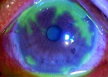
Keratitis is a condition in which the eye's cornea, the clear dome on the front surface of the eye, becomes inflamed. The condition is often marked by moderate to intense pain and usually involves any of the following symptoms: pain, impaired eyesight, photophobia, red eye and a 'gritty' sensation.

<span class="mw-page-title-main">Aciclovir</span> Antiviral medication used against herpes, chickenpox and shingles

Aciclovir (ACV), also known as acyclovir, is an antiviral medication. It is primarily used for the treatment of herpes simplex virus infections, chickenpox, and shingles. Other uses include prevention of cytomegalovirus infections following transplant and severe complications of Epstein–Barr virus infection. It can be taken by mouth, applied as a cream, or injected.

<span class="mw-page-title-main">Herpetic whitlow</span> Medical condition

A herpetic whitlow is a lesion (whitlow), typically on a finger or thumb, caused by the herpes simplex virus (HSV). Occasionally infection occurs on the toes or on the nail cuticle. Herpes whitlow can be caused by infection by HSV-1 or HSV-2. HSV-1 whitlow is often contracted by health care workers that come in contact with the virus; it is most commonly contracted by dental workers and medical workers exposed to oral secretions. It is also often observed in thumb-sucking children with primary HSV-1 oral infection (autoinoculation) prior to seroconversion, and in adults aged 20 to 30 following contact with HSV-2-infected genitals.

<span class="mw-page-title-main">Herpetic gingivostomatitis</span> Medical condition

Gingivostomatitis is a combination of gingivitis and stomatitis, or an inflammation of the oral mucosa and gingiva. Herpetic gingivostomatitis is often the initial presentation during the first ("primary") herpes simplex infection. It is of greater severity than herpes labialis which is often the subsequent presentations. Primary herpetic gingivostomatitis is the most common viral infection of the mouth.

<span class="mw-page-title-main">Corneal ulcers in animals</span> Veterinary inflammatory condition of the cornea

A corneal ulcer, or ulcerative keratitis, is an inflammatory condition of the cornea involving loss of its outer layer. It is very common in dogs and is sometimes seen in cats. In veterinary medicine, the term corneal ulcer is a generic name for any condition involving the loss of the outer layer of the cornea, and as such is used to describe conditions with both inflammatory and traumatic causes.

<span class="mw-page-title-main">Genital herpes</span> Infection by herpes simplex viruses of the genitals

Genital herpes is an infection by the herpes simplex virus (HSV) of the genitals. Most people either have no or mild symptoms and thus do not know they are infected. When symptoms do occur, they typically include small blisters that break open to form painful ulcers. Flu-like symptoms, such as fever, aching, or swollen lymph nodes, may also occur. Onset is typically around 4 days after exposure with symptoms lasting up to 4 weeks. Once infected further outbreaks may occur but are generally milder.

<i>Acanthamoeba</i> keratitis Eye infection caused by a protist

Acanthamoeba keratitis (AK) is a rare disease in which amoebae of the genus Acanthamoeba invade the clear portion of the front (cornea) of the eye. It affects roughly 100 people in the United States each year. Acanthamoeba are protozoa found nearly ubiquitously in soil and water and can cause infections of the skin, eyes, and central nervous system.

<span class="mw-page-title-main">Idoxuridine</span> Chemical compound

Idoxuridine is an anti-herpesvirus antiviral drug.

<span class="mw-page-title-main">Corneal neovascularization</span> Medical condition

Corneal neovascularization (CNV) is the in-growth of new blood vessels from the pericorneal plexus into avascular corneal tissue as a result of oxygen deprivation. Maintaining avascularity of the corneal stroma is an important aspect of corneal pathophysiology as it is required for corneal transparency and optimal vision. A decrease in corneal transparency causes visual acuity deterioration. Corneal tissue is avascular in nature and the presence of vascularization, which can be deep or superficial, is always pathologically related.

<span class="mw-page-title-main">Corneal ulcer</span> Medical condition of the eye

Corneal ulcer, also called keratitis, is an inflammatory or, more seriously, infective condition of the cornea involving disruption of its epithelial layer with involvement of the corneal stroma. It is a common condition in humans particularly in the tropics and in farming. In developing countries, children afflicted by vitamin A deficiency are at high risk for corneal ulcer and may become blind in both eyes persisting throughout life. In ophthalmology, a corneal ulcer usually refers to having an infection, while the term corneal abrasion refers more to a scratch injury.

Herpes gladiatorum is one of the most infectious of herpes-caused diseases, and is transmissible by skin-to-skin contact. The disease was first described in the 1960s in the New England Journal of Medicine. It is caused by contagious infection with human herpes simplex virus type 1 (HSV-1), which more commonly causes oral herpes. Another strain, HSV-2 usually causes genital herpes, although the strains are very similar and either can cause herpes in any location.

<span class="mw-page-title-main">Herpes simplex</span> Viral disease caused by herpes simplex viruses

Herpes simplex is a viral infection caused by the herpes simplex virus. Infections are categorized based on the part of the body infected.

<span class="mw-page-title-main">Cold sore</span> Herpes simplex virus infection of the lip

A cold sore, also known as a fever blister and herpes labialis, is a type of infection by the herpes simplex virus that affects primarily the lip. Symptoms typically include a burning pain followed by small blisters or sores. The first attack may also be accompanied by fever, sore throat, and enlarged lymph nodes. The rash usually heals within ten days, but the virus remains dormant in the trigeminal ganglion. The virus may periodically reactivate to create another outbreak of sores in the mouth or lip.

<span class="mw-page-title-main">Herpes esophagitis</span> Medical condition

Herpes esophagitis is a viral infection of the esophagus caused by Herpes simplex virus (HSV).

Deepak Shukla is an American molecular virologist with expertise in herpesviruses. He contributed to the discovery of HSV-1 entry receptors and establishing a link between the receptors and HSV-1 induced ocular diseases such as keratitis and retinitis. He has authored over 100 published papers and several book chapters on herpes viruses.

<span class="mw-page-title-main">Herpes zoster ophthalmicus</span> Shingles in the human eye

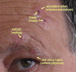
Herpes zoster ophthalmicus (HZO), also known as ophthalmic zoster, is shingles involving the eye or the surrounding area. Common signs include a rash of the forehead with swelling of the eyelid. There may also be eye pain and redness, inflammation of the conjunctiva, cornea or uvea, and sensitivity to light. Fever and tingling of the skin and allodynia near the eye may precede the rash. Complications may include visual impairment, increased pressure within the eye, chronic pain, and stroke.

Mooren's ulcer is a rare idiopathic ocular disorder that may lead to blindness due to progressive destruction of the peripheral cornea. Although the etiology of Mooren's ulcer is poorly understood, recent evidence suggests that the pathogenesis of this disease appears to be the result of an autoimmune process directed against molecules expressed in the corneal stroma.

Neurotrophic keratitis (NK) is a degenerative disease of the cornea caused by damage of the trigeminal nerve, which results in impairment of corneal sensitivity, spontaneous corneal epithelium breakdown, poor corneal healing and development of corneal ulceration, melting and perforation. This is because, in addition to the primary sensory role, the nerve also plays a role maintaining the integrity of the cornea by supplying it with trophic factors and regulating tissue metabolism.

Exposure keratopathy is medical condition affecting the cornea of eyes. It can lead to corneal ulceration and permanent loss of vision due to corneal opacity.

Peripheral Ulcerative Keratitis (PUK) is a group of destructive inflammatory diseases involving the peripheral cornea in human eyes. The symptoms of PUK include pain, redness of the eyeball, photophobia, and decreased vision accompanied by distinctive signs of crescent-shaped damage of the cornea. The causes of this disease are broad, ranging from injuries, contamination of contact lenses, to association with other systemic conditions. PUK is associated with different ocular and systemic diseases. Mooren's ulcer is a common form of PUK. The majority of PUK is mediated by local or systemic immunological processes, which can lead to inflammation and eventually tissue damage. Standard PUK diagnostic test involves reviewing the medical history and a completing physical examinations. Two major treatments are the use of medications such as corticosteroids or other immunosuppressive agents and surgical resection of the conjunctiva. The prognosis of PUK is unclear with one study providing potential complications. PUK is a rare condition with an estimated incidence of 3 per million annually.

References

  1. James, William D.; Berger, Timothy G. (2006). Andrews' Diseases of the Skin: clinical Dermatology . Saunders Elsevier. p.  370. ISBN   978-0-7216-2921-6.
  2. Carr DJ, Härle P, Gebhardt BM (May 2001). "The immune response to ocular herpes simplex virus type 1 infection". Experimental Biology and Medicine. 226 (5): 353–66. doi:10.1177/153537020122600501. PMID   11393165. S2CID   21409244.
  3. 1 2 3 4 Suresh PS, Tullo AB (September 1999). "Herpes simplex keratitis". Indian Journal of Ophthalmology. 47 (3): 155–65. PMID   10858770.
  4. 1 2 3 Myron Yanoff; Jay S. Duker (2009). Ophthalmology (3rd ed.). Mosby Elsevier. pp.  279–288. ISBN   9780323043328.
  5. Farooq AV, Shukla D (September 2012). "Herpes simplex epithelial and stromal keratitis: an epidemiologic update". Survey of Ophthalmology. 57 (5): 448–62. doi:10.1016/j.survophthal.2012.01.005. PMC   3652623 . PMID   22542912.
  6. 1 2 3 4 Basic and clinical science course (2011–2012). External disease and cornea. American Academy of Ophthalmology. pp. 105–117. ISBN   978-1615251155.
  7. Barker NH (July 2008). "Ocular herpes simplex". BMJ Clinical Evidence. 2008: 0707. PMC   2907955 . PMID   19445742.
  8. Ling Poon S, Lam Wong W, Yin Lo A, Yuan H, Chen CF, Jhanji V, Chan Y, Shih K (2021). "A systematic review on advances in diagnostics for herpes simplex keratitis". Survey of Ophthalmology. 66 (3): 514–530. doi:10.1016/j.survophthal.2020.09.008. PMID   33186564. S2CID   226946434.
  9. 1 2 3 Wilhelmus KR (January 2015). "Antiviral treatment and other therapeutic interventions for herpes simplex virus epithelial keratitis". The Cochrane Database of Systematic Reviews. 1: CD002898. doi:10.1002/14651858.CD002898.pub5. PMC   4443501 . PMID   25879115.
  10. Bhatt UK, Abdul Karim MN, Prydal JI, Maharajan SV, Fares U (November 2016). Cochrane Eyes and Vision Group (ed.). "Oral antivirals for preventing recurrent herpes simplex keratitis in people with corneal grafts". The Cochrane Database of Systematic Reviews. 2016 (11): CD007824. doi:10.1002/14651858.CD007824.pub2. PMC   6464863 . PMID   27902849.