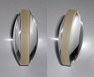
A lens is a transmissive optical device that focuses or disperses a light beam by means of refraction. A simple lens consists of a single piece of transparent material, while a compound lens consists of several simple lenses (elements), usually arranged along a common axis. Lenses are made from materials such as glass or plastic and are ground, polished, or molded to the required shape. A lens can focus light to form an image, unlike a prism, which refracts light without focusing. Devices that similarly focus or disperse waves and radiation other than visible light are also called "lenses", such as microwave lenses, electron lenses, acoustic lenses, or explosive lenses.

A corrective lens is a lens that is typically worn in front of the eye to improve daily vision. The most common use is to treat refractive errors: myopia, hypermetropia, astigmatism, and presbyopia. Glasses or "spectacles" are worn on the face a short distance in front of the eye. Contact lenses are worn directly on the surface of the eye. Intraocular lenses are surgically implanted most commonly after cataract removal but can be used for purely refractive purposes.

Near-sightedness, also known as myopia and short-sightedness, is an eye disease where light focuses in front of, instead of on, the retina. As a result, distant objects appear blurry while close objects appear normal. Other symptoms may include headaches and eye strain. Severe near-sightedness is associated with an increased risk of retinal detachment, cataracts, and glaucoma.

A dioptre or diopter is a unit of measurement with dimension of reciprocal length, equivalent to one reciprocal metre, 1 dioptre = 1 m−1. It is normally used to express the optical power of a lens or curved mirror, which is a physical quantity equal to the reciprocal of the focal length, expressed in metres. For example, a 3-dioptre lens brings parallel rays of light to focus at 1⁄3 metre. A flat window has an optical power of zero dioptres, as it does not cause light to converge or diverge. Dioptres are also sometimes used for other reciprocals of distance, particularly radii of curvature and the vergence of optical beams.
Macropsia is a neurological condition affecting human visual perception, in which objects within an affected section of the visual field appear larger than normal, causing the person to feel smaller than they actually are. Macropsia, along with its opposite condition, micropsia, can be categorized under dysmetropsia. Macropsia is related to other conditions dealing with visual perception, such as aniseikonia and Alice in Wonderland Syndrome. Macropsia has a wide range of causes, from prescription and illicit drugs, to migraines and (rarely) complex partial epilepsy, and to different retinal conditions, such as epiretinal membrane. Physiologically, retinal macropsia results from the compression of cones in the eye. It is the compression of receptor distribution that results in greater stimulation and thus a larger perceived image of an object.

Far-sightedness, also known as long-sightedness, hypermetropia, or hyperopia, is a condition of the eye where distant objects are seen clearly but near objects appear blurred. This blurred effect is due to incoming light being focused behind, instead of on, the retina wall due to insufficient accommodation by the lens. Minor hypermetropia in young patients is usually corrected by their accommodation, without any defects in vision. But, due to this accommodative effort for distant vision, people may complain of eye strain during prolonged reading. Some hypermetropes can see clear at distance, but near vision may be blurred due to insufficient accommodation. For this reason, this defect is referred as far-sightedness. If the hypermetropia is high, there will be defective vision for both distance and near. People may also experience accommodative dysfunction, binocular dysfunction, amblyopia, and strabismus. Newborns are almost invariably hypermetropic, but it gradually decreases as the newborn gets older.

Presbyopia is physiological insufficiency of accommodation associated with the aging of the eye that results in progressively worsening ability to focus clearly on close objects. Also known as age-related farsightedness, it affects many adults over the age of 40. A common sign of presbyopia is difficulty reading small print which results in having to hold reading material farther away. Other symptoms associated can be headaches and eyestrain. Different people will have different degrees of problems. Other types of refractive errors may exist at the same time as presbyopia. This condition is similar to hypermetropia or far-sightedness which starts in childhood and exhibits similar symptoms of blur in the vision for close objects.

An eyeglass prescription is an order written by an eyewear prescriber, such as an optometrist, that specifies the value of all parameters the prescriber has deemed necessary to construct and/or dispense corrective lenses appropriate for a patient. If an eye examination indicates that corrective lenses are appropriate, the prescriber generally provides the patient with an eyewear prescription at the conclusion of the exam.

Amblyopia, also called lazy eye, is a disorder of sight in which the brain fails to fully process input from one eye and over time favors the other eye. It results in decreased vision in an eye that typically appears normal in other aspects. Amblyopia is the most common cause of decreased vision in a single eye among children and younger adults.
An optical system with astigmatism is one where rays that propagate in two perpendicular planes have different foci. If an optical system with astigmatism is used to form an image of a cross, the vertical and horizontal lines will be in sharp focus at two different distances. The term comes from the Greek α- (a-) meaning "without" and στίγμα (stigma), "a mark, spot, puncture".
Anisometropia refers to a condition when two eyes have unequal refractive power. Generally, a difference in power of one diopter (1D) or more is the accepted threshold to label the condition anisometropia. Patients can tolerate 3 D of anisometropia before it becomes clinically symptomatic with headaches, asthenopia, double vision and photophobia.

Refractive error, also known as refraction error, is a problem with focusing light accurately on the retina due to the shape of the eye and or cornea. The most common types of refractive error are near-sightedness, far-sightedness, astigmatism, and presbyopia. Near-sightedness results in far away objects being blurry, far-sightedness and presbyopia result in close objects being blurry, and astigmatism causes objects to appear stretched out or blurry. Other symptoms may include double vision, headaches, and eye strain.

An eye examination is a series of tests performed to assess vision and ability to focus on and discern objects. It also includes other tests and examinations pertaining to the eyes. Eye examinations are primarily performed by an optometrist, ophthalmologist, or an orthoptist. Health care professionals often recommend that all people should have periodic and thorough eye examinations as part of routine primary care, especially since many eye diseases are asymptomatic.
Aphakia is the absence of the lens of the eye, due to surgical removal, such as in cataract surgery, a perforating wound or ulcer, or congenital anomaly. It causes a loss of ability to maintain focus (accommodation), high degree of farsightedness (hyperopia), and a deep anterior chamber. Complications include detachment of the vitreous or retina, and glaucoma.
In optics, optical power is the degree to which a lens, mirror, or other optical system converges or diverges light. It is equal to the reciprocal of the focal length of the device: P = 1/f. High optical power corresponds to short focal length. The SI unit for optical power is the inverse metre (m−1), which is commonly called the dioptre.

In optics, defocus is the aberration in which an image is simply out of focus. This aberration is familiar to anyone who has used a camera, videocamera, microscope, telescope, or binoculars. Optically, defocus refers to a translation of the focus along the optical axis away from the detection surface. In general, defocus reduces the sharpness and contrast of the image. What should be sharp, high-contrast edges in a scene become gradual transitions. Fine detail in the scene is blurred or even becomes invisible. Nearly all image-forming optical devices incorporate some form of focus adjustment to minimize defocus and maximize image quality.

Astigmatism is a type of refractive error due to rotational asymmetry in the eye's refractive power. This results in distorted or blurred vision at any distance. Other symptoms can include eyestrain, headaches, and trouble driving at night. Astigmatism often occurs at birth and can change or develop later in life. If it occurs in early life and is left untreated, it may result in amblyopia.

Emmetropia is the state of vision in which a faraway object at infinity is in sharp focus with the ciliary muscle in a relaxed state. That condition of the normal eye is achieved when the refractive power of the cornea and eye lens and the axial length of the eye balance out, which focuses rays exactly on the retina, resulting in perfectly sharp distance vision. A human eye in a state of emmetropia requires no corrective lenses for distance; the vision scores well on a visual acuity test.
The eye, like any other optical system, suffers from a number of specific optical aberrations. The optical quality of the eye is limited by optical aberrations, diffraction and scatter. Correction of spherocylindrical refractive errors has been possible for nearly two centuries following Airy's development of methods to measure and correct ocular astigmatism. It has only recently become possible to measure the aberrations of the eye and with the advent of refractive surgery it might be possible to correct certain types of irregular astigmatism.
The aim of an accurate intraocular lens power calculation is to provide an intraocular lens (IOL) that fits the specific needs and desires of the individual patient. The development of better instrumentation for measuring the eye's axial length (AL) and the use of more precise mathematical formulas to perform the appropriate calculations have significantly improved the accuracy with which the surgeon determines the IOL power.